Our
TRPV (Vanilloid); TRPM; TRPA and TRPCs have proven excellent for studying
Transient receptor potential ankyrin 1 (TRPA1) is activated by noxious cold (<17°C) and contributes to cold and mechanical hypersensitivity after inflammation and nerve injury:
Yun Sook Kim, PhD, Hoon Kap Jung, DDS, Tae Kyung Kwon, DDS, Chin Soo Kim, DDS, PhD, Jin Hyun Cho, DDS, PhD, Dong Kuk Ahn, DDS, PhD, Yong Chul Bae, DDS, PhD.Expression of Transient Receptor Potential Ankyrin in Human Dental Pulp. Journal of Endodontics. Available online 8 June 2012. doi.org/10.1016/j.joen.2012.04.024.
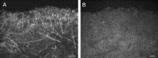
Highlights: TRPA1 was expressed in a large number of axons branching extensively in the peripheral pulp and in a few axons within the nerve bundles in the core of the coronal pulp and in the radicular pulp. Under electron microscopy, TRPA1 immunoreactivity was typically localized near the plasma membrane of unmyelinated axons in the peripheral pulp, suggesting that in these axons it may act as a functional receptor. The proportion of axons expressing TRPA1 in neurofilament 200–positive axons significantly increased in the painful pulp compared with the normal pulp. TRPA1 was also densely expressed in the processes and the cell body of odontoblasts. A large number of axons coexpressed TRPA1 and Nav1.8.
Images: Immunofluorescent staining for (A) TRPA1 in the human normal dental pulp is completely abolished by (B) preadsorption with a control peptide, proving the specificity of the TRPA1 antiserum (×200, scale bars = 50 µm). doi.org/10.1016/j.joen.2012.04.024.
Related Reagents:
All TRP Antibodies
Pain and Inflammation Research Antibodies
Neurotransmission -Neurotransmission Research Antibody Categories
Installment #1: Mesoderm Derived Stem Cells
We would like to thank our partners and customers for their input on how to best add value to our Stem Cell Research Reagents. Based on this input, we are in the process of building a new website focused on providing stem cell technical content. Better content should help catalyze better research and drug discovery related decisions.
Here're several stem cell differentiation flow charts that map the genesis of pluripotent and multipotent cells and identify the cell types that result from their differentiation.
This chart was generated using input and publications generously provided by Dr. Henry Young, Tenured Full Professor at Mercer University School of Medicine.
I anticipate the launch date for our Stem Cell Related website to occur in early Q3 2012.
In the meantime, we will posting relevant data, publications and other information here. This will include protocols and related growth factors for generating specific cell types like dopaminergic neurons from neural progenitors. Stay tuned.
Implications for treating Stress Related Depression.
Stress related disorders like Post Traumatic Stress Syndrome (PTSD), Major Depressive Disorder (MDD) and Generalized Anxiety Disorders (GAD) dysregulate neurogenesis. This disregulation can lead to disorders like chronic depression. Indeed, stress hurts.
In this study (which includes use of our Neural Progenitor Marker- Nestin), researchers show for the first time that the alpha2delta (α2δ) ligands gabapentin [1- (aminomethyl)cyclohexaneacetic acid; GBP] and pregabalin [S-[+]-3-isobutylGABA or (S)-3- (aminomethyl)-5-methylhexanoic acid; PGB] can produce a concentration-dependent increase in the number of newborn mature and immature neurons generated in vitro from adult hippocampal neural progenitor cells (NPC), and, in parallel, a decrease in the number of undifferentiated precursor cells. These effects were confirmed in vivo, since a significantly increased number of adult generated neurons was observed in the hippocampal region of mice chronically treated with PGB [10 mg/kg, i.p., 21 days] compared to vehicle-treated mice. Moreover, we demonstrated that PGB administration prevented the appearance of depression-like behaviours induced by chronic restraint stress and, in parallel, promoted hippocampal neurogenesis in adult stressed mice. Finally, we provided data suggesting the potential involvement of the α2δ1 subunit and NF-κB signaling pathway in the drug-mediated proneurogenic effects. The new pharmacological activities of α2δ ligands may help explaining their therapeutic activity as add-on therapy in major depression and on depressive symptoms in posttraumatic stress disorder and generalized anxiety disorders. Furthermore these data contribute to the identification of novel molecular pathways which may represent potential targets for pharmacological modulation in depression: Maria Maddalena Valente, Valeria Bortolotto, Bruna Cuccurazzu, Federica Ubezio, Vasco Meneghini, Maria Teresa Francese, Pier Luigi Canonico, Mariagrazia Grilli.Alpha2delta ligands act as positive modulators of adult hippocampal neurogenesis andprevent depressive-like behavior induced by chronic restraint stress. Molecular Pharmacology Fast Forward Published on May 9, 2012 as doi:10.1124/mol.112.077636.

Images: Effect of α2δ ligands on neuronal differentiation and proliferation of hippocampus-derived neural progenitor cells. (A) Representative fluorescence microscopy image of a hippocampal neurosphere immunolabelled for nestin (green) and SRY-related HMG-box gene 2 (Sox-2) (red), markers of undifferentiated NPC. Magnification = X600. Scale bar = 56 μm. (B) After 24 h in absence of growth factors, hippocampal Neural Progenitor Cells (NPC) differentiated giving rise to four different cell populations identified by double Microtubule Associated Protein-2 (MAP-2) and nestin immunolabelling: MAP-2+/nestin- mature neurons, MAP-2+/nestin+, MAP-2-/nestin+ and MAP-2-/nestin- cells. Data are expressed as mean ± S.D. of n=9 experiments, run in triplicates. Gabapentin (GBP) and pregabalin (PGB) promote neuronal differentiation of adult hippocampal NPC. GBP (C-F) and PGB (G-J) significantly increased, in a concentrationdependent manner, the percentage of MAP-2+/nestin- (C, G) and MAP-2+/nestin+ (D, H) cells and decreased the percentage of MAP-2-/nestin- cells (F, J), with no effect on MAP-2-/nestin+ cells (E, I). Data are expressed as mean ± S.D. of n = 3 experiments, run in triplicates. *, p < 0.05; **, p < 0.01; ***, p < 0.001 vs vehicle (Student’s t-test). (K-M) Representative fluorescence microscopy images of MAP-2 immunolabelling (green) in cells derived from hippocampal NPC after 24 h treatment with vehicle (K), 1 nM GBP (L) and 1 nM PGB (M). Nuclei are stained with Draq5(blue). Magnification = X400. Scale bar = 75 μm. (N) Adult hippocampal NPC were treated with vehicle or 1 nM PGB for 6, 24, 48, 72, 96 h and proliferation rate was assessed. PGB had no effect on NPC proliferation, when compared to vehicle. Data, expressed as counts per second (CPS), represent the mean ± S.D. of experiments run in triplicates.
This study is good news. Results demonstrate the new pharmacological activity of α2δ ligands may
potentially explain their efficacy as add-on therapy in MDD, as well as on depressive symptoms
in PTSD and GAD. This knowledge will help the discovery of refined therapies for these debilating disorders.
Brainstem facilitations and descending serotonergic controls contribute to visceral nociception but not pregabalin analgesia in rats.
Pregabalin is used to treat Neuropathic Pain. Here the authors show state-dependent pregabalin analgesia in neuropathy does not apply to visceral pain. Our rabbit polyclonal Mu Opioid Receptor Antibody is used to demonstrate intra-RVM Derm-SAP locally ablates a substantial proportion of MOR and serotonergic cells: Shafaq Sikandar, Kirsty Bannister, Anthony H. Dickenson. Brainstem facilitations and descending serotonergic controls contribute to visceral nociception but not pregabalin analgesia in rats. Neuroscience Letters. Volume 519, Issue 1, 21 June 2012, Pages 31–36.... For MOR staining, a rabbit polycolonal μ-opioid receptor primary antibody (1:10,000 TTBS; Neuromics, MN, USA, RA10104)...
These findings are important for understanding the limitations of Preglabin as an analgesic for viceral pain. Check out all our Opioid Publications.
Researchers frequently reference use of Neuromics' Stem Cell Markers in publications. This is an important affirmation for us as these tools are critical for determining the differentiation state of Stem Cells. In this publication, the authors use our Mouse Monoclonal Nestin Antibody to understand the mechanisms underlying neural progenitor differentiation and neuronal fate. This understanding is an important precursor for using these cells in Regenerative Medicine: S erafí Cambray, Charles Arber, Graham Little, Antonios G. Dougalis, Vincenzo de Paola, Mark A. Ungless, Meng Li and Tristan A. Rodríguez. Activin induces cortical interneuron identity and differentiation in embryonic stem cell-derived telencephalic neural precursors. Nature Communications 3, Article number: 841 doi:10.1038/ncomms1817. Received 10 January 2011 Accepted 29 March 2012 Published 15 May 2012.
In this study, the authors show that Activin provides telencephalic neural precursors with positional cues that specifically promote the acquisition of a calretinin interneuron fate by controlling the expression of genes that regulate cortical interneuron identity. This work demonstrates a novel means for regulating neuronal differentiation and specification of subtype identity.

Images: (a)immunostaining (left panels) and quantifications (right panel) indicating that Shh promotes and cyclopamine inhibits proliferation in neural precursors (Nestin+/β-III-tubulin+ cells in cyclopamine 49±4.3/35.1±1.8%, Shh 80.3±3.2/14.8±0.7% and control cultures 68.4±7.2/20.1±2.8%; n=3, mean±s.e.m.). (b) Relative expression levels of Gli1 and Ptch1 during the first 5 days of Activin or control treatment. (c) Immunoblot analysis of Gli1 and Cyclin D1 levels during the first 4 days of Activin or control treatment illustrating how Activin represses the expression of these proteins. (d) Normalized mRNA levels of Gli1 and Ptch1 after 24 h exposure to the indicated conditions illustrating how Shh induces the expression of these genes and Activin inhibits their expression (n=3, mean±s.e.m. Student's t-test. *P<0.005 and **P<0.05). ESCs were differentiated for 5 days as a monolayer, then replated into poly-D-lysine/laminin-coated dishes and cultured in NBB27 media (controls), NBB27+10 ng ml−1 Activin, NBB27+100 ng ml−1 Shh, NBB27+10 μm cyclopamine or NBB27 + 10 ng ml−1 Activin + 100 ng ml−1 Shh. Scale bar=50 μm.

Image:
Image: Model for how Activin induces the differentiation and CGE fate in telencephalic neuronal precursors.
The protocol described in this manuscript represents a method to obtain an enriched source of calretinin interneurons from both mouse and human ESCs. Therefore, our work significantly contributes to the aim of generating the diverse neuronal subtypes required for the safe and successful use of ESCs in regenerative medicine.
The capabilities of stem cells markers matter in developing protocols for use in Regenerative Medicine.
Chemotherapy can induce painful peripheral neuropathy and also have a toxic effect on peripheral nerves. The authors here show that that cisplatin produces hyperalgesia and toxicity to sensory neurons as indicated by neurochemical, morphological, and functional measures. Increasing AEA signaling at CB1 receptors not only reduced the hyperalgesia but reduced the neurotoxicity of cisplatin as well: Iryna A. Khasabova,Sergey Khasabov, Justin Paz, Catherine Harding-Rose, Donald A. Simone, and Virginia S. Seybold. Cannabinoid Type-1 Receptor Reduces Pain and Neurotoxicity Produced by Chemotherapy. The Journal of Neuroscience, 16 May 2012, 32(20): 7091-7101; doi: 10.1523/JNEUROSCI.0403-12.2012.
The authors use our guinea pig TRPV1 antibody to measure cisplatin hyperalgesia vs treated and control mice.

Images: URB597 attenuated effects of cisplatin on protein expression in DRGs. A, TRPV1- and ATF-3-ir were detected by immunofluorescence in L3–L5 DRGs from mice treated with vehicle, cisplatin, or cisplatin plus URB597. Cisplatin (1 mg/kg of body weight, daily for 7 d, i.p.) increased the occurrence of TRPV1- and ATF3-ir in neurons. Co-injection of URB597 (0.3 mg/kg daily, i.p.) with cisplatin attenuated the effect cisplatin on protein-ir. Scale bars: 10 μm (for images within each antigen). B, Quantitative summary of the effect of treatments on TRPV1-ir in neurons. Data are expressed as the mean ± SEM. aSignificantly different from each other group (p < 0.05, one-way ANOVA with Student–Newman–Keuls test; n = 4 mice/treatment). C, Quantitative summary of the effect of treatments on ATF-ir in neurons. Data are expressed as the median and 25th and 75th percentile range. *Significantly different from vehicle control and cisplatin plus URB597 groups (n = 6 mice/treatment; p < 0.001, Kruskal–Wallis ANOVA on ranks test).
Related Reagents:
VR1 N-Terminus (TRPV1)
VR1 (TRPV1)-Goat
VR1 C-Terminus (TRPV1) - mouse specific
All TRP Antibodies
Pain and Inflammation Research Antibodies
Neurotransmission -Neurotransmission Research Antibody Categories
I am pleased to feature our Guinea Pig P2X3 Antibody as a tool for studying the root causes of Neuropathic Pain.
In this study, the authors showed ablation of nonpeptidergic fibers in a chronic constriction injury model caused significant sympathetic and parasympathetic P2X3 fiber sprouting, and led to an exacerbated pain response. This was an unexpected finding, as it has been suggested that nonpeptidergic fibers play a major role in mechanical pain, and suggests that these fibers play a complex role in the development of neuropathic pain: Anna M.W. Taylora, Maria Osikowicza, Alfredo Ribeiro-da-Silva. Consequences of the ablation of nonpeptidergic afferents in an animal model of trigeminal neuropathic pain. PAIN. Volume 153, Issue 6, June 2012, Pages 1311–1319. doi.org/10.1016/j.pain.2012.03.023....Sections were then incubated for 48 hours at 4°C with a guinea pig polyclonal anti-P2X 3 antibody (1:25,000; Neuromics, Edina, MN, USA), diluted in PBS-T. Following primary antibody incubation, sections were treated with a biotin-conjugated...
Image: Example of Non-petidergic fibers expressing P2X3 receptor for ATP are present in lamina II of the contralateral dorsal horn (arrows). The P2X3-positive fibers are eliminated from the rat dorsal horn ipsilateral to the rhizotomy (arrow heads). Spinal cord segment C6. Scale bar 400 um. P2X3 antibody dilution 1:25,000. Biomédica vol.24 no.2.
Check out our related reagents categories:
I will continue to post important findings related to the root causes of Neuropathic Pain.
Our friends, Drs Mantyh and Jimenez Andrade, have published important findings on the potential root cuases of age related Arthritic Pain: Juan M Jimenez-Andrade and Patrick W Mantyh. Sensory and sympathetic nerve fibers undergo sprouting and neuroma formation in the painful arthritic joint of geriatric mice. Note: our Chicken Polyclonal NF200 Antibody was used to label primary afferent sensory nerve fibers.
Overview: Introduction: Although the prevalence of arthritis dramatically increases with age,
the great majority of preclinical studies concerning the mechanisms that drive
arthritic joint pain have been performed in young animals. One mechanism
hypothesized to contribute to arthritic pain is ectopic nerve sprouting; however,
neuroplasticity is generally thought to be greater in young versus old nerves. Here
we explore whether sensory and sympathetic nerve fibers can undergo a
significant ectopic nerve remodeling in the painful arthritic knee joint of geriatric
mice.
In other words, there is a clear root reason that the pain arthritis sufferers experience increases overtime. Here's some related data. Notice the increase in labeled nerves:

Images. Sensory nerve fiber sprouting and formation of neuroma-like
structures in the painful geriatric arthritic knee joint. Schematic of a frontal
view of a cross-sectioned mouse knee joint (A). The red square illustrates the
synovial region from which the confocal images were obtained. Representative
confocal images of calcitonin gene-related peptide (CGRP+), neurofilament 200
kDa (NF200+) sensory nerve fibers (yellow/orange) and growth associated protein
(GAP43; marker of fibers undergo regeneration, yellow/orange) and DAPI labeled
nuclei (blue) in knee joint sections (20 μm thick) of vehicle-injected (B,D,F) and
CFA-injected (C,E,G ) mice. In vehicle-injected mice, a low level, regular pattern
of innervation by CGRP+ and NF200+ fibers is observed in the synovial space of the knee joint. Twenty-eight days following the initial CFA injection, a significant
number of CGRP+ and NF200+ nerve fibers have sprouted and have a
disorganized appearance as compared to vehicle-injected mice. Note that,
CGRP+ and NF200+ sprouted nerve fibers are localized in the synovium
and are not observed in the meniscus of the joint. Furthermore, there was
formation of neuroma-like structures in the synovium of the geriatric mice injected
with CFA (G).
Related Reagents: Neuronal-Glial
Markers-Astrocytes, Glia, Microglia, Olidogodendrocytes, Progenitors
and Schwann Cell Markers.
I advertise our Primary Neurons and Astrocytes as being easy to culture, grow and maintain. We confirm this via the data/images our customers generously share and the many publications referencing use of the cells.
I would like to thank George Kenneth Todd (Patterson Lab at UC Davis) for these wonderful images of our e18 Primary Rat Hippocampal Neurons. These were taken at day 67!
Prior to staining, the cells were treated with Wnt5 for 2 min, then Wnt5 + Wnt3 for an additional 2 min during Calcium Imaging experiments. The cells were then fixed and stained for IP3R (green), Frizzled2 (blue), and B-Catenin (red), and the confocal images were captured at 10x. Notice the parallel neurites formation in image 3.
For staining culture options, check out our neuron-glial-astrocyte markers.
I consider publications important for confirming our Neuronal-Glial
Markers are working as advertised. These tools often used by our customers for lineages of differentiating Neural Progenitors. This publication references use of our Mouse Monoclonal GFAP and Rabbit Polyclonal GFAP Antibodies: Frances Y. Cheng, Xi Huang, Anuraag Sarangi, Tatiana Ketova, Michael K. Cooper, Ying Litingtung, Chin Chiang. Widespread Contribution of Gdf7 Lineage to Cerebellar Cell Types and Implications for Hedgehog-Driven Medulloblastoma Formation. PLoS ONE 7(4): e35541. doi:10.1371/journal.pone.0035541.
Abstract:The roof plate is a specialized embryonic midline tissue of the central nervous system that functions as a signaling center regulating dorsal neural patterning. In the developing hindbrain, roof plate cells express Gdf7 and previous genetic fate mapping studies showed that these cells contribute mostly to non-neural choroid plexus epithelium. We demonstrate here that constitutive activation of the Sonic hedgehog signaling pathway in the Gdf7 lineage invariably leads to medulloblastoma. Lineage tracing analysis reveals that Gdf7-lineage cells not only are a source of choroid plexus epithelial cells, but are also present in the cerebellar rhombic lip and contribute to a subset of cerebellar granule neuron precursors, the presumed cell-of-origin for Sonic hedgehog-driven medulloblastoma. We further show that Gdf7-lineage cells also contribute to multiple neuronal and glial cell types in the cerebellum, including glutamatergic granule neurons, unipolar brush cells, Purkinje neurons, GABAergic interneurons, Bergmann glial cells, and white matter astrocytes. These findings establish hindbrain roof plate as a novel source of diverse neural cell types in the cerebellum that is also susceptible to oncogenic transformation by deregulated Sonic hedgehog signaling.

Images: A subset of Gdf7-lineage cells express neural stem cell markers.(A–B″′) Many cells within the tumor tissue of Gdf7Cre/+;SmoM2 mice coexpress neural stem cell marker Nestin (red) and glial marker GFAP (green). Arrows indicate co-localization.
I will continue to post publication and customer data showing results with our Neuron-Glial Markers.
Our Neuronal-Glial Markers are used in a variety of applications. Here our P0 or P-Zero antibody is used to show myelination levels in the cochlear ganglion. Demyelination is caused by hyperglycemia and type 2 diabetes and results in hearing loss and eventually deafness. Here's the related publication and highlights: Silvia Murillo-Cuesta, Guadalupe Camarero, Águeda González-Rodríguez, Lourdes Rodríguez-de la Rosa, Deborah J Burks, Carlos Avendaño,Ángela M Valverde and Isabel Varela-Nieto1. Insulin Receptor Substrate 2 (IRS2)-Deficient Mice Show Sensorineural Hearing Loss That Is Delayed by Concomitant Protein Tyrosine Phosphatase 1B (PTP1B) Loss of Function. Online address: http://www.molmed.org. doi: 10.2119/molmed.2011.00328
Highlights: The authors objective was to study the hearing function and cochlear morphology of Irs2-null mice and the impact of PTP1B deficiency. They have studied the auditory brainstem responses and the cochlear morphology of systemic Irs2–/–Ptpn1+/+, Irs2+/+Ptpn1–/– and Irs2–/–Ptpn1–/– mice at different postnatal ages. The results indicated that Irs2–/–Ptpn1+/+ mice present a profound congenital sensorineural deafness before the onset of diabetes and altered cochlear morphology with hypoinnervation of the cochlear ganglion and aberrant stria vascularis, compared with wild-type mice. Simultaneous PTP1B deficiency in Irs2–/–Ptpn1–/– mice delays the onset of deafness.

Images: Cochlear ganglion and nerve fibers. (A–C) Cresyl violet staining of midmodiolar methacrylate sections of the cochlear ganglion at the cochlear basal turn in Irs2+/+Ptpn1+/+ (A), Irs2–/–Ptpn1+/+ (B) and Irs2–/–Ptpn1–/–mice (C) at postnatal wk 11. A slight reduction in the cellular density was more evident in both mutants (B, C) compared with wild-type (A). (D–F) Myelin P0 immunostaining of the cochlear ganglion shows less intense labeling in Irs2–/–Ptpn1+/+ (E) and Irs2–/–Ptpn1–/– (F) mice than in control mice (D). (G–I) Accordingly, myelin P0 immunostaining of nerve fibers projecting from the cochlear ganglion to the sensory cells is less intense in Irs2–/–Ptpn1+/+ and Irs2–/–Ptpn1–/– (arrows in H and I) than in wild-type mice (G). (J–L) Similarly, a weaker neurofilament 200-kDa immunostaining is observed in Irs2+/+Ptpn1–/– (K, white arrow) and Irs2–/–Ptpn1–/– (L, white arrow) mice compared with wild-type mice (J). Scale bars: A–I, 50 μm; J–L, 75 μm.
The results presented in this study demonstrate for the first time a unique tissue-specific role of IRS2 in cochlear development and hearing function; therefore, the Irs2–/– mouse could be a novel model for the in vivo study of hearing loss associated with altered glucose metabolism. The data also suggest that modulation of PTP1B activity could be a pharmacological target of interest for the sensory syndromes associated with diabetes.
Researchers require a dependable and cost-effective source for Mesenchymal Stem Cells (hMSCs). These cells can be differentiated into cartilage, bone, fat, muscle and even neural cells using our MSCGro™ Media for growth and differentiation. Better cells and media means satisfaction with your Cell Based Assays
This makes them an ideal solution for research studies that include developmental biology, regenerative medicine, cell therapy, and tissue engineering.
These cells have the capabilities of passaging a minimum of 10-fold.
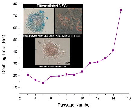 Images: MSCs (Catalog #: SC00A1) Growth in MSCGroTM, Low Serum Medium (Catalog#: SC00B1). Inset: MSCs differentiatiated using MSCGro Differentiation Media.
I will continue to update you on customer data and pubs using these cells and media. I wish you exciting and rewarding discoveries.
Implications for De-Myelinating Diseases Like MS and ALS
Dr. Juana María Pasquini and her team at the University of Buenos Aires are ongoing users of our Neural Stem Cell-Progenitor (NSC-NP) Markers. In this study, they use these markers to determine the states and fates of NSCs and NPs as they proliferate and differentiate and the related role of ApoTransferrin (aTF). Here we learn aTf exposure during differentiating conditions favours OL maturation from OPCs by promoting OL morphological development. This evidence supports a key role of Tf on the generation of OL from NSC/NPCs and highlights its potential in demyelinating disorder treatment: Silvestroff L , Franco PG , Pasquini JM (2012) ApoTransferrin: Dual Role on Adult Subventricular Zone-Derived Neurospheres. PLoS ONE 7(3): e33937. doi:10.1371/journal.pone.0033937.

Proliferation rates under different conditions are shown in A–C. BrdU incorporation (red) during proliferation (CTLP, A) or differentiation (CTLPCTLD, B). BrdU+ cells are expressed as a percentage of total nuclei for either condition in C. Free floating NS during proliferation express Nestin (D, green) and GFAP (E, green). After dissociation, NS-derived cells continue to express Nestin (F, green). PDGFRα+ (G, green) and NG2+ cells (H, green). Few MBP+ (I, green) cells were found under proliferative conditions. A large proportion of BrdU incorporating cells (J, red) co-expressed with NG2 (J, green). Some BLBP+ cells (K, green) incorporated BrdU (K, red). After differentiation (L–O), MBP+ cells were found with a highly branched and complex morphology (L, green). Cells expressed GFAP (M, green), as well as the neuronal NF200 marker (N, green). BrdU incorporating (O, red) cells were mostly NG2+ (O, green) during differentiation conditions. BrdU+ cells co-expressing NG2, as a proportion of total BrdU+ cells, are shown in P for either culture condition. A representative Western Blot membrane in Q shows how MBP levels increase in whole cell protein extracts as cells differentiate. The densitometric analysis of the MBP isoforms/GAPDH ratio of 5 independent experiments was semi-quantitated in R. All 4 MBP isoforms were pooled and considered as a single value before normalizing to GAPDH values. Blue colour in images indicates Höechst nuclear dye. Scale bar in A represents 250 µm for A and B. Scale bar in D equals 100 µm in D–I and L–N, scale bar in J equals 250 µm in J and O, and scale bar in K represents 50 µm. Bars in P represent mean values of 2 independent experiments. Bars in C and R represent Mean + SD of 4 and 5 individual cultures, respectively. Student's t Test was used to analyze data in C, while a One Way ANOVA with an SNK Post-test was used to analyze data in R. * p<0.05, ** p<0.01, *** p<0.001
Note: PDGFRα+ is a marker for oligodendrocytes (OLs).
Here's the pathway model that sumarizes authors' findings
I will keep you posted on research that could implications for the discovery of de-myelinating disorder therapies.
We want to help our customers, collaborators and friends bring their cell based assays to life! Neuromics began offering solutions in early 2011. We have extended our reach with our 3D Nanofibers. These 3-D environments are more biologically realistic leading to:
•more effective biomedical research.
•earlier breakthroughs.
•faster and cheaper time to market for drug development.
•improved stem cell expansion rates.
Here's a video showing green neurosphere (brain cancer cells) migrating along our nanofibers aligned in the vertical direction. This enables cancer researchers to see cancer in ways never before possible!
Find more videos like this on TechLounge
I will update you as data and related publications become avaialable using these solutions.
I am pleased to update you in recent publications and videos referencing use of our reagents for pain research. Check them out.
Andrew C. Eschenroeder, Allison A. Vestal-Laborde, Emilse S. Sanchez, Susan E. Robinson, Carmen Sato-Bigbee. Oligodendrocyte responses to buprenorphine uncover novel and opposing roles of μ-opioid- and nociceptin/orphanin FQ receptors in cell development: Implications for drug addiction treatment during pregnancy. Glia Volume 60, Issue 1, pages 125–136, January 2012....The MOR and NOP receptor antibodies were from Neuromics (Edina, MN) and used for immunocytochemistry (1:100)and western blotting (1:500)...
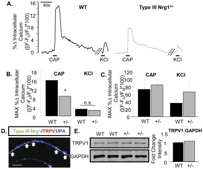 FiFigure. Sensory axons, but not soma, from Type III Nrg1+/− mice show reduced capsaicin responsiveness compared to axons from WT mice. (A) Representative traces of intracellular calcium along sensory axons in response to 1 µM capsaicin or 56 mM KCl. The change in intracellular calcium from baseline over time ([(F−F0)/F0]*100) is shown for WT (left) and Type III Nrg1+/− (right) axons. Hatched diagonal lines indicate where the time course was non-continuous. (B) Quantification of the maximum change in intracellular calcium in response to application of 1 µM capsaicin or 56 mM KCl by genotype. Averages of 5 animals per genotype were compared using a Student's t-test. Type III Nrg1+/− axons showed a significantly decreased response to capsaicin (p<0.05), but not to KCl, relative to WTs. Graph shows mean±SEM. (C) Type III Nrg1+/− sensory soma show normal response to capsaicin. Quantification of maximal change in fluorescence from baseline ([(F−F0)/F0]*100) in WT or Type III Nrg1+/− sensory neuron soma in response to 1 µM capsaicin or 56 mM KCl. Average responses from 4 WT and 4 Type III Nrg1+/− animals to application of capsaicin or KCl were compared by genotype using a Student's t-test. There was no statistically significant difference between genotypes. Graphs show mean±SEM. (D) Type III Nrg1 (green) and TRPV1 (red) are co-expressed along P21 WT cultured sensory neuron axons identified with a pan-axonal (PA) marker (blue). White arrows indicate examples where Type III Nrg1 and TRPV1 are in close proximity. Scale bar equals 10 µm. (E) P21 WT and Type III Nrg1+/− sensory neuron cultures have equivalent levels of total TRPV1 protein. Total TRPV1 protein measurement by immunoblot. The 95 kD TRPV1 band and the 35 kD GAPDH band are shown from a representative experiment comparing protein from P21 WT and Type III Nrg1+/− cultures. Quantification of fold change in intensity of TRPV1:GAPDH normalized to WT average. There was no statistically significant change in the ratio of TRPV1 to GAPDH between genotypes (WT, Type III Nrg1+/−, n = 3 animals). Genotype comparisons were made using a Student's t-test. Graph shows mean±SEM. doi:10.1371/journal.pone.0025108.g004
Our Leptin and Leptin Receptor Markers have proven effective for researchers studying root causes of obesity. In this reference the authors show an interesting twist regarding the impact of endothial vs neuronal leptin signaling: Weihong Pan, Hung Hsuchou, Germaine G. Cornelissen-Guillaume, Bhavvani Jayaram, Yuping Wang, Hong Tu, Franz Halberg, Xiaojun Wu, Streamson C. Chua Jr., and Abba J. Kastin. Endothelial leptin receptor mutation provides partial resistance to diet-induced obesity. Published online before print February 2012, doi:
10.1152/japplphysiol.00590.2011.
...chicken anti-ObRb (1:100, Neuromics)...
Highlights: Leptin, a polypeptide hormone produced mainly by adipocytes, has diverse effects in both the brain and peripheral organs, including suppression of feeding. Other than mediating leptin transport across the blood-brain barrier, the role of the endothelial leptin receptor remains unclear. We recently generated a mutant mouse strain lacking endothelial leptin receptor signaling, and showed that there is an increased uptake of leptin by brain parenchyma after its delivery by in-situ brain perfusion. Here, we tested the hypothesis that endothelial leptin receptor mutation confers partial resistance to diet-induced obesity. These ELKO mice had similar body weight and percent fat as their wildtype littermates when fed with rodent chow, but blood concentrations of leptin were significantly elevated. In response to a high fat diet, wildtype mice had a greater gain of body weight and fat than ELKO mice . As shown by metabolic chamber measurement, the ELKO mice had higher oxygen consumption, carbon dioxide production, and heat dissipation, although food intake was similar to that of the wildtype mice and locomotor activity was even reduced. This indicates that the partial resistance to diet-induced obesity was mediated by higher metabolic activity in the ELKO mice. Since neuronal leptin receptor knockout mice show obesity and diabetes, the results suggest that endothelial leptin signaling shows opposite effects from that of neuronal leptin signaling, with a facilitatory role in diet-induced obesity.
Obesity is a growing health problem and will continue to drive up costs. Research like this could help find effective and log term solutions for helping with weight loss.
Our P2X3 Receptor Antibodies are widely used and frequently published. This publication references use of our P2X3 Guinea Pig Antibody.
I like the "cool factor" in this study: Ji Z-G , Ito S , Honjoh T , Ohta H , Ishizuka T , et al. 2012 Light-evoked Somatosensory Perception of Transgenic Rats That Express Channelrhodopsin-2 in Dorsal Root Ganglion Cells. PLoS ONE 7(3): e32699. doi:10.1371/journal.pone.0032699.-"We have recently generated several transgenic lines of rat in which channelrhodopsin-2 (ChR2) transgene is driven by the Thy-1.2 promoter. In one of them, W-TChR2V4, some neurons were endowed with photosensitivity by the introduction of the ChR2 gene, coding an algal photoreceptor molecule. The DRG neurons expressing ChR2 were immunohistochemically identified using specific antibodies to the markers of mechanoreceptive or nociceptive neurons. Their peripheral nerve endings in the plantar skin as well as the central endings in the spinal cord were also examined. We identified that ChR2 is expressed in a certain population of large neurons in the DRG of W-TChR2V4. On the basis of their morphology and molecular markers, these neurons were classified as mechanoreceptive but not nociceptive. ChR2 was also distributed in their peripheral sensory nerve endings, some of which were closely associated with CK20-positive cells to form Merkel cell-neurite complexes or with S-100-positive cells to form structures like Meissner's corpuscles. These nerve endings are thus suggested to be involved in the sensing of touch. Each W-TChR2V4 rat showed a sensory-evoked behavior in response to blue LED flashes on the plantar skin. It is thus suggested that each rat acquired an unusual sensory modality of sensing blue light through the skin as touch-pressure. This light-evoked somatosensory perception should facilitate study of how the complex tactile sense emerges in the brain."
The researchers used Blue LED light to fire neurons involved somatosensory or tactile response!
Images. Distribution of ChR2V in the dorsal part of the spinal cord.
A–C. Immunohistochemical localizationof ChR2V with the cell-type specific markers, NF200 (A), CGRP (B) or P2X3 (C). Scale bars indicate 40 µm.
Note that the receptors involved in Nociceptive Pain Sensing do not overlap with ChR2V. From this the authors conclude that ChR2V is involved in mechanoreception.
This rat model should facilitate future study of how complex tactile perception, such as for texture, size and shape, is generated. We will keep you posted.
In the meantime check out our markers and antibodies for studying Neurotransmission and Synaptic Mechanisms.
"Where there's smoke there's fire" or in this study, where there's inflammation there is pain: Robert
P. Watson, Elliot Lilley, Moh Panesar, Gurdip Bhalay, Steven Langridge,
Shin-Shay Tian, Conor McClenaghan, Anna Ropenga, Fanning Zeng, Mark S. Nash.
Increased prokineticin 2 expression in gut inflammation: role in visceral pain
and intestinal ion transport. Neurogastroenterology & Motility. Volume
24, Issue 1, pages 65–e12, January 2012.... Formalin-fixed,
wax-embedded tissues with no overt signs of pathology were purchased from
Asterand (Detroit, MI, USA); informed consent had been sought and received from
all donors. All tissues were used in accordance with the Human Tissue Act 2004
(UK)...guinea pig anti-TRPV1 antibody (GP14100 – Neuromics, Edina, MN, USA)...
Images: expression of prokineticin receptors in human and rat tissues. (A) Immunohistochemical analysis of PKR1 expression in the myenteric and submucosal ganglia of human stomach, ileum, and colon. Pkr1 (B) and Pkr2(C) expression in a range of human tissues determined using qRT-PCR. (D, E) Immunohistochemical of PKR1 distribution in rat DRG showing an absence of expression in large diameter, NF200 positive neurons (D), but co-expression with TRPV1 in presumptive nociceptive sensory neurons (E). (F) Distribution of Pkr2 mRNA by in situ hybridization in rat DRG. The IHC/ISH images shown are representative of the data obtained from n = 3 donors/animals.
Key Results Prok2 gene expression was up-regulated in biopsy samples from ulcerative colitis patients, and similar elevations were observed in rodent models of inflammatory colitis. Prokineticin receptor 1 (PKR1) was localized to the enteric neurons and extrinsic sensory neurons, whereas Pkr2 expression was restricted to sensory ganglia. In rats, PROK2-increased intracellular calcium levels in cultured enteric and dorsal root ganglia neurons, which was blocked by Compound 3. Moreover, PROK2 acting at prokineticin receptors stimulated intrinsic neuronally mediated ion transport in rat ileal mucosa. In vivo, Compound 3 reversed intracolonic mustard oil-induced referred allodynia and TNBS-induced visceral hypersensitivity, but not non-inflammatory, stress-induced visceral pain.
Check out our Pain and Inflammation Research Antibodies.
Our Guinea Pig P2X3 Antibody has historically been one of our most popular Purinergic Receptor Antibodies. It has been frequently cited in publications.
We exhausted our supply of the antibody in the spring of 2011 and it took us many rounds with multiple partners and a degree of disappointing results. Well our efforts have finally yielded the positive testing results required to again provide users this important antibody. Here're related images from our internal testing:
P2X3 staining in rat Dorsal Root Ganglia.
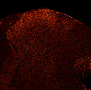
P2X3 in rat Dorsal Horn.
What causes Muscular Dystrophies like Limb girdle muscular dystrophy type 2H (LGMD2H)? Here researchers demonstrate for the first time that TRIM32 is expressed in the skeletal muscle stem cell lineage of adult mice, and that in the absence of TRIM32, myogenic differentiation is disrupted. Moreover, they show that the ubiquitin ligase TRIM32 controls this process through the regulation of c-Myc, a similar mechanism to that previously observed in neural progenitors. Importantly, they show that loss of TRIM32 function induces a LGMD2H-like phenotype and strongly affects muscle regeneration in vivo. These studies implicate that the loss of TRIM32 results in dysfunctional muscle stem cells which could contribute to the development of LGMD2H: Nicklas S , Otto A , Wu X , Miller P , Stelzer S , et al. 2012 TRIM32 Regulates Skeletal Muscle Stem Cell Differentiation and Is Necessary for Normal Adult Muscle Regeneration. PLoS ONE 7(1): e30445. doi:10.1371/journal.pone.0030445.
Stem Cell Markers like PAX7 are important for types of studies. PAX7 is a marker for muscle progenitors or stem cells. If these cells fail to differentiate the level of PAX7 expression will remain high. MyoD is an indicator for successfully differentiating cells. As a consequence, MyoD expression levels increase within the cell and the ratio of Pax7:MyoD is reduced, thus inhibiting proliferation and inducing differentiation through Myogenin expression. In the absence of TRIM32, this balance of Pax7:MyoD is dysregulated, because degradation of c-Myc is not initiated, MyoD levels do not increase and Myogenin expression is not activated.

Figure . Differentiation is strongly reduced in satellite cell progeny on TRIM32−/− myofibers. (a), (c) and (e) Immunostainings of satellite cells on freshly isolated myofibers from wild type (+/+) and TRIM32−/− (−/−) mice cultured for 24 h (a), 48 h (c) and 72 h (e) and labelled with the indicated markers (upper grey boxes). (b), (d) and (f) Diagrams showing the relative frequency of Pax7+/MyoD− cells on fibers cultured for 24 h (b), Pax7+/Myogenin− cells on fibers cultured for 48 h (d) and Pax7−/Myogenin+ cells on fibers cultured for 72 h (f). In all cases cells on myofibers from wild type mice and TRIM32−/− mice are compared. (mean ± std; *P<0.001 compared to wild type).
Check out our more publications referencing use of our PAX7.
Our Neurotrophins and Growth Factor Antibodies and Recombinant Proteins help support a wide span of research areas. These areas include: neuroscience, immunology, cardiac disease research and cancer.
I would like to update you on recent publications highlighting use of some of these reagents: Aiko Sada, Kazuteru Hasegawa, Pui Han Pin, Yumiko Saga. NANOS2 Acts Downstream of Glial Cell Line-Derived Neurotrophic Factor Signaling to Suppress Differentiation of Spermatogonial Stem Cells. DOI: 10.1002/stem.790. Copyright © 2011 AlphaMed Press... anti-GFRA1 (1:200, Neuromics, Edina, MN)...
Images: Ngn3-Cre targets GFRA1-negative cells. (A-F): At P7, Rosa-YFP; Ngn3-Cre double transgenic testes were immunostained with the indicated markers. Most of YFP-positive spermatogonia (Ngn3-lineage cells) were not stained with GFRA1. Scale bar, 100 μm. (G): Quantification of YFP-positive cells per GFRA1-positive cells at P7 and 6 weeks (N=3).
Karine Bédard, Stéphanie Segura, Stéphanie Mahaut, Catherine Tardivel, Guylaine Ferland, Bruno Lebrun, Pierrette Gaudreau. Effects of aging and caloric restriction on brainstem satiety center signals in rats. Mechanisms of Ageing and Development. dx.doi.org/10.1016/j.mad.2012.01.004...goat serum and 0.3% Triton-X-100) (Sigma–Aldrich), overnight at 4 °C with chicken anti-rat BDNF antibody (Neuromics, Edina, MN; diluted 1/200 in blocking buffer), rinsed 3× 10 min in fresh PBS and incubated for 2 h, in the dark...
Tali Kobilo, Qing-Rong Liu, Kriti Gandhi, Mohammed Mughal, Yavin Shaham and Henriette van Praag. Running is the neurogenic and neurotrophic stimulus in environmental enrichment. doi: 10.1101/lm.2283011. Learn. Mem. 2011. 18: 605-609... human recombinant BDNF (0.1 µg) monomer (Neuromics)...
Masamichi Shinoda, Masatake Asano, Daisuke Omagari, Kuniya Honda, Suzuro Hitomi, Ayano Katagiri, and Koichi Iwata. Nerve Growth Factor Contribution via Transient Receptor Potential Vanilloid 1 to Ectopic Orofacial Pain. The Journal of Neuroscience, 11 May 2011, 31(19):7145-7155; doi:10.1523/JNEUROSCI.0481-11.2011....expression immunohistochemically in TG- anti-p75 goat antibody (1:200; Neuromics)...
Images: Mandibular nerve fibers were labeled by β-NGF which was administrated into the lower lip with CFA (A) or saline (F). On day 1 after β-NGF administration into the lower lip with CFA or saline, β-NGF-positive whisker pad or lower lip TG neurons defined by FG or DiI, respectively. B, G, β-NGF-positive TG neurons. C, H, DiI-labeled TG neurons. D, I, FG-labeled TG neurons. E, J, DiI- and FG-labeled β-NGF-positive TG neurons. Mandibular nerve fibers (K) and TG neurons (L) on day 1 after labeled BSA administration into the lower lip with CFA. Open arrow, β-NGF-positive nerve fibers. Arrow, DiI-labeled β-NGF-positive TG neurons. Arrowhead, FG-labeled β-NGF-positive TG neurons. Scale bar, 50 μm. M, Frequency of β-NGF-positive neurons in FG- or DiI-labeled TG neurons after CFA or saline injection into the lower lip (n = 5 in CFA-injected group; n = 4 in saline-injected group; Student's t test).
F. R. Carreño, J. D. Walch, M. Dutta, T. P. Nedungadi, J. T. Cunningham. Brain-Derived Neurotrophic Factor-Tyrosine Kinase B Pathway Mediates NMDA Receptor NR2B Subunit Phosphorylation in the Supraoptic Nuclei Following Progressive Dehydration. Journal of Neuroendocrinology. Volume 23, Issue 10, pages 894–905, October 2011. DOI: 10.1111/j.1365-2826.2011.02209.x... TrkB receptor (Neuromics, Edina, MN, USA)...
Jie Shen, Yoko Ishii, Guihua Xu, Thanh Chung Dang, Takeru Hamashima, Takako Matsushima, Seiji Yamamoto, Yuichi Hattori, Yusuke Takatsuru, Junichi Nabekura and Masakiyo Sasahara. PDGFR-β as a positive regulator of tissue repair in a mouse model of focal cerebral ischemia. Journal of Cerebral Blood Flow & Metabolism , (28 September 2011). doi:10.1038/jcbfm.2011.136... goat polyclonal anti-PDGFR-β antibody (1:100; Neuromics, Edina)...
Thomas Karsten Kilvaer, Andrej Valkov, Sveinung W. Sorbye, Tom Donnem, Eivind Smeland, Roy Martin Bremnes,and Lill-Tove Busund. Platelet-Derived Growth Factors in Non-GIST Soft-Tissue Sarcomas Identify a Subgroup of Patients with Wide Resection Margins and Poor Disease-Specific Survival. Volume 2010 (2010), Article ID 751304, 10 pages doi:10.1155/2010/751304... PDGF-CC (goat polyclonal; GT15151; Neuromics; 1 : 80)...
I will keep you posted on new developments.
As we age, selective BDNF receptors increase in the the dorsal vagal complex (DVC), the brainstem center mediating the satiety reflex. Aging also affects the suppressor of cytokine signaling-3. This results in abnormal responses to BDNF and Leptin Signaling.
In this study, the investigators found that by restricting calories in rats, DVC BDNF immunoreactive concentrations were elevated and resulting in satiety threshold stability. This indicates functional desensitization of the DVC to these signals: Karine Bédard, Stéphanie Segura, Stéphanie Mahaut, Catherine Tardivel, Guylaine Ferland, Bruno Lebrun, Pierrette Gaudreau. Effects of aging and caloric restriction on brainstem satiety center signals in rats. Mechanisms of Ageing and Development. dx.doi.org/10.1016/j.mad.2012.01.004.
The authors used our BDNF Antibody to determine expression in the DVC.....goat serum and 0.3% Triton-X-100) (Sigma–Aldrich), overnight at 4 °C with chicken anti-rat BDNF antibody (Neuromics, Edina, MN; diluted 1/200 in blocking buffer), rinsed 3× 10 min in fresh PBS and incubated for 2 h, in the dark...
These findings could lead to potential therapies based on modulating BDNF expression in the DVC.
| 









 Posts
Posts

