Andrew C. Eschenroeder, Allison A. Vestal-Laborde, Emilse S. Sanchez, Susan E. Robinson, Carmen Sato-Bigbee. Oligodendrocyte responses to buprenorphine uncover novel and opposing roles of μ-opioid- and nociceptin/orphanin FQ receptors in cell development: Implications for drug addiction treatment during pregnancy. Glia Volume 60, Issue 1, pages 125–136, January 2012....The MOR and NOP receptor antibodies were from Neuromics (Edina, MN) and used for immunocytochemistry (1:100)and western blotting (1:500)...
Our P2X3 Receptor Antibodies are widely used and frequently published This publication references use of our P2X3 Guinea Pig Antibody: Ji Z-G , Ito S , Honjoh T , Ohta H , Ishizuka T , et al. 2012 Light-evoked Somatosensory Perception of Transgenic Rats That Express Channelrhodopsin-2 in Dorsal Root Ganglion Cells.
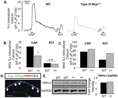
This recent publication features use out TRPV1-C guinea pig polyclonal for immunohistochemistry and TRPV1-mouse specific for Western Blotting: Sarah E. Canetta, Edlira Luca, Elyse Pertot, Lorna W. Role, David A. Talmage. Type III Nrg1 Back Signaling Enhances Functional TRPV1 along Sensory Axons Contributing to Basal and Inflammatory Thermal Pain Sensation. PLoS ONE 6(9): e25108. doi:10.1371/journal.pone.0025108...IHC: TRPV1 (guinea pig, 1:1000, GP14100 Neuromics); WB: TRPV1 (rabbit, 1:1000, RA14113 Neuromics).

FiFigure. Sensory axons, but not soma, from Type III Nrg1+/− mice show reduced capsaicin responsiveness compared to axons from WT mice. (A) Representative traces of intracellular calcium along sensory axons in response to 1 µM capsaicin or 56 mM KCl. The change in intracellular calcium from baseline over time ([(F−F0)/F0]*100) is shown for WT (left) and Type III Nrg1+/− (right) axons. Hatched diagonal lines indicate where the time course was non-continuous. (B) Quantification of the maximum change in intracellular calcium in response to application of 1 µM capsaicin or 56 mM KCl by genotype. Averages of 5 animals per genotype were compared using a Student's t-test. Type III Nrg1+/− axons showed a significantly decreased response to capsaicin (p<0.05), but not to KCl, relative to WTs. Graph shows mean±SEM. (C) Type III Nrg1+/− sensory soma show normal response to capsaicin. Quantification of maximal change in fluorescence from baseline ([(F−F0)/F0]*100) in WT or Type III Nrg1+/− sensory neuron soma in response to 1 µM capsaicin or 56 mM KCl. Average responses from 4 WT and 4 Type III Nrg1+/− animals to application of capsaicin or KCl were compared by genotype using a Student's t-test. There was no statistically significant difference between genotypes. Graphs show mean±SEM. (D) Type III Nrg1 (green) and TRPV1 (red) are co-expressed along P21 WT cultured sensory neuron axons identified with a pan-axonal (PA) marker (blue). White arrows indicate examples where Type III Nrg1 and TRPV1 are in close proximity. Scale bar equals 10 µm. (E) P21 WT and Type III Nrg1+/− sensory neuron cultures have equivalent levels of total TRPV1 protein. Total TRPV1 protein measurement by immunoblot. The 95 kD TRPV1 band and the 35 kD GAPDH band are shown from a representative experiment comparing protein from P21 WT and Type III Nrg1+/− cultures. Quantification of fold change in intensity of TRPV1:GAPDH normalized to WT average. There was no statistically significant change in the ratio of TRPV1 to GAPDH between genotypes (WT, Type III Nrg1+/−, n = 3 animals). Genotype comparisons were made using a Student's t-test. Graph shows mean±SEM. doi:10.1371/journal.pone.0025108.g004
In this study, researchers successfully transfect DRG cultures with IKAP-shRNA using our pn-Fect kit. The own regulation of IKAP in these cultures support findings that helped explain the potential pathology of Familial Dysautonomia (FD; Hereditary Sensory Autonomic Neuropathy; HSAN III): Hunnicutt BJ , Chaverra M , George L , Lefcort F , 2012 IKAP/Elp1 Is Required In Vivo for Neurogenesis and Neuronal Survival, but Not for Neural Crest Migration. PLoS ONE 7(2): e32050. doi:10.1371/journal.pone.0032050.
Related Links:
- Pain and Inflammation Research Antibodies
- Transfection Kits and Reagents-optimizing the delivery of siRNA and plasmids to neurons and the CNS.
- Primary Neurons and Astrocytes-Primary human, rat and mouse neurons and astrocytes
I will continue to post updates related to this important research area.


