Showing posts with label DRG. Show all posts

Wednesday, September 12, 2012

ASIC3 and Osteoarthritis

ASIC3 modulates pain and disease progression

Neuromics' foundation is built on providing reagents for pain researchers. I have posted the twists and turns via key publications and related data. Here's yet another success story with one of our Pain and Inflammation Research Antibodies.

Acid sensing ion channels (ASICs) are sodium-selective ion channels activated by low extracellular pH, and belong to the degenerin/epithelial Na+ channel superfamily. ASIC3  is the most sensitive to such a pH change [2,3], abundantly expressed in dorsal root ganglia (DRG) [4], and strongly correlated with pain. Here researchers show the role of ASIC3 in osteoarthritis: Masashi Izumi, Masahiko Ikeuchi, Qinghui Ji, Toshikazu Tani. Local ASIC3 modulates pain and disease progression in a rat model of osteoarthritis. Journal of Biomedical Science 2012, 19:77 doi:10.1186/1423-0127-19-77.
Highlights: OA was induced via intra-articular mono-iodoacetate (MIA) injection, and pain related behaviors were evaluated including weight bearing measured with an incapacitance tester and paw withdrawal threshold in a von Frey hair test. OA rats showed not only weight-bearing pain but also mechanical hyperalgesia outside the knee joint (secondary hyperalgesia). ASIC3 expression in knee joint afferents was significantly upregulated approximately twofold at Day 14. Continuous intra-articular injections of APETx2 inhibited weight distribution asymmetry and secondary hyperalgesia by attenuating ASIC3 upregulation in knee joint afferents. Histology of ipsilateral knee joint showed APETx2 worked chondroprotectively if administered in the early, but not late phase.

Images: Fast Blue labeling and immunohistochemistry staining for ASIC3 : (a-b) Naïve- model, (c-d) OA-model, (e-f) APETx2 administration to OA-model in early phase. Photos in each row are the same DRG. In (b),(d),(f), large arrows indicate Fast Blue labeled, ASIC3 immunoreactive (ASIC3-ir) DRG cells, while ASIC3-ir cells that were not labeled by Fast Blue are indicated by small arrowheads. More than 100 FB-labeled neurons were analyzed from 4 rats in each group. The percentage of ASIC3-ir knee joint afferents was 18 ± 3% (mean ± SD) in naïve models, 46 ± 4% in OA-models (p = 0.003), and 20 ± 5% in the early-phase APETx2 group (p = 0.006), respectively. Scale bar: 50 μm

Protocol: The [DRG] sections were blocked in 3% normal goat serum for 1 h, then incubated in primary antibody of ASIC3 (Neuromics; Edina, MN, GP 14015, 1:500) overnight in a humid chamber. The next day, the sections were incubated in the secondary antibody (Vector; Burlingame, CA, FI-7000, 1:500, FITC tagged) for 2 h. All antisera used were diluted in PBS containing 1% normal goat serum and 0.05% Triton X-100. Before, between, and after each incubation step, the sections were washed 3 times for 5 min in PBS. Finally, all sections were mounted with Vectashield (Vector, Burlingame, CA).
1. Waldmann R, Champigny G, Bassilana F, Heurteaux C, Lazdunski M: A proton-gated cation channel involved in acid-sensing. Nature 1997, 386:173–177. 2. Lingueglia E: Acid-sensing ion channels in sensory perception. J Biol Chem 2007, 282:17325–17329. note: see http://neuromics.net/weblog/post/tag/dr-eric-lingueglia/ for research using our siRNA transfectio reagent for ASIC3 gene expression analysis. 
3. Wemmie JA, Price MP, Welsh MJ: Acid-sensing ion channels: advances, questions and therapeutic opportunities. Trends Neurosci 2006, 29:578–586.
4. Voilley N, de Weille J, Mamet J, Lazdunski M: Nonsteroid anti-inflammatory drugs inhibit both the activity and the inflammation-induced expression of acid-sensing ion channels in nociceptors. J Neurosci 2001, 21:8026–8033.

Sunday, May 20, 2012

Cannabinoid Type-1 Receptors and Chemotherapy Related Pain

Chemotherapy can induce painful peripheral neuropathy and also have a toxic effect on peripheral nerves. The authors here show that that cisplatin produces hyperalgesia and toxicity to sensory neurons as indicated by neurochemical, morphological, and functional measures. Increasing AEA signaling at CB1 receptors not only reduced the hyperalgesia but reduced the neurotoxicity of cisplatin as well:  Iryna A. Khasabova,Sergey Khasabov, Justin Paz, Catherine Harding-Rose, Donald A. Simone, and Virginia S. Seybold. Cannabinoid Type-1 Receptor Reduces Pain and Neurotoxicity Produced by Chemotherapy. The Journal of Neuroscience, 16 May 2012, 32(20): 7091-7101; doi: 10.1523/​JNEUROSCI.0403-12.2012.

The authors use our guinea pig TRPV1 antibody to measure cisplatin hyperalgesia vs treated and control mice.

Images: URB597 attenuated effects of cisplatin on protein expression in DRGs. A, TRPV1- and ATF-3-ir were detected by immunofluorescence in L3–L5 DRGs from mice treated with vehicle, cisplatin, or cisplatin plus URB597. Cisplatin (1 mg/kg of body weight, daily for 7 d, i.p.) increased the occurrence of TRPV1- and ATF3-ir in neurons. Co-injection of URB597 (0.3 mg/kg daily, i.p.) with cisplatin attenuated the effect cisplatin on protein-ir. Scale bars: 10 μm (for images within each antigen). B, Quantitative summary of the effect of treatments on TRPV1-ir in neurons. Data are expressed as the mean ± SEM. aSignificantly different from each other group (p < 0.05, one-way ANOVA with Student–Newman–Keuls test; n = 4 mice/treatment). C, Quantitative summary of the effect of treatments on ATF-ir in neurons. Data are expressed as the median and 25th and 75th percentile range. *Significantly different from vehicle control and cisplatin plus URB597 groups (n = 6 mice/treatment; p < 0.001, Kruskal–Wallis ANOVA on ranks test).

Related Reagents:

VR1 N-Terminus (TRPV1)
VR1 (TRPV1)-Goat
VR1 C-Terminus (TRPV1) - mouse specific
All TRP Antibodies
Pain and Inflammation Research Antibodies
Neurotransmission -Neurotransmission Research Antibody Categories

Wednesday, February 15, 2012

Guinea Pig P2X3 Antibody-It's Back!

Our Guinea Pig P2X3 Antibody has historically been one of our most popular Purinergic Receptor Antibodies. It has been frequently cited in publications.

We exhausted our supply of the antibody in the spring of 2011 and it took us many rounds with multiple partners and a degree of disappointing results. Well our efforts have finally yielded the positive testing results required to again provide users this important antibody. Here're related images from our internal testing:
P2X3 staining in rat Dorsal Root Ganglia.
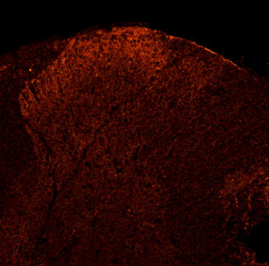
P2X3 in rat Dorsal Horn.
Check out our Purinergic Receptor Antibodies today.


Saturday, October 09, 2010

Enhancing Neurite Outgrowth

We are pleased to announce the addition of new proteins to our Axon Growth and Guidance category. These are designed to enhance neurite out growth.


Name
Catalog #TypeSpeciesSizePrice
NGF-b, NSO Derived
PR15083
ProteinH; M; R100 ug$285
NGF-b, NSO Derived, CFPR15084CFProteinH; M; R100 ug$285
S100A13, CFPR15085CF-50ProteinH; R50 ug$315
Slit1PR15075-50ProteinCh; H50 ug$315
Slit2, CHO
PR15085-50
ProteinCh; H50 ug$315

Image: Cultured chick dorsal root ganglion neurons were grown in the presence of recombinant human NGF-b with (A) or without (B) recombinant mouse Slit2. The presence of the Slit2 protein signifi cantly enhanced neurite outgrowth.