Showing posts with label Neuropathic Pain. Show all posts

Saturday, August 05, 2017

Pain and Inflammation Markers

Our TRPV1 Markers Rock
As sales of our Cell Based Assay Solutions continue to explode. We don't want you to forget that our roots firmly remain in products for Inflammation and Pain Researchers.

To verify this, you only need to check out the many publications referencing use of our TRPV1 Markers.

Image: Immunoreactivity for CXCL12 was detected in the macrophages (F4/80)

Friday, May 19, 2017

TRPV1 Channels and Neuropathic Pain

Neuromics' TRPV1 Stain Neurons and Microglia for Study

TRPV1 is mainly functional in the microglia. Its activation, beyond controlling microglia reaction per se, modulated microglia-neuron communication, by promoting release of extracellular vesicles (EVs) from microglia. Indeed, EVs are important mediators of intercellular communication between microglia and brain cells: Maria Cristina Marrone, Annunziato Morabito, Michela Giustizieri, Valerio Chiurchiù, Alessandro Leuti, Marzia Mattioli, Sara Marinelli, Loredana Riganti, Marta Lombardi, Emanuele Murana, Antonio Totaro, Daniele Piomelli, Davide Ragozzino, Sergio Oddi, Mauro Maccarrone, Claudia Verderio & Silvia Marinelli. TRPV1 channels are critical brain inflammation detectors and neuropathic pain biomarkers in mice. Nature Communications 8, Article number: 15292 (2017) doi:10.1038/ncomms15292.

(a–f) Sections of cortical tissue from WT and TRPV1−/− mice, fixed after exposure to ACSF (a,d), ACSF plus vehicle (DMSO; b,e) and ACSF plus 1μM capsaicin (c,f) and immune-processed for iba-1 to stain microglia cells (in red). INSETs are zoom images taken from an area delimited by the yellow square for each condition. (g), Bar graph of percentage of cortical microglia cell phenotype (resting, ameboid, bushy and hypertrophied), in control (grey bars), vehicle- (dark grey bars) and capsaicin- treated (red bars) cortical sections from WT mice. Capsaicin treatment causes a significant shift from ramified and bushy to hypertrophied morphology. (h), Same as in ‘g’ but in cortical sections from TRPV1−/− mice. In these tissues capsaicin fails to induce morphological changes of microglia cells. Note that microglia cells in −/− tissues are already hypertrophied in control conditions (d–f,h).
Our Pain and Inflammation Research Antibodies Continue to be widely used and frequently published.

Thursday, March 24, 2016

Epigenetics and Pain Research

i-Fect Used to Study Impacts

Our i-Fect siRNA, miRNA and shRNA Trasfection Kit was recently used to study the impact of G9a-specific siRNA (AGUAACGGGCAUCAAUGC) on Mu Opioid Receptors: Yuhao Zhang, Shao-Rui Chen, Geoffroy Laumet, Hong Chen and Hui-Lin Pan. Nerve Injury Diminishes Opioid Analgesia through Lysine Methyltransferase-Mediated Transcriptional Repression of µ-Opioid Receptors in Primary Sensory Neurons. First Published on February 25, 2016, doi: 10.1074/jbc.M115.711812... In some SNL rats, G9a-specific siRNA (4 µg) or the negative control siRNA was administered intrathecally. G9a-specific siRNA(AGUAACGGGCAUCAAUGC) or universal negative control siRNA (#SIC001, Sigma-Aldrich) was mixed with i-Fect (Neuromics, Edina, MN) to a final concentration of 400 mg/L for the intrathecal injections...

Figures: G9a knockdown with siRNA reverses the MOR expression in the DRG and the morphine analgesic effect diminished by nerve injury. (A,B) Quantitative PCR (A) and Western blotting (B) analyses show the mRNA and protein levels of MORs in the DRGs of sham and SNL rats treated with control or G9a-specific siRNA (n = 10 rats in each group). The ipsilateral L5 and L6 DRG tissues were removed 24 h after the last siRNA injection. The amount of MOR mRNA and protein was normalized to GAPDH in the same samples, and the mean value of MOR levels in sham control rats was considered to be 1. (C) Time course of the intrathecal morphine effects on the tactile and pressure withdrawal thresholds in sham and SNL rats treated with G9a-specific siRNA or negative control siRNA (n = 9 rats in each group). The withdrawal thresholds after the last siRNA injection were plotted as the baseline control (BL).

Summary: The findings provide new insight into the epigenetic mechanism regulating MOR expression in primary sensory neurons in neuropathic pain. This multidisciplinary approach provides conclusive evidence for G9a as a key chromatin regulator responsible for MOR downregulation in the DRG and the analgesic efficacy of opioids reduced by nerve injury. A better understanding of the epigenetic mechanisms underlying nerve injury-induced downregulation of MORs in primary sensory neurons could help improve the analgesic efficacy of opioids for treating chronic neuropathic pain. G9a inhibitors could be used to enhance the opioid analgesic effect and reduce opioid consumption in patients with chronic neuropathic pain.

Monday, November 17, 2014

Neuropathy Research Solutions

New Publications and Past Postings

Here're are several key postings on Neuropathy and Neuropathic Pain:
Here're some "hot of the press" publications referencing use of Neuron-Glial Markers: 
Bethany L. Johnson-Kerner, Faizzan S. Ahmad, Alejandro Garcia Diaz, J. Palmer Greene, Steven J. Gray, R. Jude Samulski, Wendy K. Chung, Rudy Van Coster, Paul Maertens, Scott A. Noggle, Christopher E. Henderson and Hynek Wichterle. Intermediate filament protein accumulation in motor neurons derived from giant axonal neuropathy iPSCs rescued by restoration of gigaxonin. Hum. Mol. Genet. (2014) doi: 10.1093/hmg/ddu556. First published online: November 4, 2014.
...MAP2 (1:2,000, Neuromics, CH22103), NF-H (1:2000, Neuromics, CH22104), NF-L (1:200, Neuromics, MO22104)...

Images: Cells grown from adult rat brain. Large cell in middle is stained with mouse monoclonal to NF-L clone DA2 (green). Another type of neuronal lineage cell was stained with rabbit polyclonal to alpha-internexin (red). These cells were mitotic but had several characteristics of neurons. Rat spinal cord homogenate showing the major intermediate filament proteins of the nervous system (lane 1). The remaining lanes show blots of this material stainted with various antibodies including: NF-H, NF-M,, NF-L,, NF66 and GFAP 

Jianfei Guo, Xudong Fu, Xia Cui, Minhua Fan. Contributions of purinergic P2X3 receptors within the midbrain periaqueductal gray to diabetes-induced neuropathic pain. The Journal of Physiological Sciences November 2014.
...An equal volume of total and membrane samples was applied to SDS-PAGE. Membranes were incubated with the rabbit anti-P2X 3 primary antibody (1:1000, Neuromics, Edina, MN, USA) and goat anti-rabbit secondary antibody (1:200, Neuromics, Edina, MN, USA).

Neuropathy Research Solutions Include:


Wednesday, September 10, 2014

Cytokines and Neuropathic Pain

I have been frequently posting results of testing blood serum of our clients suffering from Autoimmune (including Neuroimmuno) Diseases and Autistic Children. Our testing includes both standard medical testing (serum levels of toxins, metals, pathogen loads and markers like Cortisol, CRP, CDs, ALPS, NSE, S100b). In addition, we also doing testing using our Quantibody® Antibody.

Many of the clients report chronic mild to severe neuropathic pain.

By definition, this pain encompasses a series of heterogeneous conditions with some similar clinical manifestations. Peripheral examples include traumatic nerve injury, diabetic peripheral neuropathy and chemotherapy-induced peripheral neuropathy or Multiple Sclerosis. (see: Front Pharmacol. 2013; 4: 142.Published online Nov 22, 2013. doi: 10.3389/fphar.2013.00142). It is hard to unravel the cause and effects of this pain,

However, in analyzing the results from our clients' test we notice a relationship between dysregulation of some key cytokines and levels of pain (more data is required to confirm this). Here're our results (n=5)
Figure: Clients reporting pain vs healthy controls.

We will continue posting results here. We are releasing a Custom Quantibody Neuroimmuno-Pain Array which will include more bio-markers suspected to play a role in pain signaling.

Sunday, January 27, 2013

Epidermal Nerve Fibers in Neuropathic Pain Model

Re-innervation in spared nerve injury (SNI) vs normal rats
Neuromics' Pain and Inflammation Research Antibodies are frequently used to help researchers find root causes of neuropathic pain. This is a fascinating study of re-innervation patterns of epidermal and dermal nerve fibers in a rat neuropathic pain model (Note: our Guinea Pig Polyclonal P2X3 Antibody was used in this study): Liron S. Durakua, Mehdi Hossaini, Barthold N. Schüttenhelm, b, Joan C. Holstege, Martijn Baas, Tom J.H. Ruigrok, Erik T. Walbeehm. Re-innervation patterns by peptidergic Substance-P, non-peptidergic P2X3, and myelinated NF-200 nerve fibers in epidermis and dermis of rats with neuropathic pain. Experimental Neurology Volume 241, March 2013, Pages 13–24. doi.org/10.1016/j.expneurol.2012.11.029.
Non-footpad area vs. footpad.The distribution pattern between the center non-footpad area and footpad is compared for the different subgroups of sensory skin fibers. For epidermal Sub P-IR and upper dermal NF-200-IR fibers there is an equal density of fibers in the center and footpad area suggesting a homogenous distribution over the foot sole of a rat. Whereas for epidermal P2X3-IR fibers there is a significant lower number of P2X3-IR fibers in the footpads as compared to the center non-footpad area, suggesting a heterogeneous distribution in the foot sole for this type of sensory fibers. N = 5 per group. Unpaired t-test. ***: p < 0,001. Scale bar: 250 μm. doi.org/10.1016/j.expneurol.2012.11.029

Illustration of the skin innervation in normal and SNI situation.Illustration showing the innervation pattern of subgroups of sensory skin fibers in the normal situation and in the SNI model 10 weeks PO. In the naïve animals the peptidergic CGRP and non-peptidergic P2X3 fibers have a lower density in the footpads as compared to the non-footpad areas, while the Substance P and NF-200 fibers have an equal distribution over the complete foot sole. In the SNI model there is a significant increase of CGRP epidermal fibers in the medial and lateral area of the foot sole and there is a complete re-innervation of the center area. In addition the footpads in the SNI model are hyper-innervated with CGRP fibers. Substance P and P2X3 and NF-200 fibers show no increased density in the uninjured medial and lateral area after 10 weeks PO. In addition, Substance P and P2X3 fibers hardly re-innervate the center area; however, the NF-200 fibers have the same density of fibers in the denervated area as in the normal situation. In the SNI model the medial, center and lateral area have all an increase in LC's, with the center area being the most prominent one. In addition the epidermis thickness of the SNI model has decreased significantly in all the three areas after 10 weeks PO.
Related Postings: http://neuromics.blogspot.com/search/label/Neuropathic%20Pain.

Monday, December 10, 2012

Pain Research and Gene Expression Analysis

There have been multiple publications referenced here on using Neuromics' i-Fect siRNA Delivery Kit to study the effect of silencing genes known to play a role in Pain Signaling. These include: DOR,The β3 subunit of the Na+,K+-ATPase, NTS1, NAV1.8, TRPV1 NOV, β-arrestin, TRPV1, CAV1.2 and ASIC.

I would like to highlight an exciting new study referencing how knocking down Kv9.1 Potassium Channel Subunit in vivo mediates neuropathic pain: Christoforos Tsantoulas, Lan Zhu, Yasin Shaifta, John Grist, Jeremy P. T. Ward, Ramin Raouf, Gregory J. Michael, and Stephen B. McMahon. Sensory Neuron Downregulation of the Kv9.1 Potassium Channel Subunit Mediates Neuropathic Pain following Nerve Injury. The Journal of Neuroscience, 28 November 2012, 32(48): 17502-17513; doi: 10.1523/​JNEUROSCI.3561-12.2012.

Highlights: Here, we report that the potassium channel subunit Kv9.1 is expressed in myelinated sensory neurons, but is absent from small unmyelinated neurons. Kv9.1 expression was strongly and rapidly downregulated following axotomy, with a time course that matches the development of spontaneous activity and pain hypersensitivity in animal models. Interestingly, siRNA-mediated knock-down of Kv9.1 in naive rats led to neuropathic pain behaviors. Diminished Kv9.1 function also augmented myelinated sensory neuron excitability, manifested as spontaneous firing, hyper-responsiveness to stimulation, and persistent after-discharge. Intracellular recordings from ex vivo dorsal root ganglion preparations revealed that Kv9.1 knock-down was linked to lowered firing thresholds and increased firing rates under physiologically relevant conditions of extracellular potassium accumulation during prolonged activity. Similar neurophysiological changes were detected in animals subjected to traumatic nerve injury and provide an explanation for neuropathic pain symptoms, including poorly understood conditions such as hyperpathia and paresthesias. In summary, our results demonstrate that Kv9.1 dysfunction leads to spontaneous and evoked neuronal hyperexcitability in myelinated fibers, coupled with development of neuropathic pain behaviors.

n vivo RNA interference: Anesthetized rats were subjected to a thoracic laminectomy and a silastic tube was inserted subdurally to lie just rostral to L3 DRG and externalized to deliver bolus injections (one injection per day for 4 consecutive days). Animals were allowed to recover for 5 d before treatment commenced. On the day of injection, siRNA was mixed with i-Fect (Neuromics) to a final concentration of 0.2 μg μl−1, according to published protocols (Luo et al., 2005). For each treatment, 10–20 μl of Kv9.1 siRNA or scrambled control mixture was injected, followed by a 10 μl saline flush. Twenty-four hours after the fourth injection animals were killed and L5 DRGs fresh dissected for qRT-PCR analysis. A separate set of animals were PFA perfused and DRGs retrieved for IHC. Passenger strand sequences for Kv9.1 and scrambled control siRNAs were cuuggaaucuguaggauca and gaggcctaatcgatatgtt, respectively (Dharmacon; “in vivo processing” option).
Intrathecal Kv9.1 siRNA treatment induces pain behaviors in naive rats. A, qRT-PCR quantification of Kv9.1 mRNA in rat PASMC cultures transfected with one of three Kv9.1 siRNA sequences or control siRNA (control, n = 6; siRNA, n = 3 per group; *p < 0.05 vs control, one-way ANOVA with Tukey's). B, qRT-PCR showing Kv9.1 in vivo knock-down in L5 DRG, 4 d after intrathecal delivery of siRNA #1 compared with vehicle or matched scrambled control (vehicle, n = 4; scrambled, n = 5; Kv9.1, n = 7; *p < 0.05, t test). C, IHC for Kv9.1 in scrambled- and siRNA-treated DRG to determine protein knockdown. Graphs illustrate quantification of number of positive myelinated neurons and mean Kv9.1 signal intensity (scrambled, n = 4; siRNA, n = 6; **p < 0.01, ***p < 0.001, t test). D, Kv9.1 siRNA infusion inflicts a reduction in mechanical pain withdrawal thresholds (Kv9.1, n = 7; control, n = 6; *p < 0.05, **p < 0.01, ***p < 0.001 vs scrambled control or baseline, two-way repeated measurements ANOVA with Tukey's). E, There was no change in heat pain thresholds after siRNA treatment. Vertical arrows on x-axis denote siRNA injections. All data represent mean ± SEM.
Kv9.1 knock-down triggers ectopic activity and a form of peripheral wind-up in response to stimulation. A, Schematic illustrating the positions of stimulating and recording electrodes. B, Example recordings from centrally disconnected L4/L5 strands demonstrating SA in Kv9.1 siRNA-treated or nerve-injured rats, but not in control (scrambled siRNA) animals. C, Frequency-dependent SEA (denoted by double arrowheads) in Kv9.1 siRNA-treated (middle) and injured (right), but not control (left) animals. This activity is not locked in time and can be seen in between stimulation events (vertical arrows on top of 5 Hz stimulation traces, only first 5 shown). Also note the prolonged after-discharge (AD) observed in siRNA-treated and injured animals. D, Percentage of units showing SA and SEA in control (n = 269), Kv9.1 siRNA-treated (n = 369) and injured (n = 176) animals (*p < 0.05, **p < 0.01, ***p < 0.001 vs control, χ2 test). E, Firing rate of SEA units at different stimulation frequencies (mean ± SEM; control, n = 4; siRNA, n = 22; injured, n = 17; *p < 0.05 vs control, two-way ANOVA with Tukey's). F, Quantification of AD rate per SEA unit (mean ± SEM; *p < 0.05 vs control, Mann–Whitney test).

Results propose that Kv9.1 downregulation after nerve injury may be the molecular switch controlling myelinated sensory neuron hyperexcitability. Intriguingly, a recent wide-genome association screen in humans identified a Kv9.1 polymorphism associated with susceptibility to develop chronic neuropathic pain after back surgery or leg amputation (Costigan et al., 2010), suggesting that the mechanisms described in our studies will be of direct clinical relevance to human pain. Future efforts to elucidate the precise pathways involved, combined with approaches aiming to compensate loss of Kv9.1 function, may create novel therapeutic opportunities for neuropathic pain management.

Saturday, September 22, 2012

Neuron-specific class III beta-tubulin (TuJ1) Staining in hNT2.19 Neurons

Our Tuj-1 antibody is widely used and frequently published. It is proving a potent marker for confirming the differentiation of human neural progenitors to neurons.

In this study the authors use the marker for staining hNT2.19 Neurons: Mary J. Eaton, Yerko Berrocal, and Stacey Q. Wolfe. Potential for Cell-Transplant Therapy with Human Neuronal Precursors to Treat Neuropathic Pain in Models of PNS and CNS Injury: Comparison of hNT2.17 and hNT2.19 Cell Lines. Pain Research and Treatment. Volume 2012 (2012), Article ID 356412, 31 pages. doi:10.1155/2012/356412. 
The results show great promise. They show hNT2 or hNT2-derived cell lines, such as hNT2.17 and hNT2.19, have great potential to permanently reverse symptoms of neuropathic pain following PNS and CNS injuries and can offer new hope to treat these intractable conditions to significantly improve human health. This includes neuropathic pain resulting from diabetic neuropathy and Spinal Cord Injury.


Images: Comparison of graft sites of hNT2.17 and hNT2.19 in the QUIS and severe contusive-SCI models, respectively, at 6 weeks after cell transplant. (a) Sagittal section of anti-GABA-immunostained QUIS + hNT2.17 transplant lumbar spinal cord 6 weeks after grafting. Easily detectible hNT2.17 cells stain for GABA (arrows) on the pial membranes. (b) Sagittal section of anti-NuMA-immunostained QUIS + hNT2.17 transplant lumbar spinal cord 6 weeks after grafting. Easily detectible hNT2.17 cells stain for NuMA (arrows) on the pial membranes in adjacent sections. The hNT2.19 were alternately injected into the subarachnoid space two weeks after severe contusive SCI. Cell graft sites were co-localized with 5HT (c) and the human-specific marker TuJ1(d) (neuron-specific class III β-tubulin). There are many surviving hNT2.19 (d) grafted cells visible on the pial surface, which stain for TuJ1 (arrows) at the end of the experiment, 56 days after SCI and about 6 weeks after cell transplant. Adjacent sections with the same grafted hNT2.19 (c) are labeled for 5HT (arrows). Magnification bar = 20 μm.

Protocol: Modified methods for staining spinal cord sections for the human neuron-specific class III beta-tubulin (TuJ1) to identify grafted hNT2.19 neurons after grafting have previously been described [51]. The sections were washed with 0.1 M PBS pH 7.4 and permeabilized with 0.4% Triton-X-100 in 0.1 M PBS, 10% normal goat serum (NGS) for one hour. The sections were then incubated overnight at 4°C in the primary anti-TuJ1 antibody (1 : 100 DPBS), and the permeabilizing solution, followed by a one-hour incubation at room temperature with the secondary antibody solution, biotinylated mouse IgG raised in goat (Vector; 1 : 200), a Peroxidase ABC reporter in 0.1 M PBS (Vector) and “VIP” substrate (Vector). Some sections were stained in the absence of primary antibody and served as the negative controls.

I will continue to post new applications for our stem cell research reagents.

Saturday, June 09, 2012

Serotonin and Pain

Brainstem facilitations and descending serotonergic controls contribute to visceral nociception but not pregabalin analgesia in rats.

Pregabalin is used to treat Neuropathic Pain. Here the authors show state-dependent pregabalin analgesia in neuropathy does not apply to visceral pain. Our rabbit polyclonal Mu Opioid Receptor Antibody is used to demonstrate intra-RVM Derm-SAP locally ablates a substantial proportion of MOR and serotonergic cells: Shafaq Sikandar, Kirsty Bannister, Anthony H. Dickenson. Brainstem facilitations and descending serotonergic controls contribute to visceral nociception but not pregabalin analgesia in rats. Neuroscience Letters. Volume 519, Issue 1, 21 June 2012, Pages 31–36.... For MOR staining, a rabbit polycolonal μ-opioid receptor primary antibody (1:10,000 TTBS; Neuromics, MN, USA, RA10104)...

These findings are important for understanding the limitations of Preglabin as an analgesic for viceral pain. Check out all our Opioid Publications.


Thursday, May 17, 2012

Pinpointing Neuropathic Pain

I am pleased to feature our Guinea Pig P2X3 Antibody as a tool for studying the root causes of Neuropathic Pain.

In this study, the authors showed ablation of nonpeptidergic fibers in a chronic constriction injury model caused significant sympathetic and parasympathetic P2X3 fiber sprouting, and led to an exacerbated pain response. This was an unexpected finding, as it has been suggested that nonpeptidergic fibers play a major role in mechanical pain, and suggests that these fibers play a complex role in the development of neuropathic pain: Anna M.W. Taylora, Maria Osikowicza, Alfredo Ribeiro-da-Silva. Consequences of the ablation of nonpeptidergic afferents in an animal model of trigeminal neuropathic pain. PAIN. Volume 153, Issue 6, June 2012, Pages 1311–1319. doi.org/10.1016/j.pain.2012.03.023....Sections were then incubated for 48 hours at 4°C with a guinea pig polyclonal anti-P2X 3 antibody (1:25,000; Neuromics, Edina, MN, USA), diluted in PBS-T. Following primary antibody incubation, sections were treated with a biotin-conjugated...

Image: Example of  Non-petidergic fibers expressing P2X3 receptor for ATP are present in lamina II of the contralateral dorsal horn (arrows). The P2X3-positive fibers are eliminated from the rat dorsal horn ipsilateral to the rhizotomy (arrow heads). Spinal cord segment C6. Scale bar 400 um. P2X3 antibody dilution 1:25,000. Biomédica vol.24 no.2.

Check out our related reagents categories:
 I will continue to post important findings related to the root causes of Neuropathic Pain.


Monday, March 26, 2012

Pain Research-Pubs Update

I am pleased to update you in recent publications and videos referencing use of our reagents for pain research. Check them out.
Andrew C. Eschenroeder, Allison A. Vestal-Laborde, Emilse S. Sanchez, Susan E. Robinson, Carmen Sato-Bigbee. Oligodendrocyte responses to buprenorphine uncover novel and opposing roles of μ-opioid- and nociceptin/orphanin FQ receptors in cell development: Implications for drug addiction treatment during pregnancy. Glia Volume 60, Issue 1, pages 125–136, January 2012....The MOR and NOP receptor antibodies were from Neuromics (Edina, MN) and used for immunocytochemistry (1:100)and western blotting (1:500)...
TRPV1s

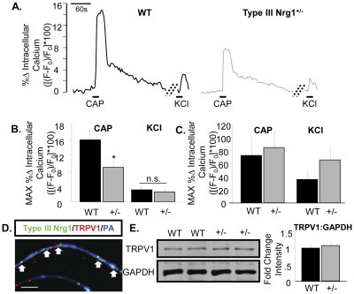
FiFigure. Sensory axons, but not soma, from Type III Nrg1+/− mice show reduced capsaicin responsiveness compared to axons from WT mice. (A) Representative traces of intracellular calcium along sensory axons in response to 1 µM capsaicin or 56 mM KCl. The change in intracellular calcium from baseline over time ([(F−F0)/F0]*100) is shown for WT (left) and Type III Nrg1+/− (right) axons. Hatched diagonal lines indicate where the time course was non-continuous. (B) Quantification of the maximum change in intracellular calcium in response to application of 1 µM capsaicin or 56 mM KCl by genotype. Averages of 5 animals per genotype were compared using a Student's t-test. Type III Nrg1+/− axons showed a significantly decreased response to capsaicin (p<0.05), but not to KCl, relative to WTs. Graph shows mean±SEM. (C) Type III Nrg1+/− sensory soma show normal response to capsaicin. Quantification of maximal change in fluorescence from baseline ([(F−F0)/F0]*100) in WT or Type III Nrg1+/− sensory neuron soma in response to 1 µM capsaicin or 56 mM KCl. Average responses from 4 WT and 4 Type III Nrg1+/− animals to application of capsaicin or KCl were compared by genotype using a Student's t-test. There was no statistically significant difference between genotypes. Graphs show mean±SEM. (D) Type III Nrg1 (green) and TRPV1 (red) are co-expressed along P21 WT cultured sensory neuron axons identified with a pan-axonal (PA) marker (blue). White arrows indicate examples where Type III Nrg1 and TRPV1 are in close proximity. Scale bar equals 10 µm. (E) P21 WT and Type III Nrg1+/− sensory neuron cultures have equivalent levels of total TRPV1 protein. Total TRPV1 protein measurement by immunoblot. The 95 kD TRPV1 band and the 35 kD GAPDH band are shown from a representative experiment comparing protein from P21 WT and Type III Nrg1+/− cultures. Quantification of fold change in intensity of TRPV1:GAPDH normalized to WT average. There was no statistically significant change in the ratio of TRPV1 to GAPDH between genotypes (WT, Type III Nrg1+/−, n = 3 animals). Genotype comparisons were made using a Student's t-test. Graph shows mean±SEM. doi:10.1371/journal.pone.0025108.g004
In this study, researchers successfully transfect DRG cultures with IKAP-shRNA using our pn-Fect kit. The own regulation of IKAP in these cultures support findings that helped explain the potential pathology of Familial Dysautonomia (FD; Hereditary Sensory Autonomic Neuropathy; HSAN III): Hunnicutt BJ , Chaverra M , George L , Lefcort F , 2012 IKAP/Elp1 Is Required In Vivo for Neurogenesis and Neuronal Survival, but Not for Neural Crest Migration. PLoS ONE 7(2): e32050. doi:10.1371/journal.pone.0032050.

Related Links:
I will continue to post updates related to this important research area.

Saturday, August 27, 2011

Is Neuropathy Really Gliopathy?

I found this excellent website from posting by Dr. Jan M. Keppel Hesselink, Professor molecular pharmacology, director Institute neuropathic pain: http://www.neuropathie.nu/. It represents a new way of understanding root causes and potential therapies for Neuropathic Pain. Here're highlights:

Gliopathic pain: is a brand new term for what we always thought to be neuropathic pain. It refers to pain related to neuropathic pain, however, the primary driver of this pain is most probably more linked to glia and asterocytes. The mechanism of gliopathic pain is the hyperactivation of glia cells, which results in neuropathic pain.

The role of Glia and Astrocytes:Glia and astrocytes play a central role in neuropathic pain, and gliopathic pain, or asteropathic pain will become new synonyms for neuropathic pain. In a recent hallmark paper the term 'Gliopathic pain' was coined.

This is a reason to put our magnifying glass on glia. Gliamodulating drugs will become a new class of neuropathic drugs, the so called gliopathic modulating drugs, and the first prototype, the endogenous fatty acid palmitoylethanolamide, has already been explored in positive proof of principle studies.

For more than a century doctors are aware of the special properties of glia in response to injury. In Germany in 1894 professor Franz Nissl decribed the reaction of glial cells in relation to the nerve fibers in the spinal cord and highlighted their morphological changes after injury. Microglia becomes mores bigger and more abundant after injury and these glial responses can be seen as a biological reponse to promote nerve repair after injury. However, this response can go berzerk and might be one of the most important mechanisms leading to neuropathic pain.

This is a short synopsis. There is a wealth of more information on the website. That said, I will be posting more on Gliopathic Pain.

Wednesday, July 06, 2011

Guinea Pig P2X3 Update-Good News

I have had to say to many customers, "our guinea pig P2x3 is on backorder". The increasing number of pubs referencing this antibody only amped demand.

We tried and tried to re-make it. The result was none of the bleeds we tested had a signal strong enough to release the antibody. We had a customer suggest re-testing several of the more promising bleeds. Thank you! We have good news on results and we are offering for 50% off. This is to acknowledge the investment required for TSA and Guinea Pig Biotinylated Antibody.


Here're the recent pubs I referenced:

Gabriela Castañeda-Corral, Héctor I. Rocha-González, Beatriz Godínez-Chaparro, Juan Miguel Jiménez-Andrade and Vinicio Granados-Soto. Role of the spinal Na+/H+ exchanger in formalin-induced nociception. Neuroscience Letters. doi:10.1016/j.neulet.2011.06.048....SP (guinea pig; 1:500; Cat# GP14110; Neuromics), CGRP (goat, 1:500; Cat# Ab36001; Abcam) and P2X3 receptor (guinea pig: 1:10,000; Cat# GP10108; Neuromics)...
Anna M.W. Taylora and Alfredo Ribeiro-da-Silva. GDNF levels in the lower lip skin in a rat model of trigeminal neuropathic pain: Implications for nonpeptidergic fiber reinnervation and parasympathetic sprouting. PAIN Volume 152, Issue 7, July 2011, Pages 1502-1510. doi:10.1016/j.pain.2011.02.035.
...Sections were then incubated for 48h at 4°C with a guinea pig polyclonal anti-P2X3 (1:25,000; Neuromics, Edina, MN)...

Thursday, September 09, 2010

Potential Therapeutic Targets for Bone Cancer Pain-P2X Receptors

Cancer pain is difficult to treat as it appears to be driven simultaneously by inflammatory, neuropathic and tumorigenic mechanisms. I have reported on multiple occasions publication referencing use of our Pain and Inflammation Research Antibodies in studying bone cancer pain.

I would like to alert you to the latest reference:

Timothy K. Y. Kaan, Ping K. Yip, Sital Patel, Meirion Davies, Fabien Marchand, Debra A. Cockayne, Philip A. Nunn, Anthony H. Dickenson, Anthony P. D. W. Ford, Yu Zhong, Marzia Malcangio, and Stephen B. McMahon Systemic blockade of P2X3 and P2X2/3 receptors attenuates bone cancer pain behaviour in rats. Brain, September 2010; 133: 2549 - 2564.

......Slides were then incubated with rabbit anti-P2X3 (1:2000, Neuromics) and sheep anti-calcitonin gene-related peptide (1:1000, Biomol...anti-beta-III-tubulin (1:4000, Promega) and guinea pig anti-P2X3 (1:100, Neuromics). The next day, after three washes with phosphate-buffered......

Summary: Pain remains an area of considerable unmet clinical need, and this is particularly true of pain associated with bone metastases, in part because existing analgesic drugs show only limited efficacy in many patients and in part because of the adverse side effects associated with these agents. An important issue is that the nature and roles of the algogens produced in bone that drive pain-signalling systems remain unknown. Here, we tested the hypothesis that adenosine triphosphate is one such key mediator through actions on P2X3 and P2X2/3 receptors, which are expressed selectively on primary afferent nocioceptors, including those innervating the bone. Using a well-established rat model of bone cancer pain, AF-353, a recently described potent and selective P2X3 and P2X2/3 receptor antagonist, was administered orally to rats and found to produce highly significant prevention and reversal of bone cancer pain behaviour. This attenuation occurred without apparent modification of the disease, since bone destruction induced by rat MRMT-1 carcinoma cells was not significantly altered by AF-353. Using in vivo electrophysiology, evidence for a central site of action was provided by dose-dependent reductions in electrical, mechanical and thermal stimuli-evoked dorsal horn neuronal hyperexcitability following direct AF-353 administration onto the spinal cord of bone cancer animals. A peripheral site of action was also suggested by studies on the extracellular release of adenosine triphosphate from MRMT-1 carcinoma cells. Moreover, elevated phosphorylated-extracellular signal-regulated kinase expression in dorsal root ganglion neurons, induced by co-cultured MRMT-1 carcinoma cells, was significantly reduced in the presence of AF-353. These data suggest that blockade of P2X3 and P2X2/3 receptors on both the peripheral and central terminals of nocioceptors contributes to analgesic efficacy in a model of bone cancer pain. Thus, systemic P2X3 and P2X2/3 receptor antagonists with central nervous system penetration may offer a promising therapeutic tool in treating bone cancer pain.

Related Reagents:

All Purinergic Receptors
Neurotransmission Research Antibodies

Friday, November 06, 2009

gp130, IL-6 and Expression and Neuropathic Pain

We wanted to post yet another publication referencing use of one of our Pain and Inflammation Research Antibodies. Here the researchers used our Mouse Monoclonal gp130/CD130 for Immunohistochemistry.

Manfred Andratsch, Norbert Mair, Cristina E. Constantin, Nadja Scherbakov, Camilla Benetti, Serena Quarta, Christian Vogl, Claudia A. Sailer, Nurcan Üceyler, Johannes Brockhaus, Rudolf Martini, Claudia Sommer, Hanns Ulrich Zeilhofer, Werner Müller, Rohini Kuner, John B. Davis, Stefan Rose-John, and Michaela Kress. A Key Role for gp130 Expressed on Peripheral Sensory Nerves in Pathological Pain. J. Neurosci., Oct 2009; 29: 13473 - 13483 ; doi:10.1523/JNEUROSCI.1822-09.2009

The results suggest that gp130 expressed in sensory nerves not only mediates chronic inflammatory pain, but also contributes significantly to complex interactions between immune cells, tumor cells, and nerves in the context of cancer-evoked pain. Moreover, we identify IL-6 activating gp130, Gab1/Gab2, PI3K, and PKC- and regulating TRPV1 as a key mechanism linking cytokine release to sensitization of pain-sensing nerves.

Related Links
Immune Response
Opioid Neuropeptides
Opioid Receptors
Neurotransmissiom Research Antibodies
GPCRs, Ligand Gated Ion Channels, Biogenic Amines and more
Purinergic Receptors
TRPV (Vanilloid); TRPM; TRPA and TRPCs
i-Fect Transfection Kit
gene silencing of DOR, NaV1.8 tetrodotoxin-resistant sodium channel, NTS2 and more in-vitro and in vivo
Primary Neurons and Astrocytes
Primary human, rat and mouse neurons and astrocytes by Category

Wednesday, July 30, 2008

TH-Marker for Diabetic Neuropathy

New Pub Referencing our Tyrosine Hydroxylase.

Blount, Andrew L. B.A.; Peled, Ziv M. M.D.; Dexter, Erica L. B.S.; Nagle, Raymond B. M.D., Ph.D.; Maloney, Christopher T. M.D.; Dellon, A Lee M.D., Ph.D. Sympathetic Nerves in the Tarsal Tunnel: Implications for Blood Flow in the Diabetic Foot. Plastic & Reconstructive Surgery. 122(1):188-191, July 2008.

...Specimens were fixed in 10% neutral buffered formalin for 3 to 4 hours and then embedded in paraffin. Three-micron sections were obtained from the block and used for immunohistochemistry, which was performed using a mouse monoclonal antibody against tyrosine hydroxylase (MO20001; Neuromics, Edina, Minn.) diluted 1:50 in Discovery Diluent (Ventana Medical Systems, Inc., Tucson, Ariz.) on a Discovery XT Automated Immunostainer (Ventana Medical Systems)...
Images: (A) Tyrosine hydroxylase immunohistochemical staining of tibial epineurium from the tarsal tunnel. Positively staining sympathetic fibers (arrows) are seen within a larger nerve bundle. (B) Tyrosine hydroxylase immunohistochemical staining of connective tissue from the tarsal tunnel. Several positively staining sympathetic fibers (arrows) are seen innervating the media of a local venule.

Thursday, July 24, 2008

SP is Back and Better than Ever

We have successfully re-made our widely used Guinea Pig Substance P antibody. It has been tested both internally and by an interested customer. The results exceeded expectations.

This antibody has proven useful for Pain/Inflammation and Diabete/Obesity Research.

Image: Immuofluorescent detection of Substance P in rat spinal cord dorsal horn (red fluorescence). DAPI (blue) was used as counter stain.

Here're are related publications:

P . Tsai , A . Alonso , M . Pinto , D . Leigh , E . Weiler , J . Fricton , M . Erickson , L . Stone , L . Kehl. Substance P is co-localized with protein gene product 9.5-immunoreactive nerve fibers in intervertebral discs from patients with painful degenerative disc disease. doi:10.1016/j.jpain.2006.01.096
...guinea pig anti-substance P (1:500; Neuromics, Edina, MN) ...
Mei Bigliardi-Qi, Claire Gaveriaux-Ruff, Katrin Pfaltz, Pierre Bady, Tommy Bauman, Theo Rufli, Brigitte L Kieffer and Paul L Bigliardi. Deletion of - and -Opioid Receptors in Mice Changes Epidermal Hypertrophy, Density of Peripheral Nerve Endings, and Itch Behavior. Journal of Investigative Dermatology (2007) 127, 1479–1488. doi:10.1038/sj.jid.5700661; published online 21 December 2006.
.. substance P (Neuromics 1:200) staining, the sections were incubated at 4°C over night in Zamboni buffer. ...

Related Reagents:
Neurokinin-1 (NK 1) Receptor
Neurokinin-1 (NK 1) Human Receptor
Neurokinin-3 (NK 3) Receptor
proNeurokinin B (proNKB or P2)
All Neuropeptides
Pain and Inflammation Antibodies
Diabetes and Obesity Antibodies
Substance P Customer Data

Thursday, June 19, 2008

Amped Up ORL-1

We have been collaborating with a customer to amp up our ORL-1. Here's an example of a resulting IHC images.

Image: ORL 1 staining in rat brain (singulate cortex). ORL1 Detection was done using anti-rabbit Cy3 conjugated antibodies (red color). DAPI was used to counterstain cell nuclei (blue color). Working dilution is 1:40-1:100.

Wednesday, June 04, 2008

TRPV1 Pub

Katrin Schnizler, Leonid P. Shutov, Michael J. Van Kanegan, Michelle A. Merrill, Blake Nichols, G. Stanley McKnight, Stefan Strack, Johannes W. Hell, and Yuriy M. Usachev. Protein Kinase A Anchoring via AKAP150 Is Essential for TRPV1 Modulation by Forskolin and Prostaglandin E2 in Mouse Sensory Neurons. J. Neurosci., May 2008; 28: 4904 - 4917 ; doi:10.1523/JNEUROSCI.0233-08.2008

......anti-TRPV1 antibody (catalog #RA14113; Neuromics, Edina, MN) and 30 mul of prewashed protein-A...mm NaCl). Probing was done using the Neuromics anti-TRPV1 antibody (1:500 dilution...with other TRPV1 antibodies either from Neuromics (N-terminus rabbit anti-TRPV1) or from......