Showing posts with label Nociceptive Pain. Show all posts

Wednesday, December 28, 2016

Dopamine and Morphine Tolerance

Dopamine Identified as Key Player

Our Mu Opioid Receptor antibody is used to show that blocking dopamine decreases morphine tolerance: Wen-Ling Dai, Feng Xiong, Bing Yan, Zheng-Yu Cao, Wen-Tao Liu, Ji-Hua Liu1, Bo-Yang Yu. Blockade of neuronal dopamine D2 receptor attenuates morphine tolerance in mice spinal cord. Scientific Reports 6, Article number: 38746 (2016). doi:10.1038/srep38746.

Images: (A) Double immunofluorescence staining showed that MOR (green) and D2DR (red) were co-localized in the mice spinal cord (20X magnification). Chronic morphine treatment increased the co-localization of MOR and D2DR in the spinal cord, and D2DR antagonist sulpiride (8 μg/10 μl, i.t.) reduced the increased co-expression of D2DR with MOR (n = 4). (B) Co-IP experiments showed that D2DR could interact with MOR, and the MOR/D2DR interactions were increased in the spinal dorsal horn after chronic morphine treatment for 7 days while D2DR antagonist sulpiride (8 μg/10 μl, i.t.) disrupted the interactions of the MOR/D2DR (n = 3).

Blockade of D2DR in spinal cord can disrupt the interactions between MOR and D2DR to attenuate morphine tolerance. These findings highlight the possibility of a new clinical strategy to prevent morphine antinociceptive tolerance.

Friday, August 28, 2015

Small Molecules/Peptides for Pain Researchers

Agonists, Antagonists, Inhibitors and Ligands

The roots of Neuromics is providing solutions for the study of nociceptive and neuropathic pain. Our tools are widely used and frequently published. We are pleased to bring you these small molecules designed to help you better understand the roots of pain and potential therapies.

Figure: Infarct volume after 3 days of reperfusion in the ipsilateral cortex and CP complex in male rats treated with vehicle saline (n=15) or 1 mg/kg per hour BRL 52537 (n=15) started at onset of reperfusion and continued for 22 hours (male-vehicle n=15; male-BRL n=15; mean±SEM). P<.0.05. Related Pub.

Name
Catalog #
Type
Size
Price
187-10
Agonist
10 mg
50 mg
$165
$665
323-10
Antagonist
10 mg
50 mg
$319
$1,319
1224-10
Modulator
10 mg
50 mg
$99
$299
1062-10
Agonist
10 mg
$299
1116-10
Modulator
10 mg
50 mg
$119
$449
3661-10
Antagonist
10 mg
50 mg
$183
$795
699-10
Ligand
10 mg
1 mg
$159
$609
1560-1
Antagonist
1 mg
$135
898-2
Antagonist
2 mg
$179
1055-5
Agonist
5 mg
$139
1056-5
Agonist
5 mg
$139
1480-10
Agonist
10 mg
10 mg
$229
$929
0754-10
Agonist
10 mg
10 mg
$199
$809
910-1
Inhibitor
1 mg
$179
1198-1
Inhibitor
1 mg
$195


Questions? Do not hesitate to contact me @ pshuster@neuromics.com or 612-801-1007. Thank you! Pete Shuster, CEO and Owner, Neuromics.

Tuesday, July 21, 2015

Neuropeptides and Stress

Neuropeptides Antagonists, Stress and Alcohol Seeking

Select Neuropeptides can help moderate stress and related Alcohol seeking behaviors. The authors of this study reference use of our ppENK and proDYN Neuropeptides: J.R. Schank,B.S. Nelson,R. Damadzic,J.D. Tapocik,M. Yao,C.E. King,K.E. Rowe,K. Cheng,K.C. Rice,M. Heili. Neurokinin-1 receptor antagonism attenuates neuronal activity triggered by stress-induced reinstatement of alcohol seeking. doi:10.1016/j.neuropharm.2015.07.009.

Image: Mouse striatum stained with D2 cell marker Enkephalin (RA14124) in green and with neuronal marker NeuN in red courtesy of Dr Heike Rebholz of City College of New York.

Our Pain/Inflammation Antibodies are widely used and frequently referenced in studies of stress and the intersection between stress and pain.

Tuesday, June 24, 2014

More Gains on Pain Research

Discovering the Related Pathways

There are multiple descriptions for chronic and acute pain: stabbing, burning, cutting, itching, numbing, crushing and more. Understanding the pathways are important to optimizing the therapies and treatments for the many different forms of pain.

The foundation of Neuromics is providing top notch pain research markers, gene expression analysis tools and cell based assays. Here I share recent publications from customers using these:
Inflammatory Joint Pain: Fiona B Carr, Sandrine M Géranton and Stephen P Hunt. Descending controls modulate inflammatory joint pain and regulate CXC chemokine and iNOS expression in the dorsal horn. Molecular Pain 2014, 10:39 doi:10.1186/1744-8069-10-39...mu opioid receptor, 1:10,000, Neuromics (RA10104)...

Images: Representative single plane confocal images of MOR immunohistochemistry in the 1.5pmole dermorphin-saporin group and saline control. Double labelling with NeuN indicated that although many MOR+ neurons are depleted at this dose, some surviving MOR- neurons remain in the region, indicated by white arrows. Scale bars indicate 25 µm

Temperature Sensation: Marics I, Malapert P, Reynders A, Gaillard S, Moqrich A (2014) Acute Heat-Evoked Temperature Sensation Is Impaired but Not Abolished in Mice Lacking TRPV1 and TRPV3 Channels. PLoS ONE 9(6): e99828. doi:10.1371/journal.pone.0099828. ...TRPV1 antibody (1/1000 dilution, Neuromics)...

Acute PainPaulino Barragán-Iglesias, Hector I. Rocha-González, Jorge Baruch Pineda-Farias, Janet Murbartián, Beatriz Godínez-Chaparro, Peter S. Reinach, Thiago M. Cunha, Fernando Q. Cunha, Vinicio Granados-Soto, Inhibition of peripheral anion exchanger 3 decreases formalin-induced pain, European Journal of Pharmacology, Available online 27 May 2014, ISSN 0014-2999, http://dx.doi.org/10.1016/j.ejphar.2014.05.029. (http://www.sciencedirect.com/science/article/pii/S0014299914003914). ...substance P (guinea pig; 1:200; Cat # GP14110; Neuromics, Edina, MN), and purinergic P2×3 receptor (guinea pig: 1:1000; Cat # GP10108 ...

We are pleased with the many publications and customer provided data proving the quality of our many pain research tools. I am always available to serve you, Pete Shuster (pshuster@neuromics.com) or direct phone: 612-801-1007.

Tuesday, January 28, 2014

Pot and Pain

The pressure for states to legalize Marijuana for medical and recreational use is building. The tax benefits are self evident.

The debate for many centers of "true medical benefits". That's why research on understanding analgesic pathways is so important. Ironically, this study was conducted by my friends at Université de Montréal and Université de Sherbrooke in Quebec Canada: J. Desroches, J.-F. Bouchard, L. Gendron, P. Beaulieu. Involvement of cannabinoid receptors in peripheral and spinal morphine analgesia ☆ Neuroscience, Volume 261, 7 March 2014, Pages 23–42. http://dx.doi.org/10.1016/j.neuroscience.2013.12.030.

These teams have proven expert is using our Opioid Receptor Antibodies in their pain research. Here's a synopsis:
•Analgesia is the most common feature shared by the cannabinoid and opioid systems.
•The role of the cannabinoid system in the morphine-induced analgesia is uncertain.
•Peripheral and intrathecal morphine analgesia is altered in cnr1KO and cnr2KO mice.
•This attenuation is neither caused by a MOP malfunction nor by its downregulation.


Images: Deletion of the CB1 or CB2 receptors has no effect on the expression of MOP in the spinal cord. Immunofluorescence of spinal MOP revealed that the expression of MOP in laminae I and II of the dorsal horn of the spinal cord did not differ between cnr1WT (A) and cnr1KO (B) mice or between cnr2WT (C) and cnr2KO mice (D).


Observations here further support the existence of interactions between the cannabinoid and opioid systems. The loss of peripheral and spinal morphine analgesia is apparently caused neither by a decrease in MOP spinal expression nor by altered binding properties or G protein coupling of this receptor in cnr1KO and cnr2KO mice. The mechanisms underlying the loss of morphine analgesia are not clear but could include the release of endogenous cannabinoids in structures along the pain pathway or a disrupted endocannabinoid tone.

It is important funding that enables researchers to understand the analgesic pathways of marijuana continues to grow. This research could yield better control of pain with reduced side effects.

Sunday, April 14, 2013

P2X3 Receptors and Migraine

P2X3 Receptors of Trigeminal Sensory Neurons and Familial Hemiplegic Migraine Type 1 (FHM-1).

Our P2X Receptor Markers continue to be used in interesting and novel ways. Here researchers use our P2X3 Receptor Antibody to study expression using primary rat ganlia cultures: Swathi K. Hullugundi,Michel D. Ferrari, Arn M. J. M. van den Maagdenberg, Andrea Nistri. Andrea Nistri. The Mechanism of Functional Up-Regulation of P2X3 Receptors of Trigeminal Sensory Neurons in a Genetic Mouse Model of Familial Hemiplegic Migraine Type 1 (FHM-1). PLoS ONE 8(4): e60677. doi:10.1371/journal.pone.0060677

Abstract: A knock-in (KI) mouse model of FHM-1 expressing the R192Q missense mutation of the Cacna1a gene coding for the α1 subunit of CaV2.1 channels shows, at the level of the trigeminal ganglion, selective functional up-regulation of ATP -gated P2X3 receptors of sensory neurons that convey nociceptive signals to the brainstem. Why P2X3 receptors are constitutively more responsive, however, remains unclear as their membrane expression and TRPV1 nociceptor activity are the same as in wildtype (WT) neurons. Using primary cultures of WT or KI trigeminal ganglia, we investigated whether soluble compounds that may contribute to initiating (or maintaining) migraine attacks, such as TNFα, CGRP, and BDNF, might be responsible for increasing P2X3 receptor responses. Exogenous application of TNFα potentiated P2X3 receptor-mediated currents of WT but not of KI neurons, most of which expressed both the P2X3 receptor and the TNFα receptor TNFR2. However, sustained TNFα neutralization failed to change WT or KI P2X3 receptor currents. This suggests that endogenous TNFα does not regulate P2X3 receptor responses. Nonetheless, on cultures made from both genotypes, exogenous TNFα enhanced TRPV1 receptor-mediated currents expressed by a few neurons, suggesting transient amplification of TRPV1 nociceptor responses. CGRP increased P2X3 receptor currents only in WT cultures, although prolonged CGRP receptor antagonism or BDNF neutralization reduced KI currents to WT levels. Our data suggest that, in KI trigeminal ganglion cultures, constitutive up-regulation of P2X3 receptors probably is already maximal and is apparently contributed by basal CGRP and BDNF levels, thereby rendering these neurons more responsive to extracellular ATP.

Images: Examples of TNFR2 and P2X3 co-exexpression in (wildtype) WT and R192Q (knockin) KI neurons. Left panel shows P2X3 expression (green), and right panel shows TNFR2 staining (red). B, Histograms quantifying % of cells co-expressing TNFR2 and P2X3: both WT and KI cultures show similar TNFR2 and P2X3 co-expression. N = 3 independent experiments (6 mice). C, Representative traces of currents induced by application of α,β-meATP (10 µM, 2 s) to WT or R192Q KI neurons in control conditions or after 4 h TNFα application. D, Histograms show average peak amplitudes of P2X3 receptor-mediated currents: WT control (open bar), n = 30; WT TNFα (stippled bar), n = 38; KI control (grey bar), n = 34; KI TNFα (stippled gray bar), n = 34; ** = p<0 .006="" i="" nbsp="" p="">doi:10.1371/journal.pone.0060677.g001.

Understanding the interplay between TNFR2 and P2X3 could lead to a better understanding of the root causes of migraines. This could open up yet more potential drug targets for this insidious condition.

Check out these related reagent categories:
All Purinergic Receptor Antibodies
Pain and Inflammation Research Antibodies 
Neurotransmission Research Antibodies
Primary Neurons and Astrocytes-Primary human, rat and mouse neurons and astrocytes

Sunday, December 16, 2012

P2X Receptor Markers-Pubs Update

2012 has been a record year for publications referencing use of our Purinergic Receptor Antibodies. These publications demonstrate use of these markers in a variety of assays and applications. Examples range from analysis of P2X2 expression in human bladder epithelial cells to showing P2X3 expressing nerve fiber boutons in rat horizontal spinal cord sections by immunofluorescence and much more.

Here's a summary of publications: F.C. Pradoa, D. Araldia, A.S. Vieiraa, M.C.G. Oliveira-Fusarob, C.H. Tambelia, C.A. Parada. Neuronal P2X3 receptor activation is essential to the hyperalgesia induced by prostaglandins and sympathomimetic amines released during inflammation. http://dx.doi.org/10.1016/j.neuropharm.2012.11.011. non-fat dry milk at room temperature, followed by incubation with P2X3 or PKCɛ rabbit polyclonal antibody (1:500; Neuromics) overnight at 4 °C, rinsed six times with TBST, and then incubated for 40 min in goat anti-rabbit IgG peroxidase conjugate...

Min Liu, PhD, MD, Yun-fei Xu, PhD, MD, Yuan Feng, PhD, MD, Fengqiang Yang, MD, Jun Luo, MD, Wei Zhai, PhD, MD, Jian-ping Che, MD, Guang-chun Wang, MD, Jun-hua Zheng, PhD. Epigallocatechin gallate attenuates interstitial cystitis in human bladder urothelium cells by modulating purinergic receptors. Journal of Surgical Research. http://dx.doi.org/10.1016/j.jss.2012.11.041
...P2X2 (Neuromics, Northfield, MN)...

Abeer W Saeed, Alfredo Ribeiro-da-Silva. Non-peptidergic primary afferents are presynaptic to neurokinin-1 receptor immunoreactive lamina I projection neurons in rat spinal cord. Molecular Pain 2012, 8:64 doi:10.1186/1744-8069-8-64Anna M.W. Taylora, Maria Osikowicza, Alfredo Ribeiro-da-Silva. Consequences of the ablation of nonpeptidergic afferents in an animal model of trigeminal neuropathic pain. PAIN. Volume 153, Issue 6, June 2012, Pages 1311–1319. http://dx.doi.org/10.1016/j.pain.2012.03.023.
Confocal images at high power obtained from horizontal spinal cord sections. In a confocal optical section from lamina I adjacent to the white matter (A), note the relatively abundant P2X3-IR fibers with varicosities (boutons). CGRP-IR fibers and boutons were considerably more abundant in this lamina. In a confocal optical section from inner lamina II (B), note the very high density of P2X3-IR fibers and varicosities, higher than that of CGRP-IR fibers in lamina I. Note that most varicosities display either P2X3 or CGRP immunoreactivity, although some co-localization is observed (yellow). Scale bar (A, B) = 20 μm.
T. Cho, V. V. Chaban. Interaction Between P2X3 and Oestrogen Receptor (ER)α/ERβ in ATP-Mediated Calcium Signalling In Mice Sensory Neurones. Journal of Neuroendocrinology Volume 24, Issue 5, pages 789–797, May 2012...with polyclonal rabbit antibody against P2X3 receptor (dilution 1 : 1000; Neuromics)...

Anna M.W. Taylora, Maria Osikowicza, Alfredo Ribeiro-da-Silva. Consequences of the ablation of nonpeptidergic afferents in an animal model of trigeminal neuropathic pain. PAIN. Volume 153, Issue 6, June 2012, Pages 1311–1319. doi.org/10.1016/j.pain.2012.03.023...Sections were then incubated for 48 hours at 4°C with a guinea pig polyclonal anti-P2X3 antibody (1:25,000; Neuromics, Edina, MN, USA), diluted in PBS-T. Following primary antibody incubation, sections were treated with a biotin-conjugated...

Ji Z-G , Ito S , Honjoh T , Ohta H , Ishizuka T , et al. 2012 Light-evoked Somatosensory Perception of Transgenic Rats That Express Channelrhodopsin-2 in Dorsal Root Ganglion Cells. PLoS ONE 7(3): e32699...guinea-pig anti-P2X3 (1:1,000, GP10108, Neuromics, Edina, MN, USA)...
Distribution of ChR2V in the dorsal part of the spinal cord. A–C. Immunohistochemical localization of ChR2V with the cell-type specific markers, NF200 (A), CGRP (B) or P2X3 (C). Scale bars indicate 40 µm. doi:10.1371/journal.pone.0032699.g003.

I will continue to post new publications and data in the coming year. Stay tuned. 

Sunday, October 21, 2012

P2X3 Receptor and CGRP Antibodies Immunostaining

Dr. Alfredo Ribeiro-da-Silva, McGill University, is a serial publisher of studies using our pain and inflammation research antibodies.

Here, use of our rabbit anti-CGRP and guinea pig anti-P2X3 is referenced. Please note the high titer of these antibodies (dilution is 1:25,000): Abeer W Saeed, Alfredo Ribeiro-da-Silva. Non-peptidergic primary afferents are presynaptic to neurokinin-1 receptor immunoreactive lamina I projection neurons in rat spinal cord. Molecular Pain 2012, 8:64 doi:10.1186/1744-8069-8-64.


Images: CGRP, IB4 and P2X3 staining in transverse spinal cord sections. A and B show low magnification confocal images of CGRP-IR and IB4 positive (A) or P2X3-IR (B) fibers. C and D represent high magnification confocal images from the middle third of the lateromedial extent of the superficial dorsal horn. In C, note that there is limited co-localization of IB4 and CGRP (in yellow). Arrowheads show axonal varicosities (boutons) from nonpeptidergic fibers in lamina I, which do not co-localize CGRP immunoreactivity. The framed regions in A and B indicate the approximate regions from where C and D, respectively, were obtained (the latter originate from other sections). CGRP (in green); IB4 (in red); P2X3 (in red). Scale bar (A, B) = 200 μm; scale bar (C, D) = 20 μm

Tissue processing: The injection site at the level of the parabrachial nucleus was examined by cutting serial, 100 μm-thick coronal sections of the relevant brain region. The dorsal aspect of the L4-L5 spinal cord segment was cut into serial, 50 μm-thick horizontal sections (n = 10), 50 μm-thick parasagittal sections (n = 4) or 50 μm-thick transverse sections (n = 4). All sections were cut using a freezing sledge microtome (Leica, Richmond Hill, Ontario) and collected as freefloating in phosphate-buffered saline (PBS) with 0.2% Triton-X 100 (PBS + T). To block unspecific staining, all spinal cord sections were incubated, for one hour, in 10% normal donkey serum (NDS) (Jackson, West Grove, PA) in PBS + T at room temperature. Subsequently, the sections were placed in primary antibodies (or conjugated lectin IB4 - see below) for 48 hours at 4 °C. We used a mixture of 2 or 4 primary antibodies (each raised in a different species), or IB4, in PBS + T containing 5% NDS. Next, the sections were washed in PBS + T and then incubated in species-specific secondary antibodies that were raised in donkey and conjugated to either AlexaFluor 488, AlexaFluor 405, Rhodamine RedX or biotin. The sections were incubated in 3 different cocktails: #1) rabbit anti-CGRP at a 1:200 dilution (Sigma, St Louis, MO) and lectin IB4 conjugated to AlexaFluor 568 at a 1:200 dilution (Molecular Probes); #2) rabbit anti-CGRP and guinea pig anti-P2X3 at a 1:25,000 dilution (Neuromics, Edina, MN); #3) goat anti-CTb at a 1:5000 dilution (List Biological), rabbit anti-NK-1r at a 1:10000 dilution (Sigma, St Louis, MO), guinea pig anti-CGRP at a 1:8000 dilution (Peninsula, San Carlos, CA) and lectin IB4 conjugated to AlexaFluor 647 at a 1:200 dilution (Molecular Probes). All the sections were washed with PBS + T and then (for #1) incubated for 2 hours at room temperature with donkey anti-rabbit AlexaFluor 488; (for #2) incubated for 90 minutes in a biotin conjugated donkey anti-guinea pig IgG (Jackson Immunoresearch, West Grove, PA, 1:200). Further signal amplification was achieved by treating the sections with 1 hour incubation in an avidin-biotin (A + B) complex (Vectastain Elite ABC kit, Vector Laboratories) followed by tyramide (Perkin-Elmer, Norwalk, CT, 1:75) for 7 minutes. Sections were then incubated in streptavidin conjugated to AlexaFluor 568 (Molecular Probes, Eugene, OR, 1:200) and donkey anti-rabbit AlexaFluor 488; or (for #3) incubated for 2 hours at room temperature with secondary antibodies: donkey anti-goat Rhodamine Red X, donkey anti-rabbit AlexaFluor 488, and donkey anti-guinea pig AlexaFluor 405. Finally, sections were washed with PBS, mounted on gelatin-subbed slides and coverslipped with an anti-fading mounting medium (Aqua Polymount; Polysciences, Warrington, PA). Slides were stored at −4 °C pending further processing.

I will continue to publish outstanding customer data/images using our natibodies/markers.

Wednesday, June 20, 2012

TRPA1 and Tooth Pain

Our TRPV (Vanilloid); TRPM; TRPA and TRPCs have proven excellent for studying

Transient receptor potential ankyrin 1 (TRPA1) is activated by noxious cold (<17°C) and contributes to cold and mechanical hypersensitivity after inflammation and nerve injury: Yun Sook Kim, PhD, Hoon Kap Jung, DDS, Tae Kyung Kwon, DDS, Chin Soo Kim, DDS, PhD, Jin Hyun Cho, DDS, PhD, Dong Kuk Ahn, DDS, PhD, Yong Chul Bae, DDS, PhD.Expression of Transient Receptor Potential Ankyrin in Human Dental Pulp. Journal of Endodontics. Available online 8 June 2012. doi.org/10.1016/j.joen.2012.04.024.


Highlights: TRPA1 was expressed in a large number of axons branching extensively in the peripheral pulp and in a few axons within the nerve bundles in the core of the coronal pulp and in the radicular pulp. Under electron microscopy, TRPA1 immunoreactivity was typically localized near the plasma membrane of unmyelinated axons in the peripheral pulp, suggesting that in these axons it may act as a functional receptor. The proportion of axons expressing TRPA1 in neurofilament 200–positive axons significantly increased in the painful pulp compared with the normal pulp. TRPA1 was also densely expressed in the processes and the cell body of odontoblasts. A large number of axons coexpressed TRPA1 and Nav1.8.


TRPA1
Images: Immunofluorescent staining for (A) TRPA1 in the human normal dental pulp is completely abolished by (B) preadsorption with a control peptide, proving the specificity of the TRPA1 antiserum (×200, scale bars = 50 µm). doi.org/10.1016/j.joen.2012.04.024.
Related Reagents:
All TRP Antibodies                       
Pain and Inflammation Research Antibodies                     
Neurotransmission -Neurotransmission Research Antibody Categories
Primary Neurons and Astrocytes-Primary human, rat and mouse neurons and astrocytes

Wednesday, July 06, 2011

Guinea Pig P2X3 Update-Good News

I have had to say to many customers, "our guinea pig P2x3 is on backorder". The increasing number of pubs referencing this antibody only amped demand.

We tried and tried to re-make it. The result was none of the bleeds we tested had a signal strong enough to release the antibody. We had a customer suggest re-testing several of the more promising bleeds. Thank you! We have good news on results and we are offering for 50% off. This is to acknowledge the investment required for TSA and Guinea Pig Biotinylated Antibody.


Here're the recent pubs I referenced:

Gabriela Castañeda-Corral, Héctor I. Rocha-González, Beatriz Godínez-Chaparro, Juan Miguel Jiménez-Andrade and Vinicio Granados-Soto. Role of the spinal Na+/H+ exchanger in formalin-induced nociception. Neuroscience Letters. doi:10.1016/j.neulet.2011.06.048....SP (guinea pig; 1:500; Cat# GP14110; Neuromics), CGRP (goat, 1:500; Cat# Ab36001; Abcam) and P2X3 receptor (guinea pig: 1:10,000; Cat# GP10108; Neuromics)...
Anna M.W. Taylora and Alfredo Ribeiro-da-Silva. GDNF levels in the lower lip skin in a rat model of trigeminal neuropathic pain: Implications for nonpeptidergic fiber reinnervation and parasympathetic sprouting. PAIN Volume 152, Issue 7, July 2011, Pages 1502-1510. doi:10.1016/j.pain.2011.02.035.
...Sections were then incubated for 48h at 4°C with a guinea pig polyclonal anti-P2X3 (1:25,000; Neuromics, Edina, MN)...

Thursday, November 18, 2010

Cancer-Induced Bone Pain

Bone crushing pain. This describes pain of the highest order. Our friend, Dr. Joseph Ghilardi, VAMC-Mpls. and his colleague, Dr. Patrick Manthy are finding the root causes of the intense and growing pain suffered by Cancer Victims. Here are highlights of a recent study:

Pain frequently accompanies cancer. What remains unclear is why this pain frequently becomes more severe and difficult to control with disease progression. Here we test the hypothesis that with disease progression, sensory nerve fibers that innervate the tumor-bearing tissue undergo a pathological sprouting and reorganization, which in other nonmalignant pathologies has been shown to generate and maintain chronic pain. Injection of canine prostate cancer cells into mouse bone induces a remarkable sprouting of calcitonin gene-related peptide (CGRP+) and neurofilament 200 kDa (NF200+) sensory nerve fibers. Nearly all sensory nerve fibers that undergo sprouting also coexpress tropomyosin receptor kinase A (TrkA+). This ectopic sprouting occurs in sensory nerve fibers that are in close proximity to colonies of prostate cancer cells, tumor-associated stromal cells and newly formed woven bone, which together form sclerotic lesions that closely mirror the osteoblastic bone lesions induced by metastatic prostate tumors in humans. Preventive treatment with an antibody that sequesters nerve growth factor (NGF), administered when the pain and bone remodeling were first observed, blocks this ectopic sprouting and attenuates cancer pain. Interestingly, reverse transcription PCR analysis indicated that the prostate cancer cells themselves do not express detectable levels of mRNA coding for NGF. This suggests that the tumor-associated stromal cells express and release NGF, which drives the pathological reorganization of nearby TrkA+ sensory nerve fibers. Therapies that prevent this reorganization of sensory nerve fibers may provide insight into the evolving mechanisms that drive cancer pain and lead to more effective control of this chronic pain state.

Image: Image:Shows rat mixed neuron/glial cultures stained with mouse monoclonal antibody to neurofilament subunit NF-L clone 7D1 (green) and chicken antibody to neurofilament NF-H. This antibody binds primarily to the phosphorylated axonal forms of NF-H, in contrast to the NF-L antibody which stains both axonal and dendritic/perikaryal neurofilaments. The NF-L antibody therefore reveals a prominent cell body in green, while the surrounding axonal profiles are orange, since the are bound by both NF-L and the chicken NF-H antibody. Blue is a DNA stain. Protocol on data sheet.

 Juan M. Jimenez-Andrade, Aaron P. Bloom, James I. Stake, William G. Mantyh, Reid N. Taylor, Katie T. Freeman, Joseph R. Ghilardi, Michael A. Kuskowski, and Patrick W. Mantyh Pathological Sprouting of Adult Nociceptors in Chronic Prostate Cancer-Induced Bone Pain. J. Neurosci., Nov 2010; 30: 14649 - 14656 ; doi:10.1523/JNEUROSCI.3300-10.2010
Here're several other pubs referencing use of our antibodies in studying bone cancer pain:

Kyle G. Halvorson, BA, Molly A. Sevcik, BA, Joseph R. Ghilardi, BS, BA, Lucy J. Sullivan, BA, Nathan J. Koewler, BS, Frieder Bauss, PhD, and Patrick W. Mantyh, PhD. Intravenous Ibandronate Rapidly Reduces Pain, Neurochemical Indices of Central Sensitization, Tumor Burden, and Skeletal Destruction in a Mouse Model of Bone Cancer. Published online 2008 April 14. doi: 10.1016/j.jpainsymman.2007.10.005
...pro-dynorphin (DYN, polyclonal guinea pig anti-rat, 1:1,000; Neuromics, Minneapolis, MN)...

Timothy K. Y. Kaan, Ping K. Yip, Sital Patel, Meirion Davies, Fabien Marchand, Debra A. Cockayne, Philip A. Nunn, Anthony H. Dickenson, Anthony P. D. W. Ford, Yu Zhong, Marzia Malcangio, and Stephen B. McMahon Systemic blockade of P2X3 and P2X2/3 receptors attenuates bone cancer pain behaviour in rats. Brain, September 2010; 133: 2549 - 2564.
......Slides were then incubated with rabbit anti-P2X3 (1:2000, Neuromics) and sheep anti-calcitonin gene-related peptide (1:1000, Biomol...anti-beta-III-tubulin (1:4000, Promega) and guinea pig anti-P2X3 (1:100, Neuromics). The next day, after three washes with phosphate-buffered......

I will keep you posted on this important topic.

Thursday, July 29, 2010

Let-7 microRNAs and Nociceptive Pain

Our Opioid Receptor Antibodies have set a potent standard for studying Nociceptive and Neuropathic Pain. Related Publications.

We want to recognize Dr. Zaijie Jim Wang and his team for being the first to use our Mu Opioid Receptor for studying the potential role of microRNAs in Nociception.

Ying He, Cheng Yang, Chelsea M. Kirkmire, and Zaijie Jim Wang. Regulation of Opioid Tolerance by let-7 Family MicroRNA Targeting the µ Opioid Receptor. The Journal of Neuroscience, July 28, 2010, 30(30):10251-10258; doi:10.1523/JNEUROSCI.2419-10.2010
Abstract: MicroRNA has emerged as a critical regulator of neuronal functions. This study aimed to test whether let-7 microRNAs can regulate the µ opioid receptor (MOR) and opioid tolerance. Employing bioinformatics, we identified a let-7 binding site in the 3'-untranslated region (UTR) of MOR mRNA, which was experimentally confirmed as a direct target of let-7. The repressive regulation of MOR by let-7 was revealed using a LNA-let-7 inhibitor to knockdown let-7 in SH-SY5Y cells. Conversely, morphine significantly upregulated let-7 expression in SH-SY5Y cells and in a mouse model of opioid tolerance. The LNA-let-7 inhibitor decreased brain let-7 levels and partially attenuated opioid antinociceptive tolerance in mice. Although chronic morphine treatment did not change overall MOR transcript, polysome-associated mRNA declined in a let-7-dependent manner. let-7 was identified as a mediator translocating and sequestering MOR mRNA to P-bodies, leading to translation repression. These results suggest that let-7 plays an integral role in opioid tolerance.

  • Western blot analysis. Western blot analysis was performed as previously described (Tang et al., 2006) using the anti-µ opioid receptor antibody (1:1000; Neuromics). The expression of β-actin was similarly determined from the same blots using a monoclonal antibody (1:10,000; Sigma).
  • For immunofluorescence analysis, the antibody for hDcp1a (Santa Cruz Biotechnology) and MOR were used at 1:500 and 1:5000 dilutions, respectively. Secondary anti-goat and anti-mouse antibodies labeled with Alexa 488 and Alexa 594 fluorochromes (Invitrogen), respectively, were used at 1:500 dilutions.

Related Reagent Links:
All Opioid Receptor Antibodies

Pain and Inflammation
       
Neurotransmission Research Antibodies
-GPCRs, Ligand Gated Ion Channels,
Biogenic Amines and more
i-Fect Transfection Kit
-gene silencing of DOR,
NaV1.8 tetrodotoxin-resistant sodium channel, NTS2 and more in-vitro and in vivo
Primary Neurons and Astrocytes
-Primary human,
rat and mouse neurons and astrocytes 


Monday, July 26, 2010

δ- and μ-opioid receptors co-expression and Nociceptive Pain

Dr. Tomas Hokfelt and his team at Karolinska Institute recently published use of our Opioid Receptor Antibodies and Substance P Antibody.They show the interplay of DOR and MOR in modulation of nociceptive afferent transmission and opioid analgesia.

Hai-Bo Wanga, Bo Zhaoa, Yan-Qing Zhonga, Kai-Cheng Li, Zi-Yan Li, Qiong Wang, Yin-Jing Lua, Zhen-Ning Zhang, Shao-Qiu He, Han-Cheng Zheng, Sheng-Xi Wu, Tomas G. M. Hökfelt, Lan Baob, and Xu Zhanga. Coexpression of δ- and μ-opioid receptors in nociceptive sensory neurons. PNAS July 20, 2010 vol. 107 no. 29 13117-13122.

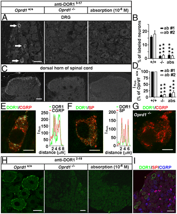
Immunostaining. Adult rats, mice, and Oprd1 exon 1-deleted mice were fixed. Cryostat sections of L4 and L5 DRGs and spinal cord segments were processed for immunofluorescence staining (13) with Rb anti-DOR13–17 (1:2,000–1:60,000; DiaSorin and 1:4,000–1:60,000; Neuromics), Rb anti-DOR12–18 (1:30,000–1:120,000; Alomone), Rb anti-DOR1358–372 (1:1,000–1:2,000; Lifespan Biosciences), Rb anti-MOR (1:1,000; Neuromics); guinea pig anti-SP (1:500; Neuromics), and mouse anti-CGRP (1:1,000; Biogenesis) antibodies. IB4-labeling was carried out with fluorescein-labeled GSL I-IB4 (1:200). The Myc-DOR1–transfected HEK293 cells and neurons were fixed and processed with mouse anti-Myc antibodies (1:500; DSHB). Nuclear DAPI staining was used to indicate HEK293 cells in control experiments.

Images: Distinct distribution patterns of DORs in subsets of DRG neurons of mice. Immunostaining with antibodies against DOR13–17 [A: 1:30,000, antibody 1 (ab #1); DiaSorin and C: antibody 2 (ab #2); Neuromics] shows DORs in small DRG neurons and afferent fibers in spinal laminae I–II. This immunostaining pattern is abolished by the antiserum preabsorption or the deletion of Oprd1 exon 1. Reduction in immunostaining is quantitatively assayed by determining the percentage of positive DRG neurons (B; n = 6) and fluorescence intensity (Ifluo.) in the laminae I–II (D; n = 5). **P < 0.01; ***P < 0.001. (Scale bars: A and C, 40 μm.). DOR labeling (anti-DOR13–17, 1:30,000; DiaSorin) associated with vesicles in peptidergic small DRG neurons (E and F) is absent in Oprd1 exon 1-deleted mice (G). Colocalization of DORs and neuropeptides is shown by correlated peaks of Ifluo. measured along lines. (Scale bar: 8 μm.) (H) Immunostaining with antibodies against DOR12–18 (1:60,000; Alomone) shows the presence of DORs on the cell surface of large DRG neurons of mice. (Scale bar: 25 μm.) This staining pattern is abolished by preabsorption and is absent in Oprd1 exon 1-deleted mice. (Scale bar: 80 μm.) (I) Triple-immunostaining shows that DOR+ large DRG neurons contain neither SP nor CGRP. (Scale bar: 80 μm.)

Immunoblotting.The samples were processed for SDS/PAGE, transferred, probed with Rb antibodies against MOR (1:500; Neuromics), phospho-DOR1 (1:1,000; Neuromics), phospho-MOR (1:1,000; Neuromics), Myc (1:500; DSHB), Flag (1:1,000; Sigma), or actin (1:50,000; Chemicon) and visualized with enhanced chemiluminescence (19).

Featured and Related Reagents:

Mu Opioid Receptor-Rabbit


Mu Opioid Receptor-Guinea Pig


Delta Opioid Receptor 3-17


Delta Opioid Receptor 358-372


Delta Opioid Receptor 358-372


Kappa Opioid Receptor


phospho-Mu Opioid Receptor (Ser375)


MOR-1C
 

ORL 1-Pure


ORL1-Whole Serum


All Opioid Receptor Antibodies


Pain and Inflammation


Neurotransmission Research Antibodies
-GPCRs, Ligand Gated Ion
Channels, Biogenic Amines and more

i-Fect Transfection Kit
-gene silencing
of DOR, NaV1.8 tetrodotoxin-resistant sodium channel, NTS2 and more
in-vitro and in vivo

Primary Neurons and Astrocytes
-Primary
human, rat and mouse neurons and astrocytes

Friday, February 19, 2010

Making Gains on Pain Research

Neuromics' Pain Research Customers continue to make gains using our Pain and Inflammation Research Antibodies and Transfection Kits. Here are the latest pubs:

Hua Zhang and A. S. Verkman. Aquaporin-1 Tunes Pain Perception by Interaction with Nav1.8 Na+ Channels in Dorsal Root Ganglion Neurons. February 19, 2010 The Journal of Biological Chemistry, 285, 5896-5906.

...chicken anti-calcitonin gene-related peptide (CGRP; 1:500, Neuromics, Edina, MN)...

Nathaniel A. Sowa, Bonnie Taylor-Blake, and Mark J. Zylka. Ecto-5'-Nucleotidase (CD73) Inhibits Nociception by Hydrolyzing AMP to Adenosine in Nociceptive Circuits. The Journal of Neuroscience, February 10, 2010, 30(6):2235-2244; doi:10.1523/JNEUROSCI.5324-09.2010.


...rabbit anti-P2X3-RA10109, Neuromics; 1:750), rabbit anti- VR1 C-Terminus (TRPV1) - mouse specific (RA14113, Neuromics; 1:750

Images: Images: Confocal images showing the effect of RTX on mu opioid receptor and TRPV1 immunoreactive DRG neurons and afferent terminals in the spinal cord. A: representative confocal images showing mu opioid receptor (green) and TRPV1 (red) immunoreactivities in DRG neurons of one vehicle- and one RTX-treated rat. Scale bar, 40 um. B: confocal images showing mu opioid receptor (green) and TRPV1 (red) immunoreactivities in afferent terminals in the spinal dorsal horn of 1 vehicle- and 1 RTX-treated rat. Scale bar, 80 um. Inset: high-magnification images (scale bar = 5 um) showing co-localization of mu opioid receptor and TRPV1 immunoreactivity in the lamina I. Co-localization of the mu opioid receptor and TRPV1 immunoreactivity is indicated in yellow when 2 images are digitally merged. All images are single confocal optical sections. Shao-Rui Chen and Hui-Lin Pan. Loss of TRPV1-Expressing Sensory Neurons Reduces Spinal mu-Opioid Receptors But Paradoxically Potentiates Opioid Analgesia. doi:10.1152/jn.01343.2005.

Neuromics' i-Fect ™ siRNA Transfection Reagent continues to be used a tool for studying expression of genes suspected to paly a role in pain . Expression studies include: DOR , The β3 subunit of the Na+,K+-ATPase, rSNSR1, NTS1. NAV1.8, CaV1.2 and more.

Here's a link to all transfection publications: Transfection Kit Pubs

Related Reagents:


Neurotransmission Research Antibodies-GPCRs, Ligand Gated Ion Channels, Biogenic Amines and more
Purinergic Receptors

Primary Neurons and Astrocytes- Primary human, rat and mouse neurons and astrocytes by Category

Monday, September 28, 2009

Prodynorphin at Work

We would like to Dr. Andrew Todd for sharing this excellent IHC image using our Guinea Pig ProDynorphin (rat) antibody.

Image: Staining of adult rat spinal cord.

The tissue is perfusion-fixed (4% freshly prepared formaldehyde) adult rat spinal cord, reacted overnight with the PPD at 1:1000 and then o/n in Alexa488 secondary (raised in donkey, Invitrogen, 1:500).

The confocal image stack was taken through a 60x oil lens (Bio-Rad Radiance confocal) - pixel size is 0.196 micrometre and this is a projection of 10 confocal optical sections at 0.5 micrometre z-spacing.
Customer Publications
Related Reagents:
proDynorphin (guinea pig)
Opioid Receptors
Pain and Inflammation Antibodies