Thursday, December 21, 2017

Cell to Cell Signaling and Depression

ATP and Your Brain
A recent article in Molecular Psychiatrydoi:10.1038/mp.2017.229 elucidates the role of ATP and Neuron-Glial interactions in depression.

This study also features the use of our excellent GFAP markers
Abstract: Extracellular ATP is a widespread cell-to-cell signaling molecule in the brain, where it functions as a neuromodulator by activating glia and neurons. Although ATP exerts multiple effects on synaptic plasticity and neuro-glia interactions, as well as in mood disorders, the source and regulation of ATP release remain to be elaborated. Here, we define Calhm2 as an ATP-releasing channel protein based on in vitro and in vivo models. Conventional knockout and conditional astrocyte knockout of Calhm2 both lead to significantly reduced ATP concentrations, loss of hippocampal spine number, neural dysfunction and depression-like behaviors in mice, which can be significantly rescued by ATP replenishment. Our findings identify Calhm2 as a critical ATP-releasing channel that modulates neural activity and as a potential risk factor of depression.

Wednesday, December 13, 2017

Big Thank You!

Big Year
We exceeded our goals for 2017. By a lot. Both in sales and new products.

I would like to personally thank our customers, collaborators, and friends for making this happen. You made it happen.

I wish Joy to you in 2018!
I wish you exciting, new discoveries, and adventures.

Pete Shuster, CEO and Owner, Neuromics.

Friday, December 08, 2017

Coming Soon Hepatocytes and CSCS

Differentiated from iPSCs

Neuromics is partnering with Javeen Biosciences to bring you Cancer Stem Cells and Hepatocytes derived from donor Induced Pluripotent Stem Cells.

Rodney Nash, Javeen CEO, and his team are working to fill a major gap in cell-based drug discoveries. The lack of cells from specific diseased or ethnic populations. This limits the ability to predict how well a drug candidate will work with a diverse population of patients.


Dr. Rodney Nash, CEO, Javeen Biosciences
Over time, we envision Javeen Biosciences as having the ability to engineer cells from donors that match tightly with the needs of drug hunters.

We ask you to "Think of 8". This video illustrates the power Javeen Biosciences' solutions

Much more to follow...

Wednesday, November 29, 2017

Need Cells? Contest is Now Live

It's Easy! 
Creatively insert your favorite human cell culture(s) anywhere into a picture of your lab for $25 Amazon Gift Card. To enter, email the picture to rose@neuromics.com with a description of the image that includes your name, the lab name and the type of cells being cultured.
First 100 entries are eligible for the gift card.

Grand prize is $500 Visa Gift Card. Only entries that include the culture of Neuromics cells will be eligible for the grand prize.

  • Official Contest Rules: 
  • Only one entry per lab. 
  • The first 100 entries are eligible. 
  • Cells cultured do not have to be Neuromics cells to be eligible for Amazon gift card. 
  • All images should be original work – you should not distribute, or reproduce in any way any copyrighted material, trademarks, or other proprietary information without obtaining the prior written consent of the owner of such rights. 
  • By entering this competition, entrants agree that Neuromics can publicize their name, their lab and their entry. 
  • Entries will be posted on Neuromics’ web page, social media channels and other promotional communications. 
  • All submissions become the sole property of Neuromics and will not be returned to the applicant. Prize restrictions may apply to certain individuals based on their institution’s respective policies; based on this, Neuromics reserves the right to decline a prize to ineligible individuals. 
  • Neuromics reserves the right to make any determinations about prizes, disqualifications, and other concerns as issues arise. 
  • Neuromics reserves the right to amend or withdraw the contest at any time.
Enjoy and good luck!

Tuesday, November 21, 2017

Need Cells? Contest

We're live; Be Creative
We are offering a $25 Amazon Gift Card to scientists who creatively places images their favorite cell cultures anywhere in a picture of their lab AND if the picture has images of our human cells, you could win a $500 VISA card.

Here's a link to learn more about our Need Cells Contest.

Please email your picture to rose@neuromics.com and we will email you a $25 Amazon Gift Card.

Thursday, November 16, 2017

Coming Soon-Need Cells? Contest

Creativity Rewarded
We are revving up another contest as our Neuromics Brain Adventure was successful and wow, were people creative.

We are expert at finding human primary and stem cells so cell cultures are central to the contest. Rules are simple. Affix an image of your favorite cell culture anywhere in your lab and we will email you a $25 Amazon gift card.

The contest will run for 3 months and the most creative entry will receive a $500 Visa Gift Card.
Simply e-mail your entry to rose@neuromics.com and we will email back the gift card. Multiple entries encouraged.

Monday, November 06, 2017

More FBS Data!

Cells+Media Supplemented with our FBS!

We continue to be pleased with the cell culture images our customers are sharing with us-https://www.neuromics.com/fbs-data

Neurons are notoriously hard to culture. Here're images of Mouse Neurons cultured in media supplemented with our FBS.
Images: Mouse cortical neurons in culture, using Neuromics heat-inactivated FBS (in plating media for two days). Data courtesy of Dr. Saif at Barrow Neurological Institute.
We continue to offer $50 Amazon Gift Card in return for data you share using any of our solutions. Just email the data to Rose Ludescher, Manager of Customer Satisfaction, rose@neuromics.com and she will email you the gift card.

Monday, October 23, 2017

3-D Retina Organoid Challenge

Proud Sponsor
The National Eye Institute has its sights set on stimulating researchers to move rapidly toward treatments for retinal diseases. We need your scientific expertise: compete in a Challenge to develop a physiologically-competent 3-D retina organoid model.
Check us out on their Sponsor's Page.


Thursday, October 12, 2017

More Neuromics' FBS Related Data

Cells Cultured in Media Supplemented with our FBS
Customers continue submitting data. Here's the latest:
Primary mouse vascular smooth muscle cells stained with smooth muscle alpha-actin in DMEM with Neuromics 10% FBS. Image courtesy of Deng-Fu Guo, University of Iowa.
HEK Cells Grown in Neuromics FBS. Image courtesy of Kavita Shah, Purdue University.
We will continue be posting new FBS related data here.

Saturday, September 30, 2017

Neuromics' FGF-2 for Expanding Human Cells

FGF-2 and Fibrochondrocytes

FGF-2 of FGF-basic is an important a growth factor for many cell-based assays. Neuromics' has rock solid FGFs for supplementing media used to grow cells. Check out publications referencing use of FGF-2.

Here's the latest publication: Yan Liang, Enaam Idrees, Stephen H. J. Andrews, Kirollos Labib, Alexander Szojka, Melanie Kunze, Andrea D. Burbank, Aillette Mulet-Sierra, Nadr M. Jomha & Adetola B. Adesida. Plasticity of Human Meniscus Fibrochondrocytes: A Study on Effects of Mitotic Divisions and Oxygen Tension. Scientific Reports 7, Article number: 12148 (2017) doi:10.1038/s41598-017-12096-x. ...Thereafter the number of viable MFCs were counted using a haemacytometer after trypan blue staining. MFCs were plated at 104 cells/cm2 and cultured in the standard medium described above supplemented with FGF-2 (5 ng/mL; Neuromics, MN, USA, Catalog#: PR80001) and TGFβ1 (1 ng/mL; ProSpec, NJ, USA, Catalog#: cyt-716) under normal oxygen tension (21% O2) at 37 °C in a humidified incubator...
Images: Immunofluorescence analysis of collagen I and collagen II in pellets derived from T1F2-expanded MFCs of four passages after 21 days chondrogenic stimulation under NRX or HYP from one representative donor (male, 20 years old). Blue (DAPI): cells, Red (Texas Red): collagen I, Green (FITC): collagen II. (A) Pellets cultured under NRX, (B) Pellets cultured under HYP from four passages. Scale bar: 100 µm
Our ISO-Kine FGF-2 is especially potent as it virtually endotoxin free.

Thursday, September 21, 2017

i-Fect Delivers Plasmids!

Important for Gene Expression Studies.
I have posted many examples of how our customers use i-FectTM  and other Transfection Solutions for Gene Manipulation Studies. There are also many publications.

Here we feature how i-Fect was used to delivery plasmids to the CNS: Sara Elramah, María José López-González, Matthieu Bastide, Florence Dixmérias, Olivier Roca-Lapirot, Anne-Cécile Wielanek-Bachelet, Anne Vital, Thierry Leste-Lasserre, Alexandre Brochard, Marc Landry & Alexandre Favereaux. Spinal miRNA-124 regulates synaptopodin and nociception in an animal model of bone cancer pain. Scientific Reports 7, Article number: 10949 (2017) doi:10.1038/s41598-017-10224-1...Intrathecal administration of miRNAs and ShRNA To over-express miR-124, we cloned the pre-miRNA sequence of miR-124 into a plasmid. To determine cells expressing this miR-124 encoding plasmid, we added a GFP-coding sequence to the construct under the control of an IRES. Thus, miR-124 over-expressing cells also express GFP. To inhibit synaptopodin expression, we cloned a ShRNA sequence directed against synaptopodin into a plasmid. To determine cells expressing this ShRNA, we added a GFP-coding sequence to the construct under the control of an IRES. Thus, ShRNA expressing cells also expressed GFP. Two micrograms of these plasmids or the corresponding controls, were solubilized in 10 µl of i-Fect reagent (Neuromics, Edina, USA), and injected intrathecally between the L5 and L6 lumbar vertebrae every two days for a total of 3 injections, according to the manufacturer’s instructions and previously published experiments...
Figures: (C and D) Immunostaining of synpo in spinal cord after miR-124 intrathecal injections: only the dorsal horn which receive nociceptive information was quantified (white dash area). Measurement of synaptopodin stained area reveals ability of miR-124 to inhibit endogenous Synpo expression (20/3 and 17/3 denotes number of sections/animals for control and miR-124-injected mice, respectively.
I am confident there will be many more positive reports regarding our Transfection Reagents.

Saturday, September 16, 2017

Dynamics of Stem Cell Differentiation

Differentiation Markers
Neuromics has excellent markers for determining stem cell differentiation.

This new publication references use of our OTX2 marker: Sumin Jang, Sandeep Choubey, Leon Furchtgott, Ling-Nan Zou, Adele Doyle, Vilas Menon, Ethan B Loew, Anne-Rachel Krostag, Refugio A Martinez, Linda Madisen, Boaz P Levi, Sharad Ramanathan. Dynamics of embryonic stem cell differentiation inferred from single-cell transcriptomics show a series of transitions through discrete cell states. eLife 2017;6:e20487. DOI: http://dx.doi.org/10.7554/eLife.20487


Figures: Single-Cell Gene Expression Profiling of mESCs during early germ layer differentiation. (A) Mouse embryonic stem cells (mESCs) were exposed to various differentiation conditions to perturb FGF, WNT, and TGF-beta signaling for up to five days of differentiation. (B) Images of immunostained mESCs undergoing differentiation show cell-to-cell variability in their expression of known germ layer marker genes. (Scale bar = 100 μm).
We will continue to post new developments.

Friday, September 01, 2017

Neuronal Markers

New Pub References 3 Markers
We are recognized for our large catalog of neuronal markers. Our strength, in this area, includes markers designed for pain researchers.

They are widely used and frequently published. This new publication references use of our Guinea Pig Substance P, Guinea Pig PGP9.5 and Chicken NF200 of NF-Heavy. andla, Jagadeesha, Lomada, Santosh Kumara, Jianninga; Kuner, Rohinia, Bali, Kiran Kumar. miR-34c-5p functions as pronociceptive microRNA in cancer pain by targeting Cav2.3 containing calcium channels. Pain: September 2017 - Volume 158 - Issue 9 - p 1765–1779 doi: 10.1097/j.pain.0000000000000971.
Neuromics' PGP9.5 Staining of  Mouse DRGs.
.Neuromics SP and NF200 Staining of Mouse DRGSs

Thursday, August 31, 2017

FBS Works Great for Cell Lines

More Customer Data
We are pleased with the continuing stream of positive feedback on our potent FBS. This includes its supplementing media for cell lines.

Here is an image courtesy of  Kavita Shah of Purdue University.
Image: HEK Cells cultured in Media Supplemented with Neuromics' FBS

We offer the best price anywhere. We will beat all competitive pricing. If you are interested, contact Rose Ludescher, Manager of Customer Satisfaction. email: rose@neuromics.com.


Monday, August 21, 2017

Data Trifecta-Cells, Media + FBS

Optimizing your Cell-Based Assays
Researchers need solid proof that the solutions the use will work in their assays. Given the time and effort it takes to culture human primary cells, it is important that the media and supplements used are optimized.

We engineer our cell media and supplements to work with our primary and stem cells. Its potency is tested every day in our cell cultures.

As further proof, we ask customers to share their data with us. We reward them by emailing a 50 USD Amazon gift card. I would like to recognize Emily Rodela from TGEN for sharing the Trifecta of Neuromics' CAFS grown in MSC-Gro Media supplemented with our FBS.
Image: Human CAFS-Pancreatic Stellates cultured in MSCGro™ supplemented with Neuromics' FBS - Fetal Bovine Serum. Courtesy of Emily Rodela, TGEN.

Monday, August 14, 2017

Neuromics' ISOKine bFGF in 3-D Cultures

Works Well in Perfusion Models

Our ISOKineTM FGF is produced in the endosperm tissue of barley grain (Hordeum vulgare), that exhibits up to 50 times less protease activity than E.coli or mammalian cells. Barley seed is void of any human or animal viral contaminants that could jeopardize your cell culture.

It is a proven solution for all cell cultures and starts at the low price of  65 USD/10 ug.

Here's a reference of its use in 3-D Cultures: Tom Kamperman, Sieger Henke, Claas Willem Visser, Marcel Karperien, Jeroen Leijten. Centering Single Cells in Microgels via Delayed Crosslinking Supports Long-Term 3D Culture by Preventing Cell Escape. DOI: 10.1002/smll.201603711.

Figure: Delayed on-chip crosslinking enables centering of single cells in microgels. a) Fluorescence confocal imaging confirmed that delayed enzymatic crosslinking enabled centering of single MSCs in Dex-TA microgels. b) On average, cell-laden microgels were only 9 µm larger than the encapsulated MSCs, effectively resulting in 3D hydrogel coatings of less than 5 µm. c) A standard microfluidic droplet generator was connected to the H2O2 diffusion-based crosslinking chip. The position of cells (white arrows) in non-crosslinking microgel precursor droplets was analyzed d) immediately after droplet generation (t1), at the start of the crosslinking chip (t2), and e) at the end of the crosslinking chip (t3). f) Cell positions within microgels produced using conventional microfluidic encapsulation systems (i.e., with coupled emulsification and gelation) are indicated with gray (i.e., references) and red (i.e., this work) data points. Cell positions within gel precursor droplets along the modular microfluidic setup are indicated with blue data points. Cell positions within delayed enzymatically crosslinked microgels are indicated with green data points. g) Cell position analyses of various combinations of distinct hydrogel materials (i.e., Dex-TA, Dex-HA-TA, PEGDA), cell types (i.e., MIN6, MSC), and crosslinking methods (i.e., enzyme-based and photo-crosslinking), revealed that delayed crosslinking consistently resulted in significantly increased cell-centering as compared to the conventional encapsulation approach where emulsification and gelation are coupled.

If you are looking for competitively priced, animal free and potent growth factors. check out our ISOkines.

Wednesday, August 09, 2017

Cancer Associated Fibroblasts in Action

Publication Reference
We introduced our Cancer Associated Fibroblasts to researchers several years ago. The demand was strong from day one and continues to grow.

We are pleased to announce the first reference in a Cancer Research Publication: Zenobia D'Costa, Keaton Jones, Abul Azad, Ruud van Stiphout, Su Y Lim, Ana L Gomes, Paul Kinchesh, Sean C Smart, W. Gillies McKenna, Francesca M Buffa, Owen J. Sansom, Ruth J. Muschel, Eric O'Neill and Emmanouil Fokas. Gemcitabine-induced TIMP1 attenuates therapy response and promotes tumor growth and liver metastasis in pancreatic cancer. Published OnlineFirst August 1, 2017 doi: 10.1158/0008-5472.CAN-16-2833...Immortalized Pancreatic CAF-Stellate Cells were obtained from Neuromics, and cultured in VitroPlus III, low serum, complete (Neuromics)...
Image: Pancreatic CAF-Stellates stained with alphaSMA

We will continue to post new developments here.


Saturday, August 05, 2017

Pain and Inflammation Markers

Our TRPV1 Markers Rock
As sales of our Cell Based Assay Solutions continue to explode. We don't want you to forget that our roots firmly remain in products for Inflammation and Pain Researchers.

To verify this, you only need to check out the many publications referencing use of our TRPV1 Markers.

Image: Immunoreactivity for CXCL12 was detected in the macrophages (F4/80)

Monday, July 31, 2017

Immune Response Research Solutions

Cytokine Markers and More
Immune and inflammatory response are elevated in most diseases. We have strong solutions for detecting these responses. These include cytokine markers and panels.

Here's a new pub referencing use of one of our Interleukin Markers. ArdoSabir and AndiSumidarti. Interleukin-6 expression on inflammed rat dental pulp tissue after capped with Trigona sp. propolis. Saudi Journal of Biological Sciences Volume 24, Issue 5, July 2017, Pages 1034-1037. doi.org/10.1016/j.sjbs.2016.12.019 ...Four rats were sacrificed at 6 h, 2 days, 4 days and 7 days respectively. The teeth and surrounding bone were resected, fixed in Bouin’s fixative for 24 h, decalcified with acetic acid/formal saline for 7 days, embedded in paraffin and sectioned serially at 6 μm thickness. The sections were stained with IL-6 monoclonal antibody (Neuromics, USA) using immunohistochemistry method and viewed by light microscopy...

Image: Interleukin-6 expression on rats dental pulp tissue. Normal dental pulp with no treatment (negative control) in groups I (A–B) and inflamed dental pulp in groups II (C–D), III (E–F), IV (G–H) and V (I–J), capped with Ethanolic Extract Propolis (EEP), Extract Flavonoid-Propolis (EFP), Extract Non-Flavonoid Propolis (ENFP), and Calcium Hydroxide (Ca(OH)2), respectively. Arrows show IL-6 expression. Immunohistochemistry method, DAB chromogen, original magnification 200×.

Monday, July 24, 2017

Quality, High Titer Neuronal Markers

You Need Them; We got Them

We are recognized for both the number and quality of Neuronal Markers. Check out our many testimonials from satisfied customers.

Here's a sampling of recent publications.

Elisabet Garcia-Pino, Nikodemus Gessele and Ursula Koch. Enhanced Excitatory Connectivity and Disturbed Sound Processing in the Auditory Brainstem of Fragile X Mice. Journal of Neuroscience 3 July 2017, 2310-16; doi.org/10.1523/JNEUROSCI.2310-16.2017. ...chicken α-MAP2 (Neuromics; dilution 1:1000)...
Anna Lisa Gündner Claas Aiko Meyera, Stefan Aigner, Klaus Christensen, Christoph Patsch, Ravi Jagasia, Karlheinz Baumann, Marc Burcin. Generation of a homozygous GBA deletion human embryonic stem cell line. Stem Cell Research. Available online 11 July 2017. https://doi.org/10.1016/j.scr.2017.07.009 ...Neuronal differentiation marker, Ch-MAP2, 1:1000, Neuromics # CH22103 RRID:AB_2314763 ....Immunocytochemistry. Cells were cultured on PO/LAM coated 96-well imaging microplates (Falcon) and fixed with 4% paraformaldehyde (15 min, room temperature). Fixed cells were permeabilized with 0.2% Triton (Sigma) in phosphate-buffered saline (PBS) for 30 min and blocked for 1 h with PBS + 5% donkey serum (Merck Millipore). Primary antibodies (in PBS) were incubated overnight at 4 °C followed by three PBS washing steps. Secondary antibodies were incubated for 2 h at room temperature. Confocal images were acquired using a Leica TCS SP5 microscope (Leica Microsystems)....
Hayk Harutyunyan, Svetlana Sharoyan, Alvard Antonyan, Sona Mardanyan. Herb Preparations Improve the Viability of Hippocampal Cells Suppressed by Amyloid Beta (1-42) Peptide. World Journal of Pharmaceutical Sciences. 2017. ISSN (Online): 2321-3086......Antibodies for immunocytochemistry were purchased from Neuromics (USA)[....]Immunocytochemistry: Hippocampal cells were cultured in Poly-D-Lysine coated Nunc EasY Flasks. On the 4th day, the adhered cells were removed by trypsinisation (0.05% trypsin, 0.5 mM EDTA, pH 8.0). Cells were fixed on microscope slide by 3.7 % paraformaldehyde in PBS followed by methanol treatment. To block a nonspecific antibody binding, samples were pre-treated with goat serum (Sigma) for 30 minutes. To determine the types of cells, constituting the obtained cell culture, the different aliquots of the culture were incubated for 60 min at room temperature with the primary antibodies against rabbit anti-rat Neurofilament (NF), chicken anti-rat GFAP and Nestin...
Images: Cells grown from adult rat brainLarge cell in middle is stained with mouse monoclonal to NF-L clone DA2 (green). Another type of neuronal lineage cell was stained with rabbit polyclonal to alpha-internexin (red). These cells were mitotic but had several characteristics of neurons. Rat spinal cord homogenate showing the major intermediate filament proteins of the nervous system (lane 1). The remaining lanes show blots of this material stainted with various antibodies including: NF-HNF-M,, NF-L,, NF66 and GFAP 
We will continue to post the latest and greatest news.

Monday, July 17, 2017

Electrical Preconditioning of Stem Cells

Cool Science

The ability to manipulate hNPCs via a conductive scaffold creates a new approach to optimize stem cell-based therapy and determine which factors (such as VEGF-A) are essential for stroke recovery: Paul M. Georgea, Tonya M. Blissb, Thuy Huab, Alex Leed, Byeongtaek Oh, Alexa Levinson, Swapnil Mehta, Guohua Sun, Gary K. Steinberg. Electrical preconditioning of stem cells with a conductive polymer scaffold enhances stroke recovery. doi.org/10.1016/j.biomaterials.2017.07.020...anti βIII-tubulin (1:500, Neuromics, Edina, MN)...

Image: Neural progenitors were labeled with anti-rat Nestin polyclonal antibody (Cat#:GT15114) and stained with conjugated donkey anti-goat secondary anti-body (green). Differentiated neurons were labeled with neuron-specific mouse anti-β-III tubulin/ Tuj1-(Cat#MO15013 monoclonal antibody (red). Nuclei were stained with DAPI (blue).
We have excellent Stem Cell Differentiation Markers. Check them out.

Tuesday, July 11, 2017

New Human RPES and BMSCS

Need Human Cells? Just Ask!

This is our cornerstone and we keep building on it. You asked for them. We are pleased to announce we now have Human Bone Marrow-Derived Stem Cells (BMSCs) and Retinal Pigment Epithelial Cells (RPES).
RPES in Culture.
BMSCS in Culture.
We will continue to post new cell offerings here.

Thursday, July 06, 2017

3D Printing the Way to Artificial Muscles

Muscles Contract Under the Control Motor Neurons

The methods used to develop the artificial muscles included use of our GDNF Protein to maintain motor neurons in the 3-D Culture-Caroline Cvetkovic, Max H. Rich, Ritu Raman, Hyunjoon Kong , Rashid Bashir. A 3D-printed platform for modular neuromuscular motor units. Microsystems; Nanoengineering 3, Article number: 17015 (2017) doi:10.1038/micronano.2017.15

Skeletal muscle cells and motor neurons were combined into a fabricated 3D co-culture system. C2C12 myoblasts were differentiated into multinucleated myotubes (a) and combined with extracellular matrix (ECM) proteins to create an engineered muscle ring tissue (b). In parallel, mouse embryonic stem cells (HBG3 mESCs) were differentiated into motor neurons (MNs) through the formation of embryoid bodies (EBs) (c and d) and then combined with the engineered muscle tissue and ECM proteins (e) on 3D-printed hydrogel devices (f and g). Once the multi-layered rings sequentially compacted and fused together, they were then placed on a stationary hydrogel skeleton (h). Scale bars, 50 μm (b and d), 500 μm (c), and 10 μm (d, inset).

Wednesday, July 05, 2017

Renal Nerves and Blood Pressure

Role of Calcitonin gene-related peptide (CGRP)

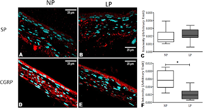
CGRP is a potent vasodilator so it is not surprising our CGRP Antibody was used in this study. Custódio AH, de Lima MC, Vaccari B, Boer PA, Gontijo JAR (2017) Renal sodium handling and blood pressure changes in gestational protein-restricted offspring: Role of renal nerves and ganglia neurokinin expression. PLoS ONE 12(6): e0179499. https://doi.org/10.1371/journal.pone.0179499

Figure. Comparative expression of SP and CGRP in the renal pelvis of 16-week-old rats. The pictures show a normal distribution of these neurokinins in NP (A and D). In LP offspring, no difference was observed in SP immunoreactivity (A, B, and C), but CGRP immunoreactivity was significantly more intense in NP (D, E, and F) compared with age-matched LP offspring.

We are always honored when our solutions are referenced in publications and will continue to post new findings here.

Tuesday, June 27, 2017

Petaka Cell Culturing System in Action

Used for Hypoxia Induction
Cells are the engine of cell-based assays. Our PetakaTM Cell Culture System knock-down barriers that stand between you and success. The system is especially designed to grow cells in an isolated, auto-modified environment, highly protected from external environmental conditions.

In this study, for hypoxia induction, Petaka G3 low oxygen transfer flasks (Neuromics) were used to culture melanoma cell lines or primary melanocytes for 48–96 h at 37°C until >80% confluent: Rachel L. G. Maus, James W. Jakub, Wendy K. Nevala, Trace A. Christensen, Klara Noble-Orcutt, Zohar Sachs, Tina J. Hieken, and Svetomir N. Markovic. Human Melanoma-Derived Extracellular Vesicles Regulate Dendritic Cell Maturation. Front Immunol. 2017; 8: 358. Published online 2017
Mar 29. doi: 10.3389/fimmu.2017.00358.

Hypoxia increases extracellular vesicles (EVs) production in metastatic melanoma cell line SKMEL28 not primary melanocytes. Melanoma cell lines and adult primary melanocytes were cultured under hypoxia or normoxia conditions for 48 h. Morphology and CD63 vesicle marker expression of SKMEL28 EVs was confirmed by immune electron microscopy (A) and vesicle size distribution and particle concentration measured by nanoparticle tracking analysis (Nanosight) [(B), representative image]. Total concentration of normoxic (gray) or hypoxic (black) vesicles isolated from three melanoma cell lines and healthy primary melanocytes was quantified by Nanosight following three replicate 30-s video captures of three EV preparations (C) (n = 3 independent experiments).

Petaka advantages include:

  • No requirement of CO2 incubator or humid incubator. 
  • Exact volume of media per culture. 
  • Cell growth on vertical walls segregates cells and debris. 
  • Negligible dehydration rates Protected against leaks or spills. 
  • Higher cell yields. 
  • Shipping and short term cell maintenance at room temperature.

Wednesday, June 21, 2017

Gap Junctions (GJs) and Glaucoma

GJs Can Offer Neuroprotection
This study references use of our GFAP Antibody,

Gap junctions (GJs), intercellular channels composed of subunit connexins, can play a major role in secondary cell death by forming conduits through which toxic molecules from dying cells pass to and injure coupled neighbors. Secondary cells like glia and astrocytes are involved in this process though the precise mechanisms have yet to be defined: Abram Akopian, Sandeep Kumar, Hariharasubramanian Ramakrishnan, Kaushambi Roy, Suresh Viswanathan, and Stewart A. Bloomfield. Targeting neuronal gap junctions in mouse retina offers neuroprotection in glaucoma. J Clin Invest. doi:10.1172/JCI91948. Copyright © 2017, The American Society for Clinical Investigation. ...anti-GFAP (1:1,000, RA22101; Neuromics)...

Figure: Reactive gliosis in retinas of microbead-injected mice is significantly reduced by GJ blockade/ablation. (A) Confocal images of retinal layers stained for GFAP, SMI32, and DAPI in control and glaucomatous retinas. Scale bar: 50 μm in all panels. Z-stack: 7 sections, 3-μm steps. (B) GFAP expression in the retinal layers of CxWT and Cx36–/– mouse retinas under different conditions (n = 6 retinas per group). (C) GFAP labeling in retinal sections from control and microbead-injected CxWT (n = 5 retinas), Cx36–/– (n = 5 retinas), and Cx36–/– Cx45–/– mice (n = 3 retinas). GFAP expression is presented as percentage of immunolabeling per area.
Our Neuron/Synapse, Astrocytes, Glia, Microglia, Oligodendrocytes, Progenitors and Schwann Cell Markers are frequently referenced and I will continue to post new developments.

Tuesday, June 13, 2017

Modeling HIV Latency

Generation of Infected Neurons
Researchers have developed a Neuronal Cell Line for the study of HSV-1 infection in humans. This line was developed by terminally differentiating human embryonic stem cells to neurons.

Our mouse monoclonal nestin antibody was used as a marker for the neural progenitor phase of this differentiation. Aldo Pourchet, Aram S. Modrek, Dimitris G. Placantonakis, Ian Mohr and Angus C. Wilson. Modeling HSV-1 Latency in Human Embryonic Stem Cell-Derived Neurons. Pathogens 2017, 6(2), 24; doi:10.3390/pathogens6020024.


Image:  In vitro derivation of human neural stem cells by differentiation of the Hes5::GFP human embryonic stem cell line. (A) Schematic showing the multistep neural induction protocol. TGFβi stands for TGF-β receptor I inhibitor (B) Bright field image of human embryonic stem cell (hESC) colonies cultured on mouse embryonic fibroblasts prior to reaching confluence. (C) Bright field image of rosette NSCs derived from dissociated hESC colonies cultured in neural induction media. (D) Phase contrast and indirect immunofluorescence images of NSC cultures grown on poly-l-ornithine/laminin-coated dishes in neural stem cell media and probed with an antibody against nestin, a neural stem cell marker. Nuclei were visualized with DAPI.
We will continue to post publications referencing use of our solutions.

Wednesday, June 07, 2017

Neuromics' Customers

How They Find Us and What they Buy

Trust is hard to maintain and easily lost. It is important to us that our customers can find us and the products required to meet their research needs.

In order to nurture trust, we follow up with each and every customer to make sure they are happy with their Neuromics' experience. We include the opportunity for them to formally rate this experience @ https://birdeye.com/neuromics-186498936.

We plan on rolling out a new website. Our goal is to make it even easier for customers to find us and present content in a way that aligns with customers' needs. This includes making sure content is configured in a way that works on all platforms used including mobile devices.

As part of the process, we recently surveyed users for areas of research, platforms used, how they find companies like ours and what the purchase. Here're rollups of some of the results.

Our customers most frequently purchase Antibodies and Markers. Cells and Cell-Based Assays are increasingly important, We plan on making it easier for users to find related protocols, publications, data and alternative product options.

Trust, for us, includes the ability for our customers to find the correct solutions and if they prove not to work, offer fixes or full refunds. As in the past, we will be posting updates, publications and customer generated data here.


Tuesday, June 06, 2017

New Website

Your Help Needed

We will be rolling out a new Neuromics' Website in August 2017. Our goal is to make it easy for you to find us and the solutions that meet your needs. Your input will help us achieve this. We have a short survey.

Tuesday, May 30, 2017

Rose Hip and Heart Disease

Helps Prevent Atherosclerotic Plaques

This study features the use of our ABCA1 Antibody. The ATP-binding cassette transporter (ABC) Protein called ABCA1 has a major impact on cellular and whole body cholesterol metabolism and is likely to play an important role in protecting against cardiovascular disease.

The authors conclude that rose hip is a suitable dietary supplement for preventing atherosclerotic plaques formation by modulating systemic blood pressure and the expression of RCT and inflammatory genes.Michele Cavalera, Ulrika Axling, Catarina Rippe, Karl Swärd, and Cecilia Holm. Dietary rose hip exerts anti-atherosclerotic effects and increases nitric oxide-mediated dilation in ApoE-null mice. The Journal of Nutritional Biochemistry. Available online 21 March 2017. http://dx.doi.org/10.1016/j.jnutbio.2017.02.017...Western blotting. Approximately 50 mg of tissue was homogenized in lysis buffer (50 mM Tris–HCl, pH 7.5, 1 mM EGTA, 1 mM EDTA, 1% NP40, 1 mM Na-orthovanadate, 40 mM NaF, 4 mM Na-pyrophosphate, 0.27 M sucrose, 1 mM DTT, 20 μg/ml leupeptin, 10 μg/ml antipain and 1 μg/ml pepstatin) and subsequently centrifuged at 12,000 g for 20 min at 4 °C. Protein concentration of the supernatant was determined using BCA-assay (Pierce) and 20 μg of proteins were resolved on NuPAGE 4–12% Bis-Tris Gel (Life Technologies – Carlsbad, CA) and electroblotted onto nitrocellulose membranes (Amersham, GE Healthcare – UK). The membranes were washed and incubated with primary and appropriate secondary antibodies. Primary antibodies used were: SR-B1 (ab3 - Abcam), beta-actin (A5441 – Sigma), Abca1 (MO13101 - Neuromics), Abcg1 (orb214917, Biorbyt), Abcg5 (orb5656, Biorbyt), Abcg8 (orb228808, Biorbyt), IL1b (orb101745, Biorbyt), IL6 (orb228222, Biorbyt), NF-kB (ab7970, Abcam) and HSP90 (BD610418, BD bioscience)...

RH supplementation modulates the reverse cholesterol transport pathway. (a) Liver qPCR analysis of cholesterol metabolism showed similar expression of genes involved in the synthesis and conversion of cholesterol into bile acids but increased expression of various RCT genes upon rose hip feeding (n=5–6).
(b) Protein levels of SR-B1, Abcg1 and Abcg5 were increased in the liver of RH-feeding mice (n=6). *P<.05, **P<.01 and ^P=.056 versus CTR.


We have an extensive catalog of Characterized, Tested and Research Ready Antibodies/Markers. Check us out.

Wednesday, May 24, 2017

Parkinson's Disease and the Striatum

Excellent Video
Researchers at the Karolinska Institute recently released a video outlining their findings regarding the root causes of Parkinson's.


Highlights

Striatal sensory responses were studied by whole-cell recordings and optogenetics
Dopamine (DA) depletion affects intrinsic and sensory properties in direct pathway neurons
The encoding of bilateral tactile stimuli is impaired following DA depletion
Administration of L-DOPA can correct sensory deficits caused by DA depletion.
for more see: https://doi.org/10.1016/j.neuron.2017.05.004
This study came to my attention the researchers used our Enkaphalin Antibody as a marker for the Dopaminergic Neurons. 
Image: Mouse striatum stained with D2 cell marker Enkephalin (RA14124) in green and with neuronal marker NeuN in red courtesy of Dr Heike Rebholz of City College of New York

Friday, May 19, 2017

TRPV1 Channels and Neuropathic Pain

Neuromics' TRPV1 Stain Neurons and Microglia for Study

TRPV1 is mainly functional in the microglia. Its activation, beyond controlling microglia reaction per se, modulated microglia-neuron communication, by promoting release of extracellular vesicles (EVs) from microglia. Indeed, EVs are important mediators of intercellular communication between microglia and brain cells: Maria Cristina Marrone, Annunziato Morabito, Michela Giustizieri, Valerio Chiurchiù, Alessandro Leuti, Marzia Mattioli, Sara Marinelli, Loredana Riganti, Marta Lombardi, Emanuele Murana, Antonio Totaro, Daniele Piomelli, Davide Ragozzino, Sergio Oddi, Mauro Maccarrone, Claudia Verderio & Silvia Marinelli. TRPV1 channels are critical brain inflammation detectors and neuropathic pain biomarkers in mice. Nature Communications 8, Article number: 15292 (2017) doi:10.1038/ncomms15292.

(a–f) Sections of cortical tissue from WT and TRPV1−/− mice, fixed after exposure to ACSF (a,d), ACSF plus vehicle (DMSO; b,e) and ACSF plus 1μM capsaicin (c,f) and immune-processed for iba-1 to stain microglia cells (in red). INSETs are zoom images taken from an area delimited by the yellow square for each condition. (g), Bar graph of percentage of cortical microglia cell phenotype (resting, ameboid, bushy and hypertrophied), in control (grey bars), vehicle- (dark grey bars) and capsaicin- treated (red bars) cortical sections from WT mice. Capsaicin treatment causes a significant shift from ramified and bushy to hypertrophied morphology. (h), Same as in ‘g’ but in cortical sections from TRPV1−/− mice. In these tissues capsaicin fails to induce morphological changes of microglia cells. Note that microglia cells in −/− tissues are already hypertrophied in control conditions (d–f,h).
Our Pain and Inflammation Research Antibodies Continue to be widely used and frequently published.

Monday, May 15, 2017

TRPV1 Antibodies

Designed for Your Success.
The roots of our TRPV1 Antibodies run deep. They have been key to our ongoing success. We have been providing them to Researchers since the inception of Neuromics (12 years ago).

We measure our success one Researcher at a time. Positive feedback includes references in many publications. There are indeed "Tested; Characterized and Research Ready"

Here's a pub hot off the presses: Noémi Bohonyi, Krisztina Pohóczky, Bálint Szalontai, Anikó Perkecz, Krisztina Kovács, Béla Kajtár, Lajos Orbán, Tamás Varga, Sarolta Szegedi, József Bódis, Zsuzsanna Helyes, Miklós Koppán. Local upregulation of transient receptor potential ankyrin 1 and transient receptor potential vanilloid 1 ion channels in rectosigmoid deep infiltrating endometriosis. Molecular Pain. First published date: May-07-2017. 10.1177/1744806917705564.
Figure: Immunohistochemical staining of TRPV1 receptor in healthy eutopic endometrium and in rectosigmoid DIE nodules. (a) Negative control using tris-buffered saline instead of the primary antibody in normal endometrial tissue. (b) Rectal myenteric ganglia, serving as positive control for TRPA1 expression. (c) Healthy eutopic endometrial tissue. (d) Rectosigmoid DIE nodule. (e) Rectosigmoid DIE nodule, glandular component. (f) Rectosigmoid DIE nodule, stromal component. (d) and (f) Sections shown on panels were taken from the same DIE patient who experienced severe, endometriosis associated pain. Background staining was performed with hematoxylin and eosin to reveal the tissue structure. Black arrow heads denote TRPV1 receptor labelling. Magnification is X400, except panel (d) where it is X100. Scale bars: 50 µm, except panel (d) where it is 200 µm.
We frequently post unique data generated by use of our antibodies.

Thursday, May 04, 2017

Culturing Stem Cells in 3-D

Requires Potent Media + Supplements

Neuromics is responding to the many challenges our clients face in building 3-D, in-vivo like, cell- based assays. We do this by offering the most potent Media plus Supplements like FGFS.

Here's a protocol for single cell 3-D assays using hMSCs and Hydrogels. It features use of our ISOKineTM FGF


Images: Cell-centering in cytocompatible microgels enables long-term single-cell 3D culture by preventing cell escape. a) Qualification of Dex-TA microgel crosslinking as a function of the microemulsion flow rate (Qemulsion) and concentration of the H2O2 feed ([H2O2]feed). Blue, green, and red indicate incomplete crosslinking, complete crosslinking, and H2O2 excess, respectively. b,c) Amplex Red assay to quantify the concentration of residual H2O2 ([H2O2]emulsion) in Dex-TA microgel precursor droplets and crosslinked microgels after their retrieval from the diffusion-based crosslinking platform. d) The microencapsulation procedure had no detrimental effect on short-term cell survival. e) Delayed crosslinking resulted in 4 ± 1% cell escape after 7 d of in vitro culture, as compared to 27 ± 5% cell escape when using coupled emulsification and gelation. f) The number of encapsulated cells per microgel tightly followed the Poisson distribution and remained similar throughout long-term (28 d) of in vitro culture, which confirmed that cell centering prevents cell escape. g–i) MSCs encapsulated in delayed enzymatically crosslinked microgels remained viable and metabolically active throughout 28 d of in vitro culture. j) Positive Oil Red O and k) Alizarin Red staining confirmed that l) more than 60% of the microencapsulated MSCs could differentiate into the adipogenic and osteogenic lineage, respectively. Black scale bars: 50 µm, white scale bars: 5 µm. DOI: 10.1002/smll.20160371.

Protocol for Cell Isolation and Expansion: Human MSCs were isolated from fresh bone marrow samples and cultured as previously described. The use of patient material was approved by the local ethical committee of the Medisch Spectrum Twente and informed written consent was obtained for all samples. In short, nucleated cells in the bone marrow aspirates were counted, seeded in tissue culture flasks at a density of 500 000 cells cm−2, and cultured in MSC proliferation medium, consisting of 10% FBS, 100 U mL−1 penicillin, 100 mg mL−1 streptomycin, 1% GlutaMAX, 0.2 × 10⁻³ m ascorbic acid, and 1 ng mL−1 bFGF (added fresh) in αMEM. Mouse insulinoma MIN6-B1 cells (provided by Dr. P. Halban, University Medical Center, Geneva, Switzerland) were cultured in MIN6 proliferation medium, consisting of 10% (v/v) FBS, 100 U mL−1 penicillin, and 100 mg mL−1 streptomycin, and 71 × 10−6 m 2-mercaptoethanol (added fresh) in DMEM. When cells reached near confluence, the cells were detached using 0.25% Trypsin-EDTA at 37 °C and subsequently subcultured or used for experimentation.

I am at your beck and call to answer questions on our Cell Based Assay Solution. Pete Shuster-CEO and Owner, direct phone: (612) 801-1007 or pshuster@neuromics.com.

Tuesday, April 25, 2017

Got Autofluorescence?

Problem Solved!

Autofluorescence muddies data and can lead to incorrect solutions. Using our FluoMateTM  ,you can trust your results. Check it out today.

FluoMute™ ready-to-use reagent to reduce autofluorescence in cells and tissue. Just incubate fixed cells of tissue sections with FluoMute™ for 30-60 min at room temperature, rinse with PBS and continue with immunofluorescence ICC or/and IHC protocols. Treatment with FluoMute™ does not affect cell morphology and the integrity of tissue antigens to be detected with primary antibodies. FluoMute™ is compatible with paraffin-embedded and frozen tissue sections, stem cells, lymphocytes and mammalian cell lines of different origin.

Wednesday, April 19, 2017

Hypoxia Induced Angiogenesis and Tumor Microenvironment

Neuromic's HUVECS Used in Study

The tumor microenvironment is essential for promoting tumor physiology, structure, function, and growth. It is important that emerging therapies eradicate both tumors and related microenvironment. This important study focuses on the formation of these microenvironments: ERICA K. SCHNETTLER. THE FUNCTIONAL ROLE OF MIR-210 IN HYPOXIA-INDUCED ANGIOGENESIS. A DISSERTATION SUBMITTED TO THE FACULTY OF THE UNIVERSITY OF MINNESOTA. FEBRUARY 2017...Primary Human umbilical vein endothelial cells (HUVEC) were purchased from Lonza (Basel, Switzerland) and Neuromics (Edina, MN)...


Figure: miR-210 sensitizes HUVECs to bFGF induced tube formation. (A)Representative images of HUVEC tube formation are shown at 10X magnification. Cells were treated with miR-210 or sc-miR, serum starved, and then plated on a Geltrex (reduced growth factor) layer in the presence or absence or bFGF to stimulate tube formation. (B) Histogram represents quantification of total tube length, number of nodes, and junctions in the tube formation assay described in A. Analysis done with ImageJ Angiogenesis Analyzer plugin.

We will continue to post our updates our primary Human Endothelial Cells as they develop.

Thursday, April 13, 2017

mGluRs Protect OPCs

Valuable for Remyelination of Damaged Neurons

We add a new publication to our mGluR Markers Category: Arthur M. Butt, Ilaria Vanzulli, Maria Papanikolaou, Irene Chacon De La Rocha, Virginia E. Hawkins. Metabotropic Glutamate Receptors Protect Oligodendrocytes from Acute Ischemia in the Mouse Optic Nerve. Neurochem Res (2017). doi:10.1007/s11064-017-2220-1. Its focus is the protective characteristics of mGluRs.


Images: Expression of mGluR in optic nerve oligodendrocytes. a RT-qPCR of mGluR subtypes in the postnatal (P8–12) and young adult (P30–35) optic nerve, compared to cortex at the same ages (inset); data are expressed as mean ± SEM ΔΔCT relative to GAPDH, ***p < 0.001 determined by ANOVA and post hoc Bonferroni’s test. b, c Oligodendrocytes in optic nerve explant cultures from P8 PLP-DsRed reporter mice after 10 DIV were immunolabelled for mGluR2/3 (d) and mGluR5 (e), illustrating single channels (Di, Dii, Ei, Eii) and the merged channel in which mGluR colocalization with PLP appears white (Diii, Eiii); scale bars 10 µm

Thursday, April 06, 2017

Need the Beat?

Check out our Cardiomyocytes!
We are pleased to announce the addition of rat and mouse myocytes to our extensive catalog of primary and stem cells.

Questions? Do not hesitate to contact me directly at pshuster@neuromics.com or 612-801-1007. Thank you. Pete Shuster, CEO and Owner

Friday, March 31, 2017

Immuno-fluorescence in 3-D

Potent Markers!

We are seeing more 3-D Cell Based Assays being used for Neuroscience Research. It is important researchers have the Neuron-Astroglial-Progenitor Markers required for staining in 3-D. This helps researchers determine the cell types present in their assays.

I have posted here published results. I would like to share the latest: Yiting Liu, Katherine S. Given, Danielle E. Harlow, Adeline M. Matschulat, Wendy B. Macklin, Jeffrey L. Bennett and Gregory P. Owens. Myelin-specific multiple sclerosis antibodies cause complement-dependent oligodendrocyte loss and demyelination. Acta Neuropathologica Communications Neuroscience of Disease 20175:25
DOI: 10.1186/s40478-017-0428-6


3D movie reconstructed by super-resolution structured illumination microscopy (SIM) imaging of live organotypic mouse cerebellar slices stained with MS#30 (red), then fixed and stained for MAG (blue) and NF-H (purple). MS#30 reactivity was on oligodendrocyte processes, including those contacting adjacent axons, and on myelinated MAG+ axons, outside of MAG layer. Scale bar: 2 μm. (MPG 90340 kb).
We will continue to post cool applications for our antibodies here.

Sunday, March 26, 2017

Targeted Delivery of Our UCB Derived hMSCs

Liver-targeting Delivery via Intravenous Injection of Cells

Check out how our UCB Human Mesenchymal Stem Cells are engineered for delivery to the liver: Hahn, Sei Kwang (Pohang-si, KR), Kim, Yun Seop (Seoul, KR), Kong, Won Ho (Pohang-si, KR),Kim, Hyemin (Daegu, KR). HYALURONIC ACID DERIVATIVES AND COMPOSITION FOR CELL-SURFACE ENGINEERING USING THE SAME. United States Patent Application 20170067012.
Images: Neuromics' hMSCS in culture
Preparation method of a hyaluronic acid derivative capable of modifying the surface of cells and also having biocompatibility, biodegradability, and liver-targeting deliver property, and use of the hyaluronic acid derivative prepared thereby as a liver-targeting cell delivery system.

Saturday, March 18, 2017

High Titer Cathepsin Antibodies

Used in Thyroid Gland Research

We strive to provide quality tools to study proteases and apoptosis. Here is a publication featuring 2 of our Cathepsin Antibodies: Jonas Weber, Joseph McInnes, Cise Kizilirmak, Maren Rehders, Maria Qatato, Eva K. Wirth, Ulrich Schweizer, Francois Verrey, Heike Heuer, Klaudia Brix. Interdependence of thyroglobulin processing and thyroid hormone export in the mouse thyroid gland. European Journal of Cell Biology. Available online 6 March 2017. doi.org/10.1016/j.ejcb.2017.02.002... goat anti-cathepsin B (GT15047; 1 in 1000, Neuromics through Acris, Herford, Germany), goat anti-cathepsin L (GT15049; 1 in 1000, Neuromics through Acris)...

Images: Protein levels of cathepsin B, D, and L in thyroids of TH transporter-deficient adult mice. Whole thyroid lysates of adult mice (5–8 months old) were separated on horizontal SDS-gels, transferred to nitrocellulose and incubated with antibodies against cathepsins B, D or L (left panels). Densitometry of the indicated bands revealed that cathepsin D protein levels were elevated significantly in Mct8/Mct10-deficient mice, whereas cathepsin B and L levels were increased in all investigated genotypes when compared to WT (right panels). 

Images: Expression and localization of cathepsin B in thyroid glands from TH transporter-deficient mice. Cathepsin B (white, red) was mainly localised in vesicles of thyrocytes of WT (A) and Mct10- (B) Mct8- (C), and Mct8/Mct10-deficient (D) animals but also associated with the apical plasma membrane domain and within the lumen of thyroid follicles (E to H, asterisks). Thyroid tissue taken from cathepsin B-deficient animals served as controls for antibody specificity, which was confirmed by lack of primary antibody reactivity (I). Vesicular staining (arrows) of cathepsin B is more pronounced and signal intensities (J) are slightly increased in both, Mct8- (G) or Mct8/Mct10-deficient (H) mouse thyroids when compared to WT (E). Representative images of 3 animals (5–8 months old) per genotype are displayed.

We will continue to post relevant stories of our markers in action.