Showing posts with label Neuronal Markers. Show all posts

Tuesday, October 15, 2019

Taste and AgRP

How Hunger Impacts Taste

Nature Communications just released a publication featuring use of our Agouti-Related Protein (AgRP) Antibody.

It examines the neuronal mechanisms regulating hunger-induced taste modification. Starved mice exhibit an increased preference for sweetness and tolerance for aversive taste. This hunger-induced taste modification is recapitulated by selective activation of orexigenic Agouti-related peptide (AgRP)-expressing neurons in the hypothalamus projecting to the lateral hypothalamus.
Ou Fu, Yuu Iwai, Masataka Narukawa, Ayako W. Ishikawa, Kentaro K. Ishii, Ken Murata, Yumiko Yoshimura, Kazushige Touhara, Takumi Misaka, Yasuhiko Minokoshi & Ken-ichiro Nakajima (2019). Hypothalamic neuronal circuits regulating hunger-induced taste modification. Nature Communications volume 10, Article number: 4560 https://doi.org/10.1038/s41467-019-12478-x

Chemogenetic activation of AgRP neurons induces changes in taste preference. a Schematic image of the brief access taste test. The number of licks is measured during 10 s from the first lick. b, c Sweet (b) or bitter (c) taste preferences in fed or fasted mice. Sucrose or denatonium–sucrose solutions were presented to fed or 23-h-fasted C57BL/6J WT mice. n = 6, F = 17.81, and P = 9.4 × 10–5 in b and n = 6, F = 4.14, and P = 0.045 in c, two-way ANOVA with Bonferroni post hoc test. d Bilateral injection of AAV encoding Cre-dependent hM3Dq-mCherry or hM4Di-mCherry into the arcuate nucleus (ARC) of AgRP-ires-Cre mouse. e Representative image showing hM3Dq-mCherry-expressing AgRP neurons (left) in the AgRP-hM3Dq mouse and hM4Di-mCherry-expressing AgRP neurons (right) in the AgRP-hM4Di mouse. f Chemogenetic activation of AgRP neurons led to acute food intake in AgRP-hM3Dq mice during the light period. n = 6, paired Student’s t test. g, h Brief access taste tests for sweet (g) or bitter (h) measured in AgRP-hM3Dq mice treated with saline or CNO (1.0 mg/kg i.p.) during the light cycle. n = 6, F = 8.783, and P = 0.0045 in g and n = 6, F = 7.929, and P = 0.0064 in h, two-way ANOVA with Bonferroni post hoc test. i Chemogenetic inhibition of AgRP neurons led to a reduction of food intake in AgRP-hM4Di mice during the dark cycle. n = 7, paired Student’s t test. j, k Brief access taste tests for sweet (j) or bitter (k) measured in AgRP-hM4Di mice treated with saline or CNO (1.0 mg/kg i.p.) during the dark cycle. n = 7, F = 4.748, and P = 0.032 in j and n = 7, F = 4.761, and P = 0.032 in k, two-way ANOVA with Bonferroni post hoc test. The experiments were carried out with 8- to 16-week-old male mice.
We have an extensive catalog of Neuronal Receptor Antibodies. Check the out today

Wednesday, May 22, 2019

Neurofilament Antibodies-Check Them Out

Widely Used and Frequently Published
Our Neurofilament Antibodies are well characterized and research ready. Here is a recent publication using one of our Neurofilament Heavy Antibodies. Jennifer M. Hahn, Kelly A. Combs, Christopher M. Lloyd, Kevin L. McFarland, Steven T. Boyce, and Dorothy M. Supp. Identification of Merkel cells associated with neurons in engineered skin substitutes after grafting to full thickness wounds. PLoS One. 2019; 14(3): e0213325. Published online 2019 Mar 5. doi: 10.1371/journal.pone.0213325.
Figures: Merkel cells in grafted ESS are associated with neurons expression neurofilament heavy (NF-M) by eight weeks after grafting. Immunochemistry with antibodies against NF-H (red) and KRT20 (green) was used to localize neurons and Merkel cells, respectively, in ESS after grafting to mice. Nuclei were counterstained with DAPI (blue; B, D, F, H). Shown are cross sections of ESS at 4 weeks (A-B), 8 weeks (C-D), 12 weeks (E-F), and 14 weeks (G-H) after grafting; each row contains images of the same section. Scale bars in A is same for all images. White arrows indicate examples of NF-H-positive nerves associated with or in proximity to Merkel cells; yellow arrows indicate NF-H-positive nerves not associated with Merkel cells.

Remember. All our antibodies come with a money back guarantee. Your satisfaction is joh #1 for us. 

Thursday, March 07, 2019

Neuron-Glial Markers

Featuring Neurofilament Antibodies
We have the honor of our Neuron-Glial Markers being referenced in many publications. Here we wanted to feature some recent data from one of our Neurofilament Antibodies.

Fig. Merkel cells in grafted ESS are associated with neurons expression neurofilament heavy (NF-M) by eight weeks after grafting. Immunochemistry with antibodies against NF-H (red) and KRT20 (green) was used to localize neurons and Merkel cells, respectively, in ESS after grafting to mice. Nuclei were counterstained with DAPI (blue; B, D, F, H). Shown are cross sections of ESS at 4 weeks (A-B), 8 weeks (C-D), 12 weeks (E-F), and 14 weeks (G-H) after grafting; each row contains images of the same section. Scale bars in A is same for all images. White arrows indicate examples of NF-H-positive nerves associated with or in proximity to Merkel cells; yellow arrows indicate NF-H-positive nerves not associated with Merkel cells. https://doi.org/10.1371/journal.pone.0213325.g009

Remember we offer 100% refunds should you not be delighted with the performance of our antibodies.

Friday, September 01, 2017

Neuronal Markers

New Pub References 3 Markers
We are recognized for our large catalog of neuronal markers. Our strength, in this area, includes markers designed for pain researchers.

They are widely used and frequently published. This new publication references use of our Guinea Pig Substance P, Guinea Pig PGP9.5 and Chicken NF200 of NF-Heavy. andla, Jagadeesha, Lomada, Santosh Kumara, Jianninga; Kuner, Rohinia, Bali, Kiran Kumar. miR-34c-5p functions as pronociceptive microRNA in cancer pain by targeting Cav2.3 containing calcium channels. Pain: September 2017 - Volume 158 - Issue 9 - p 1765–1779 doi: 10.1097/j.pain.0000000000000971.
Neuromics' PGP9.5 Staining of  Mouse DRGs.
.Neuromics SP and NF200 Staining of Mouse DRGSs

Monday, July 24, 2017

Quality, High Titer Neuronal Markers

You Need Them; We got Them

We are recognized for both the number and quality of Neuronal Markers. Check out our many testimonials from satisfied customers.

Here's a sampling of recent publications.

Elisabet Garcia-Pino, Nikodemus Gessele and Ursula Koch. Enhanced Excitatory Connectivity and Disturbed Sound Processing in the Auditory Brainstem of Fragile X Mice. Journal of Neuroscience 3 July 2017, 2310-16; doi.org/10.1523/JNEUROSCI.2310-16.2017. ...chicken α-MAP2 (Neuromics; dilution 1:1000)...
Anna Lisa Gündner Claas Aiko Meyera, Stefan Aigner, Klaus Christensen, Christoph Patsch, Ravi Jagasia, Karlheinz Baumann, Marc Burcin. Generation of a homozygous GBA deletion human embryonic stem cell line. Stem Cell Research. Available online 11 July 2017. https://doi.org/10.1016/j.scr.2017.07.009 ...Neuronal differentiation marker, Ch-MAP2, 1:1000, Neuromics # CH22103 RRID:AB_2314763 ....Immunocytochemistry. Cells were cultured on PO/LAM coated 96-well imaging microplates (Falcon) and fixed with 4% paraformaldehyde (15 min, room temperature). Fixed cells were permeabilized with 0.2% Triton (Sigma) in phosphate-buffered saline (PBS) for 30 min and blocked for 1 h with PBS + 5% donkey serum (Merck Millipore). Primary antibodies (in PBS) were incubated overnight at 4 °C followed by three PBS washing steps. Secondary antibodies were incubated for 2 h at room temperature. Confocal images were acquired using a Leica TCS SP5 microscope (Leica Microsystems)....
Hayk Harutyunyan, Svetlana Sharoyan, Alvard Antonyan, Sona Mardanyan. Herb Preparations Improve the Viability of Hippocampal Cells Suppressed by Amyloid Beta (1-42) Peptide. World Journal of Pharmaceutical Sciences. 2017. ISSN (Online): 2321-3086......Antibodies for immunocytochemistry were purchased from Neuromics (USA)[....]Immunocytochemistry: Hippocampal cells were cultured in Poly-D-Lysine coated Nunc EasY Flasks. On the 4th day, the adhered cells were removed by trypsinisation (0.05% trypsin, 0.5 mM EDTA, pH 8.0). Cells were fixed on microscope slide by 3.7 % paraformaldehyde in PBS followed by methanol treatment. To block a nonspecific antibody binding, samples were pre-treated with goat serum (Sigma) for 30 minutes. To determine the types of cells, constituting the obtained cell culture, the different aliquots of the culture were incubated for 60 min at room temperature with the primary antibodies against rabbit anti-rat Neurofilament (NF), chicken anti-rat GFAP and Nestin...
Images: Cells grown from adult rat brainLarge cell in middle is stained with mouse monoclonal to NF-L clone DA2 (green). Another type of neuronal lineage cell was stained with rabbit polyclonal to alpha-internexin (red). These cells were mitotic but had several characteristics of neurons. Rat spinal cord homogenate showing the major intermediate filament proteins of the nervous system (lane 1). The remaining lanes show blots of this material stainted with various antibodies including: NF-HNF-M,, NF-L,, NF66 and GFAP 
We will continue to post the latest and greatest news.

Wednesday, June 21, 2017

Gap Junctions (GJs) and Glaucoma

GJs Can Offer Neuroprotection
This study references use of our GFAP Antibody,

Gap junctions (GJs), intercellular channels composed of subunit connexins, can play a major role in secondary cell death by forming conduits through which toxic molecules from dying cells pass to and injure coupled neighbors. Secondary cells like glia and astrocytes are involved in this process though the precise mechanisms have yet to be defined: Abram Akopian, Sandeep Kumar, Hariharasubramanian Ramakrishnan, Kaushambi Roy, Suresh Viswanathan, and Stewart A. Bloomfield. Targeting neuronal gap junctions in mouse retina offers neuroprotection in glaucoma. J Clin Invest. doi:10.1172/JCI91948. Copyright © 2017, The American Society for Clinical Investigation. ...anti-GFAP (1:1,000, RA22101; Neuromics)...

Figure: Reactive gliosis in retinas of microbead-injected mice is significantly reduced by GJ blockade/ablation. (A) Confocal images of retinal layers stained for GFAP, SMI32, and DAPI in control and glaucomatous retinas. Scale bar: 50 μm in all panels. Z-stack: 7 sections, 3-μm steps. (B) GFAP expression in the retinal layers of CxWT and Cx36–/– mouse retinas under different conditions (n = 6 retinas per group). (C) GFAP labeling in retinal sections from control and microbead-injected CxWT (n = 5 retinas), Cx36–/– (n = 5 retinas), and Cx36–/– Cx45–/– mice (n = 3 retinas). GFAP expression is presented as percentage of immunolabeling per area.
Our Neuron/Synapse, Astrocytes, Glia, Microglia, Oligodendrocytes, Progenitors and Schwann Cell Markers are frequently referenced and I will continue to post new developments.

Sunday, June 12, 2016

Neuroscience Cell Based Markers Applications

Progenitors, Neuronal, Astroglia and PNS Markers A to Z
Our Neuron/Synapse, Astrocytes, Glia, Microglia, Olidogodendrocytes, Progenitors and Schwann Cell Markers continue to widely used and frequently published.
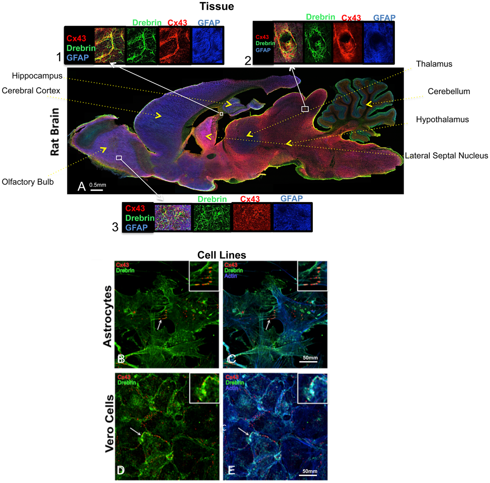
Here're some recent examples: Cinzia Ambrosi , Cynthia Ren, Gaelle Spagnol, Gabriel Cavin, Angela Cone, Elena E. Grintsevich, Gina E. Sosinsky, Paul L. Sorgen. Connexin43 Forms Supramolecular Complexes through Non-Overlapping Binding Sites for Drebrin, Tubulin, and ZO-1. Published: June 9, 2016. http://dx.doi.org/10.1371/journal.pone.0157073...chicken anti-GFAP in blue (Neuromics, Catalog # CH22102)...
Figure 1. Cx43 and drebrin colocalization analysis in brain and cellular models. (A) Rat brain transversal slice mosaic shown after multiple immunolabeling with antibodies anti-Cx43 (red), anti-drebrin (green), and anti-GFAP (blue) as astrocytes marker. White boxes localize the area enlarged in insets 1, 2, and 3 (six fold enlargement). Colocalization of drebrin and Cx43 (yellow) is especially noticeable around the blood vessels (inset 2) and in regions rich of astrocytes (insets 1 and 3). The different regions of the brain were labeled. Cultured astrocytes (B and C) and Vero cells (D and E) were immunolabeled with anti-Cx43 (red), anti-drebrin (green), and anti-actin (blue). White arrows indicate zones of colocalization of Cx43, drebrin and actin that were enlarged in the insets (white boxes, three fold enlargement).

Xiangchen Li, Yu Guo, Yaxin Yao, Jinlian Hua, Yuehui Ma, Changqing Liu, Weijun Guan.Reversine Increases the Plasticity of Long-Term Cryopreserved Fibroblasts to Multipotent Progenitor Cells through Activation of Oct4. International Journal of Biological Sciences 2016; 12(1): 53-62. doi: 10.7150/ijbs.12199...anti-GFAP and NSE (1:200,Neuromics, MN, USA)...
Save 70 USD on High Titer Neuron/Synapse Markers-Only 225/100 ul (Through June 30, 2016).
We will continue to post news on our Neuroscience Research Solutions!

Monday, May 02, 2016

New Neuronal Astroglial PNS Markers

Save 70 USD on High Titer Antibodies
The foundation of our company is built on our catalog of proven, published and high titer Neuron-Astroglia and PNS Markers. We are pleased to offer 75 USD off our latest additions.
Antibody
Type
Species Reactivity
Applications
Mouse IgG
Chicken IgY
Chicken IgY
Mouse IgG
Mouse IgG
Mouse IgG
Mouse IgG
Mouse IgG
Chicken IgY
Rabbit IgG
Mouse IgG
B; H; M; R
B; H; M; R
H; M; R
B; H; M; R
B; H; M; R
H; R
Ch; H; M; Pr; R
Ch; H; M; R
H; M; R
H; M; R
B; H; M; R
IF; WB
ICC; IF; IHC; WB
ICC; IF; IHC; WB
IF; WB
IF; WB
IF; WB
ICC;
IHC; WB
ICC; IHC; WB
ICC; IF; IHC; WB
ICC; IF; IHC; WB

Images: Left: View of mixed neuron/glial cultures stained with Aldolase-C (green) and our rabbit antibody to NeuN/FOX3 (red). MCA-4A9 antibody reveals strong cytoplasmic staining in astrocytes, while Rabbit Fox3/NeuN antibody shows nuclear and distal cytoplasmic staining in neuron cells and is complete absence of astrocytes. Blue is a DNA stain. Middle and Right: Mouse brain sections (fixed by transcardial perfusion with 4% paraformaldehyde) stained with Aldolase-C (red) and our chicken Vimentin antibody (green). In the striatum (Middle), Aldolase-C positive astrocytes are highly co-stained Vimentin, which results in yellow to gold colors. In the cerebellum (Right), however, Aldolase-C positive Purkinje cells do not express vimentin, which results in red color. Insets show a higher magnification picture of MCA-4A9 single labeling in red. Nuclei are labeled with DAPI (blue).
Our clients often use these antibodies for double and triple labeling like the above example.


We plan on aggressively be adding more and more of these type of antibodies. Stay tuned.

Tuesday, October 21, 2014

Neurite Outgrowth Assays

Cells, Media and Markers

I have previously posted use of our Neurons in Live Content Assays for the study of Neurite Outgrowth: Neurons-Live Content Assays. These assays are critical for the study of repair and regeneration.

A recent publication featured several of our Neuron MarkersSerena Quarta, Bastian E. Baeumer, Nadja Scherbakov1, Manfred Andratsch, Stefan Rose-John, Georg Dechant3, Christine E. Bandtlow, and Michaela Kress: Peripheral Nerve Regeneration and NGF-Dependent Neurite Outgrowth of Adult Sensory Neurons Converge on STAT3 Phosphorylation Downstream of Neuropoietic Cytokine Receptor gp130. The Journal of Neuroscience, 24 September 2014, 34(39): 13222-13233; doi: 10.1523/JNEUROSCI.1209-13.2014.
Live labeling of neuron cultures: After 20 or 48 h, neurons were live-labeled with α-gp130 antibody diluted in cold TNB medium for 30 min on ice. After washing, neurons were incubated with the secondary antibody diluted in cold TNB medium for 30 min and washed with PBS. Cells were fixed either with 4% PFA for 20 min at room temperature (RT) or with methanol at −20°C for 2 min. After permeabilization with 0.01% TX-100 (Pierce) unspecific binding was blocked for 30 min with 10% normal goat serum (Sigma-Aldrich) in PBS. Cells were incubated with the first antibody for 1 h, washed three times for 10 min with PBS and incubated with the appropriate secondary antibody for 30 min, counterstained with 4′, 6-diamidino-2-phenylindole (1:10,000; Sigma-Aldrich) and embedded in Mowiol (Calbiochem). As primary antibodies, α-gp130 (1:50; Neuromics), α-β-III-tubulin clone TuJ-1 (1:1000; R&D Systems), and α-neurofilament-H (α-NF-H; 1:200; Neuromics) were used. Secondary antibodies used were α-goat AlexaFluor 594 (1:1000; Invitrogen), chicken α-mouse AlexaFluor 594 or donkey α-mouse AlexaFluor 488 (1:1000; Invitrogen), and goat α-chicken AlexaFluor 568 (1:10,000; Invitrogen) for fluorescence microcopy.

Images: Reduced density of TuJ-1+ nerve endings in the epidermis in SNS-gp130−/− mice after lesion. A, Representative cross sections of hindpaw glabrous skin of naive and 12 dpl gp130fl/fl and SNS-gp130−/− mice stained with the pan neuronal marker TuJ-1. The dotted line indicates the border between dermis and epidermis. Scale bar, 40 μm. B, Quantification of the total number of TuJ-1+ fibers (NE) per 1000 μm2 of epidermal area shows a significant decrease in density in SNS-gp130−/− mice after lesion compared with control animals (*p < 0.05; n = 4 for each group). Data are presented as mean ± SEM and analyzed by Mann–Whitney U test. C, 3D reconstruction of the deeper layer of the dermis shows fewer nerve bundles in SNS-gp130−/− dermis compared with controls. D, No NF-H+ proprioceptive fibers were detectable in the epidermis of gp130fl/fl animals at 12 dpl. Scale bar, 40 μm.

If you want to learn more about our Neuron-Glial-Astrocyte based assay solutions do not hesitate to contact me (612-801-1007) or pshuster@neuromics.com. Pete Shuster, Owner and CEO, Neuromics.

Wednesday, July 16, 2014

Potents Tools for Neuroscience Based Toxicology Assays

Neuromics' Offers Best in Class Cell and Markers

I am always on the hunt for proof that are tools work in the many different applications required by Researchers Studying Neurotoxicology. Success is confirmed to us through Customer Data/Pubs and Testimonials.

I would like to feature here some examples:


Figures: Neurons stained with Neuromics' MAP2 antibody to determine Neurite Damage.

We guarantee results. If you would like to learn more, please contact me directly at pshuster@neuromics.com or direct phone line: 612-801-1007. Thank you.

Tuesday, June 25, 2013

Protocol for Flow Cytometric Sorting of Enriched Neuronal Cultures from iPSCs

Surface molecule profiles undergo dynamic changes in physiology and pathology, serve as markers of cellular state and phenotype and can be exploited for cell selection strategies and diagnostics. The isolation of well-defined cell subsets is needed for in vivo and in vitro applications in stem cell biology. In this technical report, the authors present an approach for defining a subset of interest in a mixed cell population by flow cytometric detection of intracellular antigens. They have developed a fully validated protocol that enables the co-detection of cluster of differentiation (CD) surface antigens on fixed, permeabilized neural cell populations defined by intracellular staining. Determining the degree of co-expression of surface marker candidates with intracellular target population markers (nestin, MAP2, doublecortin, TUJ1) on neuroblastoma cell lines (SH-SY5Y, BE(2)-M17) yielded a combinatorial CD49f-/CD200high surface marker panel. Its application in fluorescence-activated cell sorting (FACS) generated enriched neuronal cultures from differentiated cell suspensions derived from human induced pluripotent stem cells. Our data underlines the feasibility of using the described co-labeling protocol and co-expression analysis for quantitative assays in mammalian neurobiology and for screening approaches to identify much needed surface markers in stem cell biology: Turaç G, Hindley CJ, Thomas R, Davis JA, Deleidi M, et al. (2013) Combined Flow Cytometric Analysis of Surface and Intracellular Antigens Reveals Surface Molecule Markers of Human Neuropoiesis. PLoS ONE 8(6): e68519. doi:10.1371/journal.pone.0068519


Figure 1. Experimental outline. Schematic illustrating the research strategy of identifying novel surface marker combinations on a target population in neural and other stem cell differentiation systems for which intracellular, standard immunocytochemical markers are well established. Following harvesting, the resulting single cell suspension is subject to surface antigen candidate staining, followed by gentle fixation, permeabilization and subsequent co-staining with known intracellular markers. CD markers co-labeling the target population serve as positive markers, those absent on the target population serve as negative markers. In a separate, subsequent step, a combination of the identified positive and/or negative CD markers enables the flow cytometric enrichment of the viable population of interest from a heterogeneous cell suspension for further study and biomedical applications. doi:10.1371/journal.pone.0068519.g001.
Figure 2. Accurate detection of intracellular antigens with optimized fixation-permeabilization conditions preserving surface antigens. Flow cytometric detection of TUJ1, MAP2 and nestin antigens in BJ fibroblasts and the neural SH-SY5Y cell line (A). TUJ1 and nestin are present in both cell lines, while the mature neuronal marker MAP2 was only detected in SH-SY5Y cells (arrows). Note stable fluorescent levels of the negative population, indicating low background staining using this protocol. Representative experiment of three independent repeats shown. (B) Corresponding validation by immunofluorescence analysis. (C) Quantitation of TUJ1, MAP2 and nestin intracellular antigen detection (n=3). Error bars indicate standard deviation. (D) Response of TUJ1 and MAP2 intracellular antigen expression to 6 DIV of 10 µM retinoic acid (RA) treatment of SH-SY5Y cells. Note disappearance/reduction of subsets negative for these markers (upward shift, green arrows), as well as a shift toward CD184low expression with differentiation (blue arrows). doi:10.1371/journal.pone.0068519.g003.

Please note: In addition to the our Human Mouse Monoclonal Nestin Antibody used in the study, we have an extensive catalog of stem cell solutions. I will continue to post highlights of new applications.

Monday, April 29, 2013

Neuron-Glial Cultures-Setting a Higher Bar!

Improved Methods for Long Term, High Denisty Cultures
 
Dr. Randen Patterson and his team at UC Davis have developed new culturing techniques using our e18 Rat Primary Hippocampal Neurons. They have developed a protocol that allows for culturing of E18 hippocampal neurons at high densities for more than 120 days. These cultured hippocampal neurons are (i) well differentiated with high numbers of synapses, (ii) anchored securely to their substrate, (iii) have high levels of functional connectivity, and (iv) form dense multi-layered cellular networks. We propose that our culture methodology is likely to be effective for multiple neuronal subtypes–particularly those that can be grown in Neurobasal/B27 media. This methodology presents new avenues for long-term functional studies in neurons. This is good news indeed: Todd GK, Boosalis CA, Burzycki AA, Steinman MQ, Hester LD, et al. (2013) Towards Neuronal Organoids: A Method for Long-Term Culturing of High-Density Hippocampal Neurons. PLoS ONE 8(4): e58996. doi:10.1371/journal.pone.0058996.
 
Protocol Highlights: 
Substrate Preparation
1. On the day of plating, prepare 25 mm coverslips by removing them from 70% ethanol storage solution and propping them up at an angle in each well of a 6-well culture plate to allow drying. [No more than 5 plates (30 coverslips) should be dried simultaneously for 15–25 minutes in culture hood to avoid over-drying.]
2. Once dry, shake slips down flat into their respective wells and coat with 1 mL 0.1% poly-D-lysine, taking care to form a liquid meniscus on each slip. Carefully transfer coverslips into incubator, taking care to preserve meniscus.
3. Incubate for 1 hr at 37°C. [Keeping poly-D-lysine meniscus on top of coverslip is important; this serves to avoid poly-D-lysine coating under coverslip surface that may lead to problematic flotation of coverslip.] After incubation, remove poly-D-lysine and rinse each coverslip three times with 2 mL sterile deionized water. Take care to ensure coverslips do not completely dry at any point during the rinse. After the third and final rinse, leave coverslips in 2 mL sterile deionized water for at least 1 hr. Remove water just before plating, again, make sure to avoid over-drying. [This critical step requires attention. Take care to aspirate off all sterile water remaining from the final rinse, but also use caution as not to over-dry the coverslips. Ultimately, the coverslip must be mostly dry as to allow for the meniscus formation during plating (Fig. 1, Step 12), whereas over-drying can result in the neurons peeling off the glass coverslips days to weeks after plating.]
Fig 1: Neuron-Glial Culturing Steps
Preparation of Isolated Neurons (Numbers in Fig. 1 correspond to numbers below).
1. Store tissue at 4°C until ready to use. If dissecting your own cultures, upon isolation of the tissue, store in an appropriate storage media.
2. When ready to plate, make 2 mL of enzymatic solution without B27. In our case, we used Hibernate E-Ca, containing 4 mg (2 mg/mL) of papain. If making your own solution, use a commercially available papain dissociation kit. Make sure to sterile filter solution with 0.2 micron filter after adding papain if source of enzyme is not sterile.
3. Remove the storage media from the dissected tissue and transfer into sterile 15 mL screw-cap tube; be careful not to disturb or remove tissue from original tube. Save the storage media, do not discard.
4. Add 2 mL of media made in Step 2 to tissue (in our case, Hibernate E-Ca containing 2 mg/mL of papain). Incubate for 35 min at 37°C. [Be sure to add Hibernate E-Ca containing papain slowly as to avoid disturbing tissue.]
5. Remove enzymatic solution from tissue, again, take care not to disturb or remove tissue. Add back 1 mL of storage media saved in 15 mL tube.
6. Using a 1 mL pipettor with a sterile plastic pipette tip (tissue can adhere to glass pipettes), aspirate the tissue with the medium into the pipette and immediately dispense contents back into same container. Take care not to create bubbles. [This is another critical step that requires attention. Take care to make sure pipette tip remains in a stable position (as shown in Fig. 1, Step 6). Maintain slow, steady speed when both drawing in and re-dispensing media containing tissue.]
7. Repeat this trituration step 10–12 times or until most all the tissue is dissociated and the cells are dispersed. [Under close examination cell dispersion is highly visible. Stop pipetting immediately upon cell dispersion.]
8. Slowly transfer contents of the tissue tube into a new sterile 15 mL screw-cap tube.
9. Use the remainder of storage medium saved in Step 3 and rinse the interior of the tissue tube before adding it to the sterile 15 mL screw cap tube containing dispersed cells from step 7. [This step helps ensure minimal wastage, as any remaining cells should be saved with this extra rinse.]
10. Spin dispersed cells at 1,100 rpm (200Xg) for 1 min.
11. Discard the supernatant while being careful not to remove any of the cells from cell pellet.
12. Flick tube a few times to loosen the cell pellet. Re-suspend pellet in 2.4 mL of pre-warmed B27/Neurobasal/0.5 mM glutamine medium. Re-suspend by gently pipetting up and down. For E18 Hippocampus, medium includes 25 µM glutamate.
13. Plate cells within a meniscus (approx. 10 mm diameter) at a minimum of 40 µL per 25 mm coverslip. Take care not to disturb meniscus. [Periodically pipette up and down throughout plating process (no more than once every plate per 6 coverslips) to help maintain equal cell density. Again, plating with meniscus formation is critical.]
14. Incubate plated cells at 37°C with 5% CO2 and/or 9% or 20% oxygen for 1 hr.
15. Add 1.5 mL per well of pre-warmed 1:1 ACM/NbActiv4. [Slow and steady media addition rate and proper pipette position are necessary for successful plating density consistency. Position pipette tip at 45° angle along middle of 6-well interior sidewalls, dispense 1.5 mL as slowly and steadily as possible (see Steps 1–14).]
16. Incubate cells at 37°C with 5% CO2 and/or 9% or 20% oxygen.
17. Add Cytosine β-D-arabinofuranoside (Ara-C) to a final concentration of 5 µM, 5–6 days after plating to curb glial proliferation. [Remove 1/3 of media from each well and replace with equal volume containing final concentration of Ara-C]
18. After 4 days or longer, neurons are well differentiated. If further culture is desired, change 1/3 of medium with fresh, pre-warmed 1:1 ACM/NbActiv4 every 7–8 days.
Images: 40X Confocal Images of 30 DIV Hippocampal Cultures. Immunofluorescence detection of MAP-2 (green) and GFAP (red) in 30 DIV (A–C) cultured E18 hippocampal cells using a 40X objective. These images (A and B) clearly depict the intimate physical contact between glia processes and dendritic arbors. Under closer examination (CI and CII), it is clear that the dendrites have grown both bellow (blue arrows) and above (white arrows) glial processes, forming a highly interconnected three-dimensional network by 30 DIV. doi:10.1371/journal.pone.0058996.g004.
All Primary Neuron Assay Customer Publications
 
Related Content: If you have any questions on optimizing your cell cultures, do not hesitate to contact me @ pshuster@neuromics.com or 612-801-1007

Thursday, April 26, 2012

Primary Hippocampal Neurons Performing!

I advertise our Primary Neurons and Astrocytes as being easy to culture, grow and maintain. We confirm this via the data/images our customers generously share and the many publications referencing use of the cells.

I would like to thank George Kenneth Todd (Patterson Lab at UC Davis) for these wonderful images of our e18 Primary Rat Hippocampal Neurons. These were taken at day 67!


Prior to staining, the cells were treated with Wnt5 for 2 min, then Wnt5 + Wnt3 for an additional 2 min during Calcium Imaging experiments. The cells were then fixed and stained for IP3R (green), Frizzled2 (blue), and B-Catenin (red), and the confocal images were captured at 10x. Notice the parallel neurites formation in image 3.


For staining culture options, check out our neuron-glial-astrocyte markers.

Thursday, October 20, 2011

Immunostaining Neurons and Glia

I would like to thank Dr. Gerry Shaw, University of Florida for his excellent work with our Primary Neurons and Astrocytes and Neuronal-Glial Markers. Here's an example image with many more to follow:

Image: E18 hippocampal neurons stained with MAPT (red) and Doublecortin (green). The two proteins overlap in the proximal dendrites, but doublecortin is more abundant in the growth cones and periphery. As a result, the periphery appears green while the more proximal regions of the cells are yellow. The single longer process of this cell, presumably an axon, has a low doublecortin content and so appears red. Blue staining is the nuclear DNA. Protocol on datasheet.

Thursday, December 16, 2010

Markers for Medial Superior Olivary Neurons

This is an excellent reference for researchers looking for immunohistochemistry images of slice preparations of the Neurons in the medial superior olive (MSO). It also references use of our widely used and frequently published MAP2 (Microtubule associated protein 2).

Kiri Couchman, Benedikt Grothe and Felix Felmy. Medial Superior Olivary Neurons Receive Surprisingly Few Excitatory and Inhibitory Inputs with Balanced Strength and Short-Term Dynamics. The Journal of Neuroscience, December 15, 2010, 30(50):17111-17121; doi:10.1523/JNEUROSCI.1760-10.2010.

Summary: Neurons in the medial superior olive (MSO) process microsecond interaural time differences, the major cue for localizing low-frequency sounds, by comparing the relative arrival time of binaural, glutamatergic excitatory inputs. This coincidence detection mechanism is additionally shaped by highly specialized glycinergic inhibition. Traditionally, it is assumed that the binaural inputs are conveyed by many independent fibers, but such an anatomical arrangement may decrease temporal precision. Short-term depression on the other hand might enhance temporal fidelity during ongoing activity. For the first time we show that binaural coincidence detection in MSO neurons may require surprisingly few but strong inputs, challenging long-held assumptions about mammalian coincidence detection. This study exclusively uses adult gerbils for in vitro electrophysiology, single-cell electroporation and immunohistochemistry to characterize the size and short-term plasticity of inputs to the MSO. We find that the excitatory and inhibitory inputs to the MSO are well balanced both in strength and short-term dynamics, redefining this fastest of all mammalian coincidence detector circuits.

Related Reagents:
Neuronal-Glial Markers
-Astrocytes, Glia,
Microglia, Olidogodendrocytes, Progenitors and Schwann Cell Markers
Stem Cell Research Antibodies

Stem Cell Research Reagents

Primary Neurons and Astrocytes
-Primary
human, rat and mouse neurons and astrocytes.

Tuesday, December 29, 2009

Tuj-1-Neuronal Differentiation Marker

Our Tuj-1 antibodies are widely used and frequently referenced in customer publications. They are proven markers for Neural Progenitor and Neuronal Differentiation. Here's the latest reference:


...Tuj 1 (Neuron-specific class III beta-tubulin)-Mouse (MO15013, Neuromics Antibodies, Edina, MN)...

Immunofluorescence Method:

Cells grown on coverslips were fixed for 5 min in 4% paraformaldehydecontaining 4% sucrose in phosphate buffer saline (PBS) at 37ºC. Cells were then permeabilized with 0.2% Triton X-100 in PBS during 5 min at room temperature. After blocking (5% bovine serum albumin in PBS for 1 h), cells were incubated with the corresponding primary antibodies, and immunoreactivity was detected with the suitable fluorophore-conjugated secondary antibody before mounting on slides with Mowiol4-88 (Harland Co., UK). Confocal images were acquired using an inverted Leica TCS SP5 laser confocal microscope with a 63X Plan- Achromatic oil immersion objective and processed with LAS AF Leica Application Suite and Adobe Photoshop CS2 (Adobe Systems
Inc., CA). All images correspond to the projection of sections from a ~50μm z-stack, except for colocalization analysis where they correspond to 0.5-0.7μm single sections.

Image: Rat hippocampal neurons were fixed at 1.5 DIV and immunostained for the neuronal marker βIII-Tubulin/Tuj1 (blue).