Wednesday, December 28, 2016

Dopamine and Morphine Tolerance

Dopamine Identified as Key Player

Our Mu Opioid Receptor antibody is used to show that blocking dopamine decreases morphine tolerance: Wen-Ling Dai, Feng Xiong, Bing Yan, Zheng-Yu Cao, Wen-Tao Liu, Ji-Hua Liu1, Bo-Yang Yu. Blockade of neuronal dopamine D2 receptor attenuates morphine tolerance in mice spinal cord. Scientific Reports 6, Article number: 38746 (2016). doi:10.1038/srep38746.

Images: (A) Double immunofluorescence staining showed that MOR (green) and D2DR (red) were co-localized in the mice spinal cord (20X magnification). Chronic morphine treatment increased the co-localization of MOR and D2DR in the spinal cord, and D2DR antagonist sulpiride (8 μg/10 μl, i.t.) reduced the increased co-expression of D2DR with MOR (n = 4). (B) Co-IP experiments showed that D2DR could interact with MOR, and the MOR/D2DR interactions were increased in the spinal dorsal horn after chronic morphine treatment for 7 days while D2DR antagonist sulpiride (8 μg/10 μl, i.t.) disrupted the interactions of the MOR/D2DR (n = 3).

Blockade of D2DR in spinal cord can disrupt the interactions between MOR and D2DR to attenuate morphine tolerance. These findings highlight the possibility of a new clinical strategy to prevent morphine antinociceptive tolerance.

Friday, December 16, 2016

Neuromics Cornerstone

Human Primary + Stem Cells and 3-D Tissue Like Models

We are pleased by growing demand for our "hard to find" primary cells and 3-D BBB model. These are proving to be engines for drug discovery and toxicology assays.

We have have the capabilities isolate cells from healthy human donors. We then grow and characterize the cells and cryo-preserve in vials of 500,000 cells. They are now ready to culture.

We are increasingly determining types of cells to add to our growing catalog by meeting the specific demand of our clients. For example, our addition of primary human schwann cells and microglia happened, because they were needed for a specific client study.
Human Schwann Cells
Our 3-D Blood Brain Barrier Model is also working well in the hands of our clients. We are receiving positive input and this is supported by repeat orders. It is designed for users to test the ability of compounds, molecules and oligos to pass the BBB and penetrate into the brain.

Our cornerstone: hard to find human primary/stem cells + 3-D tissue like assays.
We can say, "Need cells or 3-D tissue like models?  Just ask us!". Pete Shuster, CEO and Owner-direct phone: 612-801-1007 or pshuster@neuromics.com

Tuesday, December 13, 2016

Need Cells From Diseased Donors?

Just Ask
Some of our customers have expressed the need for Cells that have the phenotype of a given disease. This is especially true for researchers doing drug discovery for neuro-diseases like Alzheimer's and Parkinson's.

Deriving "hard to find" cells from tissue is one of our cornerstones.
If you have need for any type of human cell(s), give us a shot. I can be reached at 612-801-1007 or pshuster@neuromics.com. Thank you. Pete Shuster,CEO and Owner, Neuromics.


Monday, December 05, 2016

Fatty Acids and IBD and Colitis

The Dangers of a High Fatty Acid Diets
Our westernized high-fat diet (HFD) is associated with the development of inflammatory bowel disease (IBD), Colitis and other intestinal related diseases and cancers. Here one the root causes is identified to elevated production of pro-inflammatory cytokine-IL-1β by macrophages. It is suspected that HFD sets off an autoimmune like cascade.

This cascade is triggered by NLRP3 inflammasomes. Our Apoptosis Kit-Magic Red-Real Time Cathepsin B is used to demonstrate these inflammasomes are activated via Cathepsin B Release: Shengnan Zhao, Zizhen Gong1, Jiefei Zhou1, Chunyan Tian, Yanhong Gao, Congfeng Xu, Yingwei Chen, Wei Cai and Jin Wu1Deoxycholic Acid Triggers NLRP3 Inflammasome Activation and Aggravates DSS-Induced Colitis in Mice. Front. Immunol., 28 November 2016 | http://dx.doi.org/10.3389/fimmu.2016.00536 ...Lipopolysaccharide-primed J774A.1 cells were incubated with or without DCA (100 μM, 24 h); then, the cells were stained with Lyso Tracker Green DND-26 (Invitrogen) or magic red cathepsin B fluorogenic substrate z-Arg-Arg cresyl violet (Neuromics) for 1 h, followed by Hoechst staining for half an hour. Fluorogenic signals were captured by inverted fluorescence microscope (Leica)...

Image: Lipopolysaccharide-primed J774A.1 cells were incubated with or without DCA (100 μM, 24 h); then, the cells were stained with Lyso Tracker Green DND-26 (Invitrogen) or Magic RedTM Cathepsin B fluorogenic substrate z-Arg-Arg cresyl violet (Neuromics) for 1 h, followed by Hoechst staining for half an hour. Fluorogenic signals were captured by inverted fluorescence microscope (Leica). Front. Immunol., 28 November 2016 http://dx.doi.org/10.3389/fimmu.2016.00536
We will continue to post how our customers are using our Apoptosis Kits for their discoveries here.

Friday, December 02, 2016

Need Cells?

Kick of Your Holiday Season with a 25 USD Gift Card
All cell orders this month will include a 25 USD Amazon Gift Card. This is to help celebrate the Holiday Season.
Here are  your options.
Human Cells
Isolated from healthy human brain tissue
Stable, Potent and Well Characterized
NeuroNet Pure Human Neurons Discovery and HTS Kits
Pure Population of hPSC Derived Neurons
Human spinal nerve derived primary cells
Derived from Cortex
Solutions for studying tumor growth, angiogenesis and metastasis.
hMSCs Derived from Umbilical Cord Blood
Human Endothelial Cells-New
Artery, Microvascular, Vein &Umbilical Cord Derived
Human Fibroblasts-New
Pancreas and Neonatal Dermal Derived
Human Cardiomyocytes-New
Derived from hMSCs
Mouse and Rat Astrocytes and Neurons

We wish you a Joyous Holiday Season.

Monday, November 21, 2016

New Schwann Cells Now $695/500,000 Cells

Priced to Buy and Try!
Schwann cells play important roles in the development, function, and regeneration of peripheral nerves. When an axon is dying, the Schwann cells surrounding it aid in its digestion, leaving an empty channel formed by successive Schwann cells, through which a new axon may then grow from a severed end.

We want to make it easy for you to buy and try these verdant cells. Also, we have a full refund or replacement should they not meet your expectations.
Schwann Cells in culture
Questions? I am available to help-Pete Shuster, CEO and Owner-612-801-1007 or pshuster@neuromics.com.

Tuesday, November 15, 2016

Infection of Mesenchymal Stem Cells

UCB derived huMSCs infected with RSVs

Here's cool customer data of our Human Mesenchymal Stem Cells.

Figure. RSV infects and replicates in human MSCs. Human MSCs grown on coverslips, infected with 1 MOI of RSV, and immunostained at 48 or 72 hours post-infection with a monoclonal antibody to RSV tagged with FITC (green), Evan’s blue dye (red) and DAPI (blue). Total magnification of images is 200X (inset 1000X) and scale bar is 50μm (A). RSVN transcripts detected in MSCs at 6, 12, 24 and 72 hours post infection normalized to mock (B). RSV titers (PFU/ml) isolated from the culture medium of infected and mock-infected MSCs is shown (C). Results representative of at least duplicate experiments. doi.org/10.1371/journal.pone.0163709.

Your data is wanted. Just email rose@neuromics.com and claim your 50 USD reward. #datawanted!

Wednesday, November 09, 2016

Detecting Expression of CAS9

Gain Trust That Your Gene Editing is on Target CRISPR/Cas9 is a hot genome-editing technology. However, the high frequency of off-target activity is a major issue.
One way to make sure CAS9 is being properly expressed is using markers against related proteins.

We are please to introduce our CAS9 markers. We plan on adding to these in the near future.

Image: Transfected Hek293 cells overexpressing the N-terminal 1-608 amino acids of S. pyogenes Cas9 . The cells were stained with MO22174 in red, and these cells also appear yellow since we counterstained with our rabbit Cas9 antibody, RA22126 to the same construct in green, giving a yellow color. The N-terminal construct contains a nuclear localization sequence and so is predominantly nuclear in localization. Most Hek293 cells in this field are not transfected so only the nuclei of these cells can be visualized with the blue DAPI DNA stain.
Name
Catalog #
Type
Applications
MO22174
Mouse IgG
ICC; IF; WB
RA22126
Rabbit IgG
ICC; IF; WB
MO22175
Mouse IgG
ICC; IF; IHC; WB
We will post new CAS9 marker additions here.

Friday, November 04, 2016

We Want Your Data!

50 USD Reward
Our Customer Data program is progressing nicely. I am pleased to present the latest addition from Dr. Vimal Swarup, University of Utah. To qualify for the reward just email Rose Ludescher-rose@neuromics.com.


Hippocampalneuron-based guidance assay. Image of micro-contact patterned chondroitin sulfate proteoglycans (pre-mixed with AlexaFluor 594) purified from astrocyte conditioned media (CM). b) Immuno-stain of the spot in (a) with anti-CSPG antibody CS56. c) Overlay of Embryonic day-18 primary rat hippocampal neurons (Cat# PC35101, Neuromics Inc.) and the underlying surface containing μC patterned CM after 48 h of growth. d) Overlay of neurons and underlying surface containing μC patterned CSPGs purified from CM. e) Average neurite length on pLL and stamped regions after 48 h. f) Average neurite length on plain field surfaces of μC patterned CM, CSPG and pLL after 48 h. Results from this experiment show that neurons avoid CM or CSPGs purified from CM while elongating. Swarup et al. JACS. 2013

Submit your data today and earn you reward.

Tuesday, October 25, 2016

Co-Cultures and Tox Assays

Data End Points Differ
Our clients continue to use our cells and kits for co-culturing. The goal is to develop the most in vivo like results possible.

They find that single cell vs multi cell type co-cultures drive different data sets.

At each dose, the results vary and as the dose of Bis1 increases this variation increases.

We welcome your questions, input and feedback on our co-cultures. Pete Shuster, Owner, pshuster@neuromics.com or 612-801-1007.

Friday, October 14, 2016

"Hard to Find" Cells

You can Trust Your Results
Cells form the foundation of your cell based assays. We work hard to provide the cells you need and, more importantly that they are healthy and happy.

We are pleased to announce that we are recognized for having primary human cells that comply with federal and international regulations. This is especially important in drug discovery, because if the cells yield targets that result in future therapies this validation becomes increasingly valuable.
Human Brain Microvascular Endothelial Cells in Culture

Looking for "hard to find cells"? Check us out today.
Cancer-Associated Fibroblasts (CAFs) and Cells
Solutions for studying tumor growth, angiogenesis and metastasis.
Human Brain Astrocytes-New
Derived from Cortex
Immortalized Human Brain Microglia Cells
Isolated from healthy human brain tissue
Human Schwann Cells - New
Human spinal nerve derived primary cells
hN2™ Neuron Discovery Kits
Derived from H9 (WA09) ECSs-Consistent, Easy to Use & Cost Effective
Human alpha Motor Neurons Kit
hMSC Derived-includes 100 ml of media to optimize your cultures
Human Cardiomyocytes-New
Derived from hMSCs
Human Chondrocytes-New
Derived from hMSCs
Human Endothelial Cells-New
Artery, Microvascular, Vein & Umbilical Cord Derived + Culture Media
Human Fibroblasts-New
Pancreas and Neonatal Dermal Derived
NeuroNet Pure Human Neurons Discovery and HTS Kits
Pure Population of hPSC Derived Neurons
Human Osteoblasts-New
Derived from hMSCs
Human Mesenchymal Stem Cells (hMSCs)
hMSCs Derived from Umbilical Cord Blood
Stressed Selected Stem Cells
They Survive to Thrive
Human Pericytes
Brain and Retinal Derived
3-D Cell Based Assay Solutions
Nanofibers, Hydrogels and Extracellular Matrix (ECM) Proteins

Thursday, October 06, 2016

Canniboids and Pain Relief

Could The CB1-TRPV1 Crosstalk be a Target
Our pain marker-TRPV1-was recently referenced in an interesting study. Here researchers suggest that spatial distribution of the CB1 receptor and TRPV1 contributes to the complexity of their functional interaction.

They conclude: "Therefore, in order to utilise the analgesic potential of endogenous anandamide fully in PSN, ways to interfere with the mechanisms, through which the CB1 receptor promotes anandamide-responsiveness and sensitisation of TRPV1, must be identified. Due to the co-expression of TRPV1 and the CB1 receptor in various areas of the brain, the CB1 receptor – TRPV1 crosstalk we characterised in the present study could also have relevance in brain signal processing."


Chen J, Varga A, Selvarajah S, Jenes A, Dienes B, Sousa-Valente J, Kulik A, Veress G, Brain SD, Baker D, Urban L, Mackie K, Nagy I. Spatial Distribution of the Cannabinoid Type 1 and Capsaicin Receptors May Contribute to the Complexity of Their Crosstalk. Sci Rep. 2016 Sep 22;6:33307. doi: 10.1038/srep33307.

The SDS-FRL method reveals two major types of CB1 receptor and TRPV1 spatial distribution in DRG neurons. (A,B) Double immunogold labelling for TRPV1 (12 nm particles; arrows) and the CB1 receptor (5 nm; arrowheads) reveals that the immunoreactivity for both proteins is moderate (A) to strong (B) on the protoplasmic face (P-face) but not on the exoplasmic face (E-face) of plasma membrane of PSN. In some patches of putative somatic membrane of PSN, the great majority of immunoparticles for TRPV1 and CB1 are isolated from each other (A), whereas on other patches of the membrane immunoparticles for TRPV1 and CB1 form co-clusters (arrow + arrowhead in B). Note the occasional co-clustering of particles labelling TRPV1 (double arrows in B). Scale bars = 0.2 μm
Methods: Sodium dodecyl sulfate (SDS)-digested freeze-fracture replica immunolabeling Immunohisto-chemical labeling for electron microscopy was performed. Briefly, adult (150–200 g) male Sprague-Dawley rats (n = 4) were anesthetized with sodium pentobarbital (50 mg/kg, i.p.), and perfused transcardially with 25 mM PBS, followed by 2% paraformaldehyde and 15% saturated picric acid in 0.1 M phosphate buffer (PB). Sections from DRGs were cut at a thickness of 110 μm. The slices were cryoprotected in a solution containing 30% glycerol in 0.1 M PB and then frozen by a high-pressure freezing machine (HPM 100; Leica, Austria). Frozen samples were inserted into a double replica table and fractured into two pieces at −130 °C and replicated by carbon deposition (5 nm thick), platinum (2 nm), and carbon (15 nm) in a freeze-fracture replica machine (BAF 060; BAL-TEC, Lichtenstein). They were digested in a solution containing 2.5% SDS and 20% sucrose in 15 mM Tris-HCl at 80 °C for 18 h then at 60 °C for 18 h. The replicas were washed in 25 mM PBS containing 0.05% bovine serum albumin (BSA; Roth, Germany) and then incubated in a blocking solution containing 5% BSA for 1 h. Subsequently, replicas were incubated in a mixture of primary antibodies (TRPV1 raised in guinea pig against the 22 amino acids of the C terminus (YTGSLKPEDAEVFKDSMVPGEK; Neuromics, USA), the dilution was 1:1000 and CB1 receptor (Prof. K. Mackie, raised in goat; Veress et al. 2014), dilution was 1:4000) in 50 mM Tris-buffered saline (TBS) containing 1% BSA. After several washes, replicas were reacted with a mixture of gold-coupled donkey anti-guinea pig (for TRPV1; 12 nm) and rabbit anti-goat (for CB1, 5 nm) secondary antibodies (1:30; BioCell Research Laboratories, Cardiff, UK) in 25 mM TBS containing 5% BSA. The anti-TRPV1 antibody from Neuromics provided better staining in the replicas than then the anti-TRPV1 antibody from Chemicon, which we tried in a pilot study. They were then washed and picked up on 100-mesh grids. The specificity of the antibodies was tested on tissues dissected from TRPV1 and CB1 knock out mice.

We will keep you posted as we discover new research on targeting CB1 for pain. That said, could this crosstalk be central to medical marijuana and its ability to ameliorate certain forms of pain?

Tuesday, October 04, 2016

Your Brain Trust

Trust is hard to gain and easy to lose. My focus over the past 12 years has been to earn the trust of all our clients. This means that we do everything possible to insure our solutions work. When we come up short, we strive to resolve your issues the same day, and we have one of the easiest replacement policies in the market today. Here's proof.
Trust and Testimonials: We use BirdEye, a 3rd party, to collect and manage feedback from our clients.
5on BirdEye, Aug 29, 2016
C. H.
The order arrived quickly. The service was great!
Product Name: Human Brain Microvascular Endothelial Cells, 500,000+ Cells, - (Cat# HEC02)

Human Brain Microvascular Pericytes (HBMPVCs), 500,000+ Cells, - (Cat# HMP104)
http://bit.ly/2bzQlPn
ENDO-Growth Media, - (Cat#MED001)
http://www.neuromics.com/endo-growth-media
Pericyte-Growth Media, - (Cat# PGB001)
http://www.neuromics.com/pericyte-growth-media
Organization: UCSF

HBMPVCs in Culture
Trust and Publications: There are hundreds of publication referencing use of our products. Want to see if a product of interest is published? Just Ask
Need hard to find human cells? Please contact me directly pshuster@neuromics.com or 612-801-1007. We can together determine your needs and desired outcomes and provide a statement of work with pricing, project milestones and delivery.
We wish you a productive October filled with new and exciting discoveries.

Monday, September 19, 2016

Your Neuron-Glia Co-Cultures Deserve the Best

There is a lot of cross talk between neurons and glia. In neuro-immuno and neuro-degenerative diseases this cross talk is dysregulated. Robust neuron-glia co-cultures are needed for drug discovery and basic research.Here is a study that references use of co-culturing to study the interactive effects of the viral protein HIV-1 Tat lipopolysaccharide (LPS) on enteric glia and neurons.

Here is a study that references use of co-culturing to study the interactive effects of the viral protein HIV-1 Tat lipopolysaccharide (LPS) on enteric glia and neurons. In order to optimize the co-culturing our Media, Coatings and GDNF growth factor are used. Joy Guedia, Paola Brun, Sukhada Bhave, Sylvia Fitting, Minho Kang, William L. Dewey, Kurt F. Hauser; Hamid I. Akbarali. HIV-1 Tat exacerbates lipopolysaccharide-induced cytokine release via TLR4 signaling in the enteric nervous system. Scientific Reports 6, Article number: 31203 (2016) doi:10.1038/srep31203.


Figures: (A) Representative confocal microscopic images of enteric β-III tubulin (green) and glial fibrillary protein (GFAP- red) showing the presence of neurons and glia isolated from the adult mouse LMMP. (B) IL-6 release from isolated mouse neuron/glia co-culture treated with 100 nM Tat, 100 ng/ml LPS or Tat+ LPS for 16 h measured by ELISA. (C) IL-1β release from neuron/glia co-culture treated with 100 nM Tat, 100 ng/ml LPS and Tat+ LPS for 16 h measured by ELISA (D) mRNA expression of TNF-α of mouse LMMP treated with 100 nM Tat, 100 ng/ml LPS and Tat+ LPS for 16 h. (E) TNF-α release from mouse LMMP treated with 100 nM Tat, 100 ng/ml LPS or Tat+ LPS for 16 h measured by ELISA.

Protocol: Myenteric neurons and glia were isolated as described recently41. Briefly, after euthanizing mice, the ileum was immediately removed and placed in ice-cold Krebs solution (118 mM NaCl, 4.6 mM KCl, 1.3 mM NaH2PO4, 1.2 mM MgSO4, 25 mM NaHCO3, 11 mM glucose and 2.5 mM CaCl2) bubbled with carbogen (95% O2/5% CO2). The ileum contents were discarded by passing Krebs solution using a syringe. The ileum was then divided into short segments which were threaded longitudinally on a plastic rod through the lumen and the longitudinal muscle containing the myenteric plexus (LMMP) strips were obtained using a cotton-tipped applicator. LMMP strips were rinsed three times in 1 ml Krebs and gathered by centrifugation (350 × g, 30 sec). LMMP strips were then minced with scissors and digested in 1.3 mg/ml collagenase type II (Worthington) and 0.3 mg/ml bovine serum albumin in bubbled Krebs (37 °C) for 1 h, followed by 0.05% trypsin for 7 min. Following each digestion, cells were triturated and collected by centrifuge (350 x g for 8 min). Cells were then plated on laminin (BD Biosciences) and poly-D-lysine coated coverslips in Neurobasal A media containing B-27 supplement, 1% fetal bovine serum, 10 ng/ml glial cell line-derived neurotrophic factor (GDNF, Neuromics, Edina, MN), and penicillin/streptomycin. Half of the cell media was replaced every 2–3 days with fresh complete neuron media.

We welcome your questions input on co-culturing. Pete Shuster, CEO and Owner , Neuromics-pshuster@neuromics.com or 612-801-1007.

Tuesday, September 13, 2016

Perky ERKies and Depression

ERK1/2 Signaling and Depression

Extracellular signal-regulated kinase 1/2- (ERK1/2-) mediated cellular signaling plays a major role in synaptic and structural plasticity. Although ERK1/2 signaling has been shown to be involved in stress and depression. Phosphorylation of the receptor is important for maintaining healthy signaling, reduction of oxidative stress and induction of growth factors.

We are pleased that our ERK1/2 markers have been published in these type of studies. Here's a recent one: MARIA CONCETTA OLIANAS, Simona Dedoni, and PIER LUIGI ONALI. LPA1 mediates antidepressant-induced ERK1/2 signalling and protection from oxidative stress in glial cells. Published online before print September 7, 2016, doi: 10.1124/jpet.116.236455. ...Membranes were incubated overnight at 4 °C with one of the following primary antibodies: anti-phospho-ERK1 (Thr202/Tyr204) / ERK2(Thr185/Tyr187) (RA 15002) (1:15,000) (Neuromics, Northfield, MN, USA.

We will continue to post updates on this important topic

Sunday, September 04, 2016

Multiplexed Protein Array for Immunologists

Assay 1,631 Antibodies in Under 8 Hours!
The RayBio® Immunome Protein Arrays are multiplex protein immunoassays for numerous research applications. Correctly folded, functional proteins are immobilized onto a solid glass slide surface. The slides can then be probed with serum or small molecules and used in protein-antibody studies, small-molecule inhibition of kinase studies, protein-protein studies, methylation studies or DNA binding studies. Our main immunome array product offering contains 1,631 proteins. Smaller custom arrays are also available.

Each 75mm x 25mm glass slide is spotted with 4 identical protein arrays (also called “subarrays”), each with 1631 correctly folded and functional spotted proteins! Each protein is attached to the streptavidin-coated slide exclusively via a biotinylated affinity tag, specifically the biotin carboxyl carrier protein (BCCP) domain of the E. coli acetyl CoA carboxylase. This affinity tag was chosen because the BCCP domain must be folded into its native 3-dimensional structure in order to become biotinylated. If the expressed protein mis-folds the BCCP tag will also mis-fold, preventing biotinylation. If the protein folds correctly then folding of the BCCP tag should be unhindered and biotinylation will result. This property allows only correctly folded proteins to be bound to the slide.


Research Applications

  • Screening protein-antibody interactions 
  • Performing small-molecule-kinase inhibition experiments 
  • Screening protein-protein interactions 
  • Performing DNA-binding experiments Performing methylation assays
How it Works
Questions? Please contact me directly-Pete Shuster, CEO and Owner. 612-801-1007 or pshuster@neuromics.com.

Tuesday, August 30, 2016

In Depth Analysis of Differentiating Neural Stem Cells

Foundation for Future Therapies

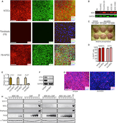
Here we share an example of this analysis. Results from worldwide research is now being made available via hPSCreg: Ilyas Singec, Andrew M. Crain, Junjie Hou, Brian T.D. Tobe, Maria Talantova, Alicia A. Winquist, Kutbuddin S. Doctor, Jennifer Choy, Xiayu Huang, Esther La Monaca, David M. Horn,5 Dieter A. Wolf, Stuart A. Lipton, Gustavo J. Gutierrez, Laurence M. Brill, and Evan Y. Snyder. Quantitative Analysis of Human Pluripotency and Neural Specification by In-Depth (Phospho)Proteomic Profiling. Stem Cell Reports (2016), http://dx.doi.org/10.1016/j.stemcr.2016.07.01.

Images: MDK Expression in Pluripotent and Differentiated Cells and Disruption of Neural Conversion by a Monoclonal Antibody against MDK (A) Representative immunocytochemical analysis of MDK expression by hESCs (WA09), skin fibroblasts (FB, line HS27), and fibroblasts (HS27) after reprogramming to hiPSCs. Scale bars represent 100 μm. (B) Western blot showing that MDK was not detected in HS27 fibroblasts but was expressed in WA09 hESCs and hiPSCs from HS27 fibroblasts, and was upregulated in differentiating cells (EBs and hNSCs from hESCs). Probing for GAPDH demonstrated similar loading of the lanes. (C) A monoclonal antibody against MDK did not impair pluripotent self-renewal of hESCs. Antibody concentrations are shown. (D) The percentage of pluripotent cells expressing OCT4 and NANOG did not differ significantly between anti-MDK and isotype control antibody treatment (3.0 μg/mL). Error bars represent SEM; n = 5. (E) qRT-PCR showed that a monoclonal antibody against MDK (1.5 μg/mL) inhibits neural gene expression during 6-day DAP treatment. Error bars represent mean + SEM, n = 3. ∗p less than  0.05. (F) Western blot analysis confirmed the inhibitory effect of anti-MDK on early neural marker expression (OTX1, PAX6), despite DAP treatment, in contrast to the isotype control. (G) Immunocytochemical evidence that neural conversion is inhibited by the anti-MDK antibody (1.5 μg/mL) during 6-day DAP treatment. Few cells induced PAX6 expression in the presence of anti-MDK antibody (right panel). Scale bar represents 200 μm. (H) Western blot analysis and time course of hESCs differentiated for 6 days with recombinant MDK alone (100 ng/mL) or a combination of the DAP cocktail and MDK (25 or 100 ng/mL) suggest that MDK specifically promotes neural commitment of hPSCs. SOX17, MIXL1, and Brachyury were not induced by MDK. In contrast, controls treated with fetal bovine serum (FBS) for 6 days, which stimulates multi-lineage differentiation of hPSCs, induced these non-neural markers.
This level of basic research yield knowledge that is important for the development of future cell based therapies for Neuro-related Diseases. We will continue to post updates here.

Tuesday, August 23, 2016

Human Mesenchymal Stem Cells Conjugates

Targeted Delivery of Stem Cells
Here's a recent publication referencing use of our Human Mesenchymal Stem Cells to form conjugates that could have application for drug delivery and cell therapies: Yun Seop Kim, Won Ho Kong, Hyemin Kim, Sei Kwang Hahn. Targeted Systemic Mesenchymal Stem Cell Delivery Using Hyaluronate - Wheat Germ Agglutinin Conjugate. http://dx.doi.org/10.1016/j.biomaterials.2016.08.027


Images: Fluorescence microscopic images of dissected livers and lungs from normal rats 4 h after intravenous injection of PBS, WGA-FITC/hMSC, and HA-WGA-FITC/hMSC (Filter for FITC). (b) Quantification of hMSCs in dissected organs estimated from ROI (n = 3). **P versus the WGA/hMSC group. (c) Representative fluorescence microscopic images of WGA-FITC/hMSC and HA-WGA-FITC/hMSC in the liver and lung (green = FITC, blue = DAPI for nucleus). Arrows indicate the location of hMSCs (scale bar = 200 μm). http://dx.doi.org/10.1016/j.biomaterials.2016.08.027
Protocol: Cultured MSCs were trypsinized, washed, and resuspended at 1 × 10⁶ cells/mL in PBS. HA-WGA conjugate at 10 μg/mL of WGA was added to MSCs in suspension, and incubated in an ice bath for 10 min with mild mixing. To remove the unbound HA-WGA conjugate to MSCs, cell suspension was washed with PBS and collected by centrifugation (1000 × g, 3 min at 4 °C). After surface modification with HA-WGA conjugate, HA-WGA/hMSC were resuspended in the cell culture medium on 96-well plates (1 × 10⁴ cells per well). At the predetermined time, cell viability of HA-WGA/hMSC was measured using an EZ-cytox cell viability assay kit according to the manufacturer’s instructions. Zeta potentials were analyzed using a Zetasizer Nano (Malvern Instruments, UK) to assess the change of surface charge after the surface modification.
Conclusions: Researchers successfully developed a target-specific systemic delivery system of MSCs to the liver using HA-WGA conjugate. HA-WGA conjugate was synthesized by the coupling reaction of HA-aldehyde with amine group of WGA. GPC analysis revealed the successful synthesis of HA-WGA conjugate. CD analysis corroborated that the secondary structure of WGA remained stable even after conjugation to HA. HA-WGA conjugate appeared to bind to the surface of MSCs and remain stably for up to 1 h. After cell surface modification, most of HA-WGA/MSC complex could be systemically delivered to the liver 4 h after intravenous injection, whereas MSCs were trapped mainly in the lung. This new strategy to target-specifically deliver MSCs to the liver using HA-WGA conjugate might be successfully exploited for treatment of various liver diseases.

We will continue to post new applications developed by users of our Stem Cells.

Friday, August 19, 2016

More on Epigenetics and Pain

Great Research Tools!

Our Markers and i-FectTM  Transection Kit are  used to study Epigenetics and pain. Here's yet another example: M. Leinders, b, N. Üçeyler, R.A. Pritchard, C. Sommer, L.S. Sorkin. Increased miR-132-3p expression is associated with chronic neuropathic pain. Experimental Neurology. Volume 283, Part A, September 2016, Pages 276–286...The inhibitor and mimetic were administered to awake rats via the it catheters. Prior to injection, active or mismatch inhibitors were mixed with (1:5 w/v) i-Fect™ in vivo transfection reagent(Neuromics, Edina, USA) to final doses of 5, 2 and 1 μg in 10 μl...

Figure: Inhibition of MOR. Stained with Dorsal Horn Neuromics' Rabbit Polyclonal Antbody.
Spinal administration of miR-132-3p antagonists via intrathecal (i.t.) catheters dose dependently reversed mechanical allodyina  and eliminated pain behavior in the place escape avoidance paradigm (p < 0.001). Intrathecal administration of miR-132-3p mimetic dose-dependently induced pain behavior in naïve rats (p < 0.001). Taken together these results indicate a pro-nociceptive effect of miR-132-3p in chronic neuropathic pain.

Finding like these could pave the way for an miRNA like therapy for pain.

Saturday, August 13, 2016

Protein Purification Solutions

Strep-tag®Tools + Strep-Tactin Columns

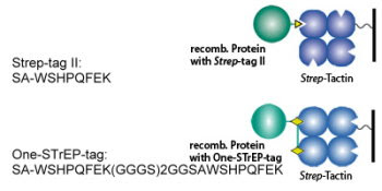
We are seeing a notable uptick in use of our Protein Purification Tools. This includes vectors and purification columns. We are especially delighted with the increased use of of our Strep-Tag + Strep-Tactin Columns.
Figure: Strep-Tag+Strep-Tactin Methods
Here's an example of researchers using the technology to purify heme proteins for the study of the diptheria pathogen: Courtni E. Allen and Michael P. Schmitt. Utilization of Host Iron Sources by Corynebacterium diphtheriae: Multiple Hemoglobin-Binding Proteins Are Essential for the Use of Iron from the Hemoglobin-Haptoglobin Complex. J. Bacteriol. February 2015 vol. 197 no. 3 553-562. doi: 10.1128/JB.02413-14...Cell debris was removed by centrifugation at 12,000 × g at 4°C, and the supernatant fraction containing the soluble protein was purified on Strep-Tactin columns (Neuromics) for Strep-tagged constructs...

Figure: (A) HtaA and derivatives were assessed for the ability to bind Hb-Hp by ELISA. GST was included as a negative control. Experiments were repeated at least three times, with similar results. Results of a representative experiment are shown. (B) Binding of HtaA and various derivatives to Hb-Hp at a 200 nM concentration is shown. Binding of HtaA-Y361A and CR2-Y361A to Hb-Hp was significantly different from binding with HtaA-wt and CR2, respectively (**, P less than 0.0001). Binding of CR1 to Hb-Hp was significantly different from that with HtaA-wt (***, P less than 0.05). *, HtaA does not bind Hp (HtaA-Hp). Values represent the means from three independent experiments (±SD).

Looking for protein purification solutions? Do not hesitate to contact me: 612-801-1007 or pshuster@neuromics.com.

Friday, July 29, 2016

Large Intestine Endothelial Cells

"Hard to Find", Potent and Pure

We asked one of our Large Biopharma users of our Large Intestine Endothelial cells, "Why Neuromics" and she told us, "It is hard to find these cells. Let alone ones that work as well as yours!"


We have many options for endothelial cells and pericytes. We also have the ability to microdissect out virtually any type of human cell. If you have need, just ask me. Direct cell: 612-801-1007 or pshuster@neuromics.com . Pete Shuster, CEO and Owner, Neuromics.