Friday, September 29, 2006

ESC to Neural Progenitors-Role of MOR-1C

Features use of Neuromics' MOR-1C

Eunhae Kim, Amy L. Clark , Alexi Kiss, Jason W. Hahn, Robin Wesselschmidt. MU AND KAPPA OPIOIDS INDUCE THE DIFFERENTIATION OF EMBRYONIC STEM CELLS TO NEURAL PROGENITORS
JBC Papers in Press. Published on September 1, 2006 as Manuscript M603862200


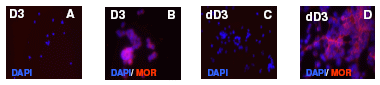
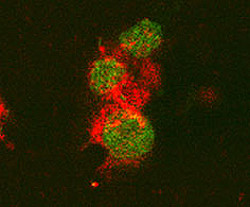
Image: A-D-Staining of Differentiated Stem Cells. D3 ES cells were maintained in serum/LIF/ME induced self-renewal conditions (undifferentiated cells, D3). Upon treatment with 1 µM RA they undergo differentiation (dD3). Cells were fixed and permeabilized. Treatments were followed by overnight incubation with polyclonal MOR-1 (C-terminus, 1:2500)

Tuesday, September 19, 2006

New TRPV1-C Publication

Amol M. Patwardhan, Nathaniel A. Jeske, Theodore J. Price, Nikita Gamper, Armen N. Akopian, and Kenneth M. Hargreaves. The cannabinoid WIN 55,212-2 inhibits transient receptor potential vanilloid 1 (TRPV1) and evokes peripheral antihyperalgesia via calcineurin. PNAS 103 (July 2006): 11393-11398.


Figure: WIN activates calcineurin in TG neurons and dephosphorylates TRPV1. (A) Calcineurin subunits are coexpressed with TRPV1. The colocalization of calcineurin A subunit (Upper) and B subunit (Lower) with TRPV1isdemonstratedin the respective panels.(B) The effect of various treatments on the nuclear translocation of NFATc4 (calcineurin activation). Cultured TG neuronswere exposed to vehicle, ionomycin (1 M), capsaicin (1 M), WIN (25 M), or WINCAIP(25 M50 M), and immunohistochemistry was performed by using an antibody againstNFATc4. (C) Graphical representation of the percent of NFATc4 positive neurons showing nuclear translocation of NFAFc4 after treatment with VEHVEH, VEHWIN (25 M), CAIP (50 M)WIN, VEHCAP (1 M), or CAIP(50 M)CAP (n 4 independent cultures assessed by blinded observer; n 152–180 cells per condition; **, P<0.01,anovawith n="3">

Saturday, September 09, 2006

4 New Interleukins

These Interleukins have been tested for IHC and WB apps and recommended for use by Diabetes/Obesity, Cancer, Neurodegenerative Disease and Pain/Inflammation Researchers

IL-1Alpha/IL-F1


Image: IL-1α staining of blood lymphocytes. Cells were stained using anti-gat-Cy3 (red) and counterstained with Fluoro Nissl Green


IL-1Beta


Image: IL-1 beta staining (red color) in mouse splenocytes stimulated with Con A. Cell nuclei counterstained green.


IL-1 R1


Image: IL-1R staining of mouse thymus. Staining was done using HRP-DAB (brown) detection. Tissue was counterstained with haematoxylin (blue).


IL-6


Image: IL-6 staining of mouse T cells. Cells were stained with goat anti-rat IL-6 (red) and nuclei counterstained with Fluoro Nissl Green.