Saturday, July 29, 2006

New P2X3 Data

P2X3 Catalog#: RA10109

Data Courtesy of Dr. Yan Sun University of Maryland School of Medicine Baltimore


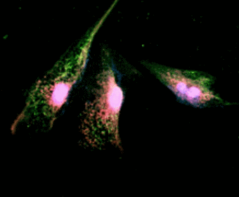
Image: Double immunofluorescence staining in cultured human bladder urothelial cells (BUCs). BUCs show cytokeratin (red areas) and P2X3 (green areas) expression.

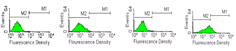
Figure: Flow cytometry and FACS analysis of P2X3 expression in human BUCs. P2X3 expression in BUCs were compared among in vitro stretched samples and unstretched samples in both normal and interstitial cystitis (IC) patients. A, unstretched normal BUCs. B, stretched normal BUCs. C, unstretched IC BUCs. D, stretched IC BUCs. M1 region represents P2X3 localization detected.

Monday, July 24, 2006

New AD Research Antibodies

We continue to add to our catalog of Neurodegenerative Disease Reagents:

BACE-1
Cleavage of amyloid precursor protein (APP) into 40 or 42 amino acid amyloid β (aβ) peptides is a result of the action of two proteases β- and γ-secretase. The beta-site APP cleaving enzyme, or BACE-1, is a transmembrane aspartic protease that is localized in the golgi and endosomes. Low levels of BACE-1 mRNA are found in most adult tissues with a higher level of expression in pancreas.


Image: Detection of BACE-1 in human cerebellum. Stained with HRP-DAP (brown) and counter-stained with hemtoxylin (blue).


Abeta 40 (beta amyloid 1-40) and Abeta 42 (beta amyloid 1-42)
Amyloid beta-protein (Abeta) is associated with neuronal injury and death in Alzheimer's disease. These proteins are the main components of amyloid plaques. Abeta 42 plays a bigger role in deneurogeneration and is more strongly associated with Alzheimer's disease than Abeta 40.


Image: Detection of Abeta 40 on 5 ng of peptide per lane using NB 300-225. Lane 1: Abeta-40, lane 2: Abeta-42, lane 3: Abeta-40 and -42 mix

Saturday, July 22, 2006

TRPV1-N and C Data

Some interesting results from:

Sharif Naeini R, Witty MF, Seguela P, Bourque CW (2006) An N-terminal variant of Trpv1 channel is required for osmosensory transduction. Nature Neuroscience. (1):93-8.


Figure: SON Neurons express an N-terminal variant(s) of TRPV1 (a) Simplified structure of mRNA (numbered boxes show exons of TRPV1 gene) encoding TRPV1 (above). Gray arrow tails and lines illustrate mRNA regions detected by primer sets A and B. RT-PCR analysis of mRNAs expressed in DRG and SON (below). Note the lack of signals for the N terminus of TRPV1 (lane 2, exons 2-6, primer set A) in SON. (b) Immunocytochemical staining confirmed the presence of full length TRPV1 in DRG neurons. In contrast, only the C-terminus of the TRPV1 protein (C-TRPV1) was detected in the AVP positive SON neurons. (c) Whole-cell recordings reveal that DRG neurons are sensitive to capsaicin ('Cap,' 10 uM, 5s) but that SON neurons are insensitive.