Friday, December 23, 2011

Salivary Neuropeptides Y2s and Satiety

A possible new slant for combating obesity

The researchers in this study acheived a sustained increased PYY3-36 (a neuropeptide) expression via viral vector-mediated gene delivery targeting salivary glands and the good news: this increase resulted in a significant long-term reduction in food intake (FI) and body weight (BW). This is evidence for new functions of the previously characterized gut peptide PYY3-36 suggesting a potential simple and efficient alternative therapeutic approach for the treatment of obesity: Andres Acosta1, Maria D. Hurtado1, Oleg Gorbatyuk, Michael La Sala, David Duncan, George Aslanidi, Martha Campbell-Thompson, Lei Zhang, Herbert Herzog, Antonis Voutetakis, Bruce J. Baum, Sergei Zolotukhin. Salivary PYY: A Putative Bypass to Satiety. PLoS ONE 6(10): e26137. doi:10.1371/journal.pone.0026137. Received: July 22, 2011; Accepted: September 20, 2011; Published: October 10, 2011...Rabbit anti-Y2R (Dilution: 1:3000) using TSA...

Images: A) Immunolocalization of Y2R-positive cells in the hippocampus of C57Bl/6J mouse (WT), a (+) control. (B) Immunolocalization of Y2R in the tongue epithelia of Y2R KO mouse, a (-) control. VEG – von Ebner's gland. (C) Immunolocalization of Y2R-positive cells in the CV area of the tongue of a C57Bl/6J mouse. (D) close-up of (C). (E), and (F) close ups of (D), top and bottom rectangles, respectively. doi:10.1371/journal.pone.0026137.g003

These findings could prove a first step in identifying new targets for obesity fighting therapies. Increasing PYY salivary output would provide satiety with less food intake. This would give a whole new perspective on dieting. If we are satsified with less food intake is this really dieting? Stay tuned.

Saturday, December 03, 2011

Opioid Addiction During Pregnancy-Implications for Neuro-development

I would like to thank Dr. Carmen Sato-Bigbee, Virginia Commonwealth University School of Medicine, for kindly sharing this important study. The publication also references use of our Mu Opioid and Nociceptin/Orphanin FQ Receptor Antibodies: Andrew C. Eschenroeder, Allison A. Vestal-Laborde, Emilse S. Sanchez, Susan E. Robinson, Carmen Sato-Bigbee. Oligodendrocyte responses to buprenorphine uncover novel and opposing roles of μ-opioid- and nociceptin/orphanin FQ receptors in cell development: Implications for drug addiction treatment during pregnancy. Glia Volume 60, Issue 1, pages 125–136, January 2012.

Highlights: Oligodendrocytes are responsible for making myelin in the CNS. The authors have shown We have shown previously that rat brain myelination is significantly altered by buprenorphine, an opioid analogue currently used in clinical trials for managing pregnant opioid addicts. In this study, perinatal exposure to low levels of this drug induced accelerated and increased expression of myelin basic proteins (MBPs), cellular and myelin components that are markers of mature oligodendrocytes. In contrast, supra-therapeutic drug doses delayed MBP brain expression and resulted in a decreased number of myelinated axons. We have now found that this biphasic-dose response to buprenorphine can be attributed to the participation of both the l-opioid receptor (MOR) and the nociceptin/orphanin FQ receptor (NOP receptor) in the oligodendrocytes. This is the first study showing the potential role of the NOP receptor in myelination.
High levels of opiate exposure could negatively disrupt the normal interplay between these two systems altering the developmental pattern of brain myelination. Understanding this pathway, could help researchers find ways to favorably modulate myelination and protect neuro-development of fetuses exposed to high levels of opiates during pregnancy.

Related Data:
Direct treatment of immature oligodendrocytes with buprenorphine alters MBP expression in a dose-specific manner. Cells isolated from 9-day-old rat brains were incubated for 4 days in CDM with or without 0.25, 0.5, 1.0, and 3.0 lM buprenorphine. MBP levels were determined by western blotting using b-actin levels as loading controls. Figures correspond to representative experiments. Results in the bar graph are expressed as percentage of controls (0 lM buprenorphine) 6 SEM from five experiments and correspond to the combined scanning of the four major MBP isoforms. **P <0.005 and ***P<0.0001.
Pre-oligodendrocytes express both MOR and the NOP receptor. Cells isolated from 9-day-old rat brain were allowed to fully attachon the culture plates by overnight incubation and stained by double
immunocytochemistry with O4 (green) together with anti-MOR or anti-NOP receptor antibodies (red). Scale bar: 20 lm. The western blot shows MOR and NOP receptor expression in two different samples of developing oligodendrocytes directly isolated from 9-day-old rat brains.

I will be posting future studies investigating the molecular mechanisms by which buprenorphine and methadone affect myelination and neuron-glial interactions. These should provide deeper understanding into these developmental processes and new and better strategies for the managing of both pregnant addicts and drug addiction in adolescence.

Friday, November 25, 2011

IF Staining of Human Primary Neurons

Primary Neurons are inputs or raw materials for cell based assays. When cells do not work as promised, there are multiple costs including lost time and potentially flawed data. Neuromics strives to provide easy to culture, potent and cost effective cells. Proving these capabilities is an ongoing activity for us. This includes testing these cells using our markers.

I wanted to share new immunofluorescence images. Here is a link to the protocol: staining primary neurons.

hN2 cells stained with our chicken polyclonal antibody to Vimentin, in red. Islands of Hn2 cells form after 4 days in culture forming beautiful flower like structures. Vimentin is a well established marker of early differentiating neuronal lineage cells. Taken with a 10X objective lens. Blue staining is the nuclear DNA.
hN2 cells grown in culture for 4 days and stained with our chicken polyclonal to MAP2, a marker of neurons. Differentiating cells show strong cytoplasmic staining for MAP2 . Blue stain is DAPI and reveals cell nuclei of some non neuronal cells in this culture.
hN2 cells grown in culture for 4 days and stained with our chicken polyclonal to Neurofilament light or low molecular weight chain NF-L, a marker of neurons. Many of the differentiating cells show strong cytoplasmic and clearly fibrillar staining for NF-L. Blue stain is DAPI and reveals cell nuclei of some non neuronal cells in this culture.

Sunday, November 20, 2011

Fear Changes Hippocampus Neuropns

Fear in mice catalyzes rapid accumalation of EphrinB2 pyramidal neurons of the CA1 area.

This study suggests that rapid accumulation of EphrinB2 in hippocampal CA1 neurons is involved in the behavioural and cellular modifications induced by contextual fear conditioning. A similar mechanism does not appear to occur in lateral amygdala neurons, in spite of the robust behavioural and cellular modifications induced in such structure by cued fear conditioning: Antonio Trabalzaa, Sandra Colazingaria, Carmelo Sgobiob, Arturo Bevilacqua. Contextual learning increases dendrite complexity and EphrinB2 levels in hippocampal mouse neurons. Behavioural Brain Research. doi:10.1016/j.bbr.2011.11.008.

Friday, November 11, 2011

Diabetic retinopathy blindness-root causes

Diabetic retinopathy is a leading cause of acquired blindness. This publication from our friends at University of Buenos Aires touches on potential root causes: Diego C. Fernandez, Laura A. Pasquini, Damián Dorfman, Hernán J. Aldana Marcos, Ruth E. Rosenstein. Early Distal Axonopathy of the Visual Pathway in Experimental Diabetes. doi:10.1016/j.ajpath.2011.09.018
" In animals that had been diabetic for 6 weeks, a large increase in astrocyte reactivity occurred in the distal (but not the intraorbital) portion, which coincided with significant axon loss. Moreover, profound myelin alterations and altered morphologic features of oligodendrocyte lineage were observed at the distal (but not the proximal) optic nerve portion. The present results suggest that axoglial alterations at the distal portion of the optic nerve could be the first structural change in the diabetic visual pathway."
The authors used our PDGFR Alpha/CD140A Marker to Study the change in Oligodendrocyte Lineage precursor cells. Expression of the protein was increased in these cells with the presence of disorganized and hypertrophic cells. This could disrupt formation of myelin resulting the pathological alteration at the distal portion.

Tuesday, November 08, 2011

Opioid Induced Itch

Our widely used and frequently published Opioid Receptors Antibodies are used for a spectrum of pain research. Here is an interesting study on the root causes of opioid induced itch.

Xian-Yu Liu, Zhong-Chun Liu, Yan-Gang Sun, Michael Ross1, Seungil Kim, Feng-Fang Tsai, Qi-Fang Li, Joseph Jeffry, Ji-Young Kim, Horace H. Loh, Zhou-Feng Chen. Unidirectional Cross-Activation of GRPR by MOR1D Uncouples Itch and Analgesia Induced by Opioids. Cell, Volume 147, Issue 2, 14 October 2011, Pages 261-262.


Root Causes: Spinal opioid-induced itch, a prevalent side effect of pain management, has been proposed to result from pain inhibition. We now report that the μ-opioid receptor (MOR) isoform MOR1D is essential for morphine-induced scratching (MIS), whereas the isoform MOR1 is required only for morphine-induced analgesia (MIA). MOR1D heterodimerizes with gastrin-releasing peptide receptor (GRPR) in the spinal cord, relaying itch information. We show that morphine triggers internalization of both GRPR and MOR1D, whereas GRP specifically triggers GRPR internalization and morphine-independent scratching. Providing potential insight into opioid-induced itch prevention, we demonstrate that molecular and pharmacologic inhibition of PLCβ3 and IP3R3, downstream effectors of GRPR, specifically block MIS but not MIA. In addition, blocking MOR1D-GRPR association attenuates MIS but not MIA. Together, these data suggest that opioid-induced itch is an active process concomitant with but independent of opioid analgesia, occurring via the unidirectional cross-activation of GRPR signaling by MOR1D heterodimerization.

Wednesday, October 26, 2011

Mouse epiSCs Into Myelinating Cells

This study published recently in Nature Methods hit my radar scope becaused it referenced use of our widely used and frequently published stem cell marker Tuj 1 (Neuron-specific class III beta-tubulin): Fadi J Najm, Anita Zaremba, Andrew V Caprariello, Shreya Nayak, Eric C Freundt, Peter C Scacheri, Robert H Miller & Paul J Tesar. Rapid and robust generation of functional oligodendrocyte progenitor cells from epiblast stem cells. Nature Methods (2011) doi:10.1038/nmeth.1712.

Dr. Paul Tesar and his team at Case Western University demonstrated the ability to convert pluripotent epiblast stem cells into pure populations of myelinating cells, called oligodendrocyte progenitor cells (OPCs). First, stem cells in a petri dish are treated with molecules to direct them to become the most primitive cells in the nervous system. To produce OPCs, these primitive cells are treated with a defined set of proteins. The cells were cultured on laminin and treated withh apporopriate growth factors. The OPCs were nearly homogenous and could be multiplied to obtain more than a trillion cells.

The OPCs were treated with thyroid hormone, which is key to regulating the transition of the OPCs to oligodendrocytes. The result was the OPCs stopped proliferating and turned into oligodendrocytes within four days.

These methods could used to potentially produce stable and pure populations of human OPCs in a significant enough number to treat patients with demyelinating diseases such as multiple sclerosis and cerebral palsy.

Thursday, October 20, 2011

Immunostaining Neurons and Glia

I would like to thank Dr. Gerry Shaw, University of Florida for his excellent work with our Primary Neurons and Astrocytes and Neuronal-Glial Markers. Here's an example image with many more to follow:

Image: E18 hippocampal neurons stained with MAPT (red) and Doublecortin (green). The two proteins overlap in the proximal dendrites, but doublecortin is more abundant in the growth cones and periphery. As a result, the periphery appears green while the more proximal regions of the cells are yellow. The single longer process of this cell, presumably an axon, has a low doublecortin content and so appears red. Blue staining is the nuclear DNA. Protocol on datasheet.

Sunday, October 09, 2011

BDNF and Exercise Study-Running Mice

Get fit and get smart. There is increasing evidence that vigorous exercise increases secretion of Brain-Derived Neurotrophic Factor (BDNF) Protein.  BDNF is a catalyst of processes that increase growth of neurons especially in the hippocampus.

In this study researchers show new cell proliferation, survival, neuron number, and neurotrophin levels were enhanced only when running was accessible to mice. They conclude that exercise is the critical factor mediating increased BDNF levels and adult hippocampal neurogenesis: Tali Kobilo, Qing-Rong Liu, Kriti Gandhi, Mohammed Mughal, Yavin Shaham and Henriette van Praag. Running is the neurogenic and neurotrophic stimulus in environmental enrichment. doi: 10.1101/lm.2283011. Learn. Mem. 2011. 18: 605-609... human recombinant BDNF (0.1 µg) monomer (Neuromics)...
BDNF, CF Recombinant Protein
Related Reagents:
Neuron-Glial Expressed-Includes Neurotrophin Proteins
Neurotrophins and Growth Factor Antibodies
Primary Neurons and Astrocytes-Primary human, rat and mouse neurons and astrocytes.

Keep your brain healthy.

Monday, September 19, 2011

Immune-Inflammatory Response and Pain Research

Our Pain and Inflammation Related Research Antibodies are increasingly being used to study the root causes of immune/inflammatory related pain induction. Here're related publications: Lintao Qu, Pu Zhang, Robert H. LaMotte, Chao Ma. Neuronal Fc-gamma receptor I mediated excitatory effects of IgG immune complex on rat dorsal root ganglion neurons. Brain, Behavior, and Immunity. Volume 25, Issue 7, October 2011, Pages 1399-1407......rabbit-anti-TRPV1, 1:1000, Neuromics...

Highlights: Pain often accompanies antigen-specific immune-related disorders though little is known of the underlying neural mechanisms. A common feature among these disorders is the elevated level of antigen-specific immunoglobulin (Ig) G in the serum and the presence of IgG immune complex (IC) in the affected tissue. We hypothesize that IC may directly activate the Fc-gamma receptor type I (FcγRI) expressed in nociceptive dorsal root ganglion (DRG) neurons and increase neuronal excitability thus potentially contributing to pain. Immunofluorescent labeling indicated that FcγRI, but not FcγRIIB or FcγRIII, was expressed in a subpopulation of rat DRG neurons including those expressing nociceptive markers. Calcium imaging revealed that the IC, but neither of the antibody (IgG) or antigen alone, produced an increase in intracellular calcium. This effect was abolished by the removal of the IgG Fc portion in the IC or the application of an anti-FcγRI antibody, suggesting a key role of the FcγRI receptor. Removal of extracellular calcium or depletion of intracellular calcium stores prevented the IC-induced calcium response. In whole-cell current-clamp recordings, IC depolarized the resting membrane potential, decreased the rheobase, and increased the number of action potentials evoked by a depolarizing current at 2× rheobase. In about half of the responsive neurons, IC evoked action potential discharges. These results suggest that a subpopulation of nociceptive neurons expresses functional FcγRI and that the activation of this receptor by IC increases neuronal excitability.

B. Huanga, X. Zhaoc, L.-B. Zhengb, L. Zhanga, B. Nia. Different expression of tissue inhibitor of metalloproteinase family members in rat dorsal root ganglia and their changes after peripheral nerve injury. Neuroscience, Volume 193, 13 October 2011, Pages 421-428....anti-P2X3 (rabbit, Neuromics, MN, USA)...

Thursday, September 15, 2011

Xona Microfluidics and Neurons

I am impressed with these Video from the Jeon Lab at UC Irvine. It represents a novel method for neuro-drug discovery:
Preparing E18 Cortical Rat Neurons for Compartmentalization in a Microfluidic Device.

This technology represents a way to separate axon from cell bodies.

Tuesday, August 30, 2011

Opioid Receptor Antibodies Trifecta

This publication proposes a role for opioid receptors in treating cancers. It also references use of our μ, δ, and κ opioid receptor antibodies.

Kohei Yamamizu1, Sadayoshi Furuta, Shiori Katayama, Michiko Narita, Naoko Kuzumaki, Satoshi Imai, Hiroshi Nagase, Tsutomu Suzuki, Minoru Narita, and Jun K. Yamashita. The κ opioid system regulates endothelial cell differentiation and pathfinding in vascular development. Blood July 21, 2011 vol. 118 no. 3 775-785.

Highlights: The opioid system is, thus, a new regulator of vascular development that simultaneously modifies 2 distinct vascular properties, EC differentiation and vascular pathfinding. We confirmed that KOR, but not MOR, was highly expressed in various ECs such as HUVECs (data not shown), suggesting that KOR agonists could directly act on tumor ECs to suppress VEGF receptor expression, similar to the effects observed in embryonic ECs. If so, a combination therapy including an MOR agonist, morphine, and a KOR agonist (such as TRK820, a clinically approved drug in Japan for uremic pruritus) may prove useful for cancer therapy through the suppression of tumor angiogenesis by dual inhibition of VEGF ligands and receptors, extending the therapeutic benefits beyond pain relief.

Images: KOR was highly expressed in Flk1+ vascular progenitors. (A) RT-PCR showing mRNA expression of MOR, DOR, and KOR in ES cells, Flk1+ cells, cells after 1 or 3 days of Flk1+ cell culture (Flk-d1 or Flk-d3), CD31-positive cells (ECs) and CD31-negative cells (MCs) at Flk-d3. (B) Fluorescent staining for MOR, DOR, and KOR at Flk-d1. Nuclei are stained with DAPI (blue). Left, MOR; middle, DOR; right, KOR. Scale bars, 100 μm. (C) Double fluorescent staining for MOR, DOR, and KOR with CD31 (red) at Flk-d3. Nuclei are stained with DAPI (blue). Top, opioid (green) receptors (green); middle, CD31 (red); bottom, merged. Scale bars, 100 μm.

Saturday, August 27, 2011

Is Neuropathy Really Gliopathy?

I found this excellent website from posting by Dr. Jan M. Keppel Hesselink, Professor molecular pharmacology, director Institute neuropathic pain: http://www.neuropathie.nu/. It represents a new way of understanding root causes and potential therapies for Neuropathic Pain. Here're highlights:

Gliopathic pain: is a brand new term for what we always thought to be neuropathic pain. It refers to pain related to neuropathic pain, however, the primary driver of this pain is most probably more linked to glia and asterocytes. The mechanism of gliopathic pain is the hyperactivation of glia cells, which results in neuropathic pain.

The role of Glia and Astrocytes:Glia and astrocytes play a central role in neuropathic pain, and gliopathic pain, or asteropathic pain will become new synonyms for neuropathic pain. In a recent hallmark paper the term 'Gliopathic pain' was coined.

This is a reason to put our magnifying glass on glia. Gliamodulating drugs will become a new class of neuropathic drugs, the so called gliopathic modulating drugs, and the first prototype, the endogenous fatty acid palmitoylethanolamide, has already been explored in positive proof of principle studies.

For more than a century doctors are aware of the special properties of glia in response to injury. In Germany in 1894 professor Franz Nissl decribed the reaction of glial cells in relation to the nerve fibers in the spinal cord and highlighted their morphological changes after injury. Microglia becomes mores bigger and more abundant after injury and these glial responses can be seen as a biological reponse to promote nerve repair after injury. However, this response can go berzerk and might be one of the most important mechanisms leading to neuropathic pain.

This is a short synopsis. There is a wealth of more information on the website. That said, I will be posting more on Gliopathic Pain.

Thursday, August 25, 2011

Primary Neurons and Cell Based Assays

The feedback I receive from Neuroscientists is consistent. To paraphrase, "gives us healthy, consistent and potent primary cells. I understand the hard work it takes to generate meaningful and publishable results from cell based assays. Our Primary Neurons and Astrocytes are merely inputs for these assays. The real cost is the time invested in culturing and time lost if they don't work.

I have numerous postings on success: Primary Neurons Postings. I wanted to share more data and feedback.

Primary DRGs-Culturing these can be tricky. I make it a point to work with labs to make sure the protocol options best match the desired outcome for assays. This includes replacing cells to make sure we can accurately troubleshoot. This approach insures I can pin point the issues and make sure they are all resolved in round two. Here's a representative testimonial: "Thanks for following up, the DRGs worked great and we were able to get excellent data from them. Thanks so much for working with us." Adam Ross, Dr. Chengji Zhou Lab, UC Davis

Image: DRGs cultured on Calf Skin Collagen.

Primary Hippocampal Neurons-I would like to thank Vimal Swarup, University of Utah for this excellent image.


The cells have been fixed after 48 hrs, they were grown over poly-lysine coated coverslips in the media supplied by Neuromics. Cells were imaged in phase contrast mode with 40x objective.

Put our primary cells to the test!


Monday, August 22, 2011

MOR and NMDAR Interplay-Implications in Pain Control

Our Opioid Receptor Antibodies continue to be referenced in publications by Pain Researchers. Many of these studies provide a greater understanding of how opioids alleviate pain and what modulates this ability.

For example, the capacity of opioids to alleviate inflammatory pain is negatively regulated by the glutamate-binding N-methyl-D-aspartate receptor (NMDAR). This study drills down into the specifics of this regulation and references use of Neuromics' MOR1C Antibody: María Rodríguez-Muñoz, Pilar Sánchez-Blázquez, Ana Vicente-Sánchez, Esther Berrocoso and Javier Garzón. María Rodríguez-Muñoz, Pilar Sánchez-Blázquez, Ana Vicente-Sánchez, Esther Berrocoso and Javier Garzón. The Mu-Opioid Receptor and the NMDA Receptor Associate in PAG Neurons: Implications in Pain Control. Neuropsychopharmacology , (3 August 2011) | doi:10.1038/npp.2011.155.

Abstract: The capacity of opioids to alleviate inflammatory pain is negatively regulated by the glutamate-binding N-methyl-D-aspartate receptor (NMDAR). Increased activity of this receptor complicates the clinical use of opioids to treat persistent neuropathic pain. Immunohistochemical and ultrastructural studies have demonstrated the coexistence of both receptors within single neurons of the CNS, including those in the mesencephalic periaqueductal gray (PAG), a region that is implicated in the opioid control of nociception. We now report that mu-opioid receptors (MOR) and NMDAR NR1 subunits associate in the postsynaptic structures of PAG neurons. Morphine disrupts this complex by protein kinase-C (PKC)-mediated phosphorylation of the NR1 C1 segment and potentiates the NMDAR–CaMKII, pathway that is implicated in morphine tolerance. Inhibition of PKC, but not PKA or GRK2, restored the MOR–NR1 association and rescued the analgesic effect of morphine as well. The administration of N-methyl-D-aspartic acid separated the MOR–NR1 complex, increased MOR Ser phosphorylation, reduced the association of the MOR with G-proteins, and diminished the antinociceptive capacity of morphine. Inhibition of PKA, but not PKC, CaMKII, or GRK2, blocked these effects and preserved morphine antinociception. Thus, the opposing activities of the MOR and NMDAR in pain control affect their relation within neurons of structures such as the PAG. This finding could be exploited in developing bifunctional drugs that would act exclusively on those NMDARs associated with MORs.

I will continue to post these studies. They give hope for pain sufferers as many propose potential new druggable targets.

Thursday, August 18, 2011

Plasma netrin-1 is a diagnostic biomarker of human cancers

I am pleased to report broadening application for our Stem Cell Markers as a diagnostic for cancers. This publication references use of our Netrin-1 antibody.

Ganesan Ramesh, Arthur Berg, and Calpurnia Jayakumar. Plasma netrin-1 is a diagnostic biomarker of human cancers. Biomarkers. Author manuscript; available in PMC 2011 July 26. Published in final edited form as: Biomarkers. 2011 March; 16(2): 172–180. Published online 2011 February 8. doi: 10.3109/1354750X.2010.541564.
Objectives: To determine whether plasma netrin-1 can be used as a diagnostic biomarker of human cancer.

Materials and Methods: A total of 300 cancer plasma samples from breast, renal, prostate, liver, meningioma, pituitary adenoma, glioblastoma, lung, pancreatic and colon cancer patients were compared against 138 control plasma samples. Netrin-1 levels were quantified by ELISA and immunohistochemistry.

Results: Plasma netrin-1 levels were significantly increased in breast, renal, prostate, liver, meningioma, pituitary adenoma, and glioblastoma cancers as compared to control samples.

Discussion and Conclusion: Our results suggest that plasma netrin-1 can be used as a diagnostic biomarker for many human cancers.

Image: Immunohistochemical localization of netrin-1 in renal cell carcinoma (RCC) tissues. A. Secondary antibody control showing no staining. B. Normal adjacent tissues do not show any staining for netrin-1. C. Stage I RCC shows staining for netrin-1. D. Stage II RCC shows staining for netrin-1. E-F.


Netrin-1 Immunohistochemistry: Stage I–III renal cell carcinoma and normal tissue section (Tissue Array) was obtained from Biomax to immunolocalize netrin-1, as described previously (29). Briefly, tissue sections were dewaxed and rehydrated with graded ethanol (100%, 90%, 70% and 30%) and then washed with PBS. Antigen retrieval was carried out using citrate buffer and steamer. The tissue section was permeabilized with 0.2% Triton X-100 in PBS, and washed and blocked with PBS containing 5% donkey serum and 1% BSA. Primary antibodies included a chicken anti-netrin-1 polyclonal antibody (Neuromics cat # CH23002). Primary antibodies were detected using secondary antibodies conjugated with biotin, which was followed by incubation with streptavidin-horseradish peroxidase (Pierce). Slides were mounted in permount and photographed using an Olympus microscope attached to a CCD camera.
Potent diagnostic tools for cancers save lives. This is especially true of markers that can diagnose them in early stages. I will post these important studies as the cross my radar scope.

Friday, August 12, 2011

SOX2 and Initiation of Breast Tumors

I consider it a feather in Neuromics' cap when our reagent(s) are referenced in a Nature Journal. More importantly it enables me to keep the pulse on novel and important discovery.

This pub references use of one of our SOX2 Antibody and comes from Dr. Angel García Martín and his Team at INBIOMED: O Leis, A Eguiara, E Lopez-Arribillaga, M J Alberdi, S Hernandez-Garcia, K Elorriaga, A Pandiella, R Rezola and A G Martin. Sox2 expression in breast tumours and activation in breast cancer stem cells. Oncogene , (8 August 2011) | doi:10.1038/onc.2011.338.

The important insight from this study is: "Over-expression of Sox2 increased mammosphere formation, effect dependent on continuous Sox2 expression; furthermore, Sox2 knockdown prevented mammosphere formation and delayed tumour formation in xenograft tumour initiation models. Induction of Sox2 expression was achieved through activation of the distal enhancer of Sox2 promoter upon sphere formation, the same element that controls Sox2 transcription in pluripotent stem cells. These findings suggest that reactivation of Sox2 represents an early step in breast tumour initiation, explaining tumour heterogeneity by placing the tumour-initiating event in any cell along the axis of mammary differentiation."

Could these findings ultimately lead to a better diagnostic for Breast Cancer? I'll keep you posted.

Lab Highlights: Breast cancer is the most frequent cancer in women making up to 20% of all tumours diagnosed in women, with 1 million new cases diagnosed every year worldwide (16,000 new cases only in Spain). Worlwide it causes over 350,000 deaths with an increasing tendency. Breast cancer stem cells show the phenotype CD44+/CD24low/-Lin- though only a fraction of this population has the capacity to initiate tumours. Therefore a complete and precise description of the breast cancer stem cells is lacking.

The focus of this laboratory is to identify, isolate and culture breast cancer stem cells from natural breast tumours and compare at the molecular level with normal mammary stem cells. This research involves the stablishment of both in vitro and in vivo functional assays and the molecular characterization (both genomic and proteomic) of breast cancer stem cells to define the mechanisms responsible for its transformed phenotype.

Sunday, August 07, 2011

High Content and High Throughput Toxicity Screening

Kits designed for Drug Discovery and Development

Our customers have been impressed with the capablities of our in vivo and in vitro apoptosis and toxicity kits. Here's a recent pub referencing use of one of our FLICA™ in vitro Caspase Kits: Giovanna Grandinetti, Nilesh P. Ingle, and Theresa M. Reineke. Interaction of Poly(ethylenimine)–DNA Polyplexes with Mitochondria: Implications for a Mechanism of Cytotoxicity. Mol. Pharmaceutics, Article ASAP Publication Date (Web): June 23, 2011 Copyright © 2011 American Chemical Society...inhibitor of caspases (FLICA) specific for caspase-9 (Neuromics, Inc)...
Images: Jurkat cells were treated with 1 µM staurosporine for 3 hours to induce caspase 9 activity (top), or were treated with a control (bottom). Both populations were incubated with ICT’s green FAM-LEHD-FMK FLICA™ caspase 9 reagent. DIC images were taken of both samples. Almost all cells in the induced sample (top) fluoresce green therefore they have activated caspase 9. None of the control cells (bottom) fluoresce green, therefore they do not have activated caspase 9 Courtesy of Dr. Brian Lee, ICT

Neuromics is pleased to announce the addition of HemoGenix® Predictive in vitro Toxicity and Apoptosis Kits:

Solutions for predictive in vitro toxicity and apoptosis are now important than ever. These kits enable you to do high throughput and high content screening of stem cells, progenitors and primary cells.
Kit options include:

LumiSTEM™-96 iPS and LumiSTEM™-iPS HT Assays to Study Induced Pluripotent Stem Cells (iPS) and Toxicity to iPS Cells and Cells Derived from iPS Cells.

LUMENESC™-Tox HT (LUMENESC™-96 Tox and LUMENESC™-384 HT). A Toxicity Screening and Testing Platform for Cells of the Mesenchymal Stem/Stromal Cell System.

HALO®-Tox HT Predictive Hemotoxicity Platform using HALO®-96 Tox and HALO®-384 HT. A Highly Predictive, In Vitro Stem and Progenitor Cell Hemotoxicity Screening and Testing Platform for all Stages of Drug Development and Xenobiotic.

I will continue to post regarding progress.

Saturday, July 30, 2011

TRPV1 and Diabetic Neuropathy

Thermal hyperalgesia is a common sympton of Diabetic Periperal Neuropathy (DPN). It is one of most difficult types of pain to treat. The development of tolerance, inadequate relief and potential toxicity of classical antinociceptives warrant the investigation of the newer agents to relieve this pain.

The elevated expression of Transient receptor potential vanilloid 1 (TRPV1) suspected as a transmitter of this pain. Dr. Louis Premkumar and his team at SIU have recently published results that further demonstrate the role of TRPV1: Mahendra Bishnoi, Christine A Bosgraaf, Mruvil Abooj, Linlin Zhong, Louis S Premkumar. Streptozotocin-Induced Early Thermal Hyperalgesia is independent of Glycemic State of Rats: Role of Transient Receptor Potential Vanilloid 1(TRPV1) and inflammatory mediators. Molecular Pain 2011, 7:52 doi:10.1186/1744-8069-7-52. Published: 27 July 2011.


Figure 4. Altered TRPV1 staining in spinal cord dorsal horn of STZ-treated rats. A. Representative images of TRPV1 staining from a vehicle-treated, STZ-HG and STZNG rats. An enlarged segment has also been shown. B. Average gray values/10,000 μm2 area of TRPV1 staining in dorsal horn was significantly increased (p<0.05) in both STZ-HG and STZ-NG rats as compared to vehicle-treated rats. Asterisk (*) represents p < 0.05. Scale bar is 200 μm and 50 μm for upper and lower panels, respectively.

Conclusions: From these results, it is concluded that TRPV1 is an integral component of initiating and maintaining inflammatory thermal hyperalgesia, which can be alleviated by intrathecal administration of RTX. Further, the results suggest that enhanced expression and inflammation-induced sensitization of TRPV1 at the spinal cord may play a role in central sensitization in STZ-induced neuropathy.

Therapies that downregulate or silence TRPV1 expression could be the key to better treatments for the Thermal Algesia cause by diabetes. I will keep you posted.

Wednesday, July 27, 2011

Potent and Cost Effective Cell Based Assays

I have had many conversations with basic and drug discovery researchers on improving cell based assays. Here's the wish list:
  • More potent cells/media
  • More accurate analytic tools-quatititative and reproducible results
  • Ability to use cells and tools in high throughput/high content screening.
  • Cost effectiveness
This wish list is front and center in determining the cells/media and related tools we add to Neuromics' offerings. We are pleased to announce the addition of our Hemogenix's Bioluminomics™ In-Vitro Cell Assays, MSCGro™ Mesenchymal Stem Cell Media and Umbilical Cord Blood derived hMesenchymal Stem Cells.

These provide quantitation, not subjectivity. It includes assay calibration and standardization. It means assay validation. It produces results you can trust and rely on. It means innovation and flexibility. It is advanced technology that is fast to learn, easy to use and above all, cost effective.

Assays options:
Available Cells:
Primary Neurons and Astrocytes-Primary human, rat and mouse neurons and astrocytes
STEMEZTM Human Neural Progenitor Neuron Discovery Kits-Derived from H9 (WA09) ECSs-Consistent, Easy to Use & Cost Effective
Human Mesenchymal Stem Cells (hMSCs-hMSCs derived from pancreas and umbilical cord blood
Mammalian Cell Lines

Media:
STEMEZ(TM) hN2 Human Neurons Culture Media
MSCGro™ Mesenchymal Stem Cell Media
NbActiv4

I will continue to post customer input and related data on Neuromics' Cell Based Assay Tools.


Saturday, July 23, 2011

Differential healing properties of human ACL and MCL Stem Cells

Autologous Stem Cell therapies for human injury and disease are gaining momentum. Understanding the properties of Stem Cell Colonies that have potential for these therapies is key to optimizing treatments. This study provides knowledge on the properties and their impact on future therapies for anterior cruciate ligament (hACL) and medial collateral ligament (hMCL) of the knee joint.
Jianying Zhang, Tiffany Pan, Hee-Jeong Im, Freddie H Fu and James HC Wang. Differential properties of human ACL and MCL stem cells may be responsible for their differential healing capacity. Differential properties of human ACL and MCL stem cells may be responsible for their differential healing capacity. BMC Medicine 2011, 9:68doi:10.1186/1741-7015-9-68.

Background: The (hACL) and medial collateral ligament (hMCL) of the knee joint are frequently injured, especially in athletic settings. It has been known that, while injuries to the MCL typically heal with conservative treatment, ACL injuries usually do not heal. As adult stem cells repair injured tissues through proliferation and differentiation, we hypothesized that the hACL and hMCL contain stem cells exhibiting unique properties that could be responsible for the differential healing capacity of the two ligaments.

Methods: To test the above hypothesis, we derived ligament stem cells from normal hACL and hMCL samples from the same adult donors using tissue culture techniques and characterized their properties using immunocytochemistry, RT-PCR, and flow cytometry.

Images:The expression of stem cell markers in hACL-SCs and hMCL-SCs. At passage 5, hACL-SCs had already become highly elongated in confluent culture, a typical fibroblast phenotype (A). In contrast, even at passage 13, confluent hMCL-SCs remained cobblestone-like (B). Moreover, hACL-SCs no longer expressed nucleostemin (C) or SSEA-4 (E) at passages > 5, whereas hMCL-SCs expressed both stem cell markers at passage 13 (D, F). Note, however, that hMCL-SCs at this high passage exhibited a lesser degree of nucleostemin expression compared to the cells at passage 1 (see Figure 3). The results shown here were obtained from a male donor of 27 years oldTo test the above hypothesis, we derived ligament stem cells from normal hACL and hMCL samples from the same adult donors using tissue culture techniques and characterized their properties using immunocytochemistry, RT-PCR, and flow cytometry.

Results: We found that both hACL stem cells (hACL-SCs) and hMCL stem cells (hMCL-SCs) formed colonies in culture and expressed stem cell markers nucleostemin and stage-specific embryonic antigen-4 (SSEA-4). Moreover, both hACL-SCs and hMCL-SCs expressed CD surface markers for mesenchymal stem cells, including CD44 and CD90, but not those markers for vascular cells, CD31, CD34, CD45, and CD146. However, hACL-SCs differed from hMCL-SCs in that the size and number of hACL-SC colonies in culture were much smaller and grew more slowly than hMCL-SC colonies. Moreover, fewer hACL-SCs in cell colonies expressed stem cell markers STRO-1 and octamer-binding transcription factor-4 (Oct-4) than hMCL-SCs. Finally, hACL-SCs had less multi-differentiation potential than hMCL-SCs, evidenced by differing extents of adipogenesis, chondrogenesis, and osteogenesis in the respective induction media.
Conclusions: This study shows for the first time that hACL-SCs are intrinsically different from hMCL-SCs. We suggest that the differences in their properties contribute to the known disparity in healing capabilities between the two ligaments.
I will be posting more on autologous stem cell therapies research.

Thursday, July 21, 2011

Understanding Rett Syndrome Pathologies

Dr Jeffrey Neul and his team at Baylor Medical College have been studying the root causes of pathologies associated with Rett Syndrome.

This disease is a neurodevelopmental disorder caused by mutations in methyl-CpG-binding protein 2 (MECP2), a transcriptional regulator. In addition to cognitive, communication, and motor problems, affected individuals have abnormalities in autonomic function and respiratory control. Sufferers often die young due to these abnormalities.

They found that MeCP2 is necessary within the brainstem and spinal cord for normal lifespan, normal control of heart rate, and respiratory response to hypoxia. Here's the exciting news: restoration of MeCP2 in a subset of the cells in this same region is sufficient to rescue abnormal heart rate and abnormal respiratory response to hypoxia. Furthermore, restoring MeCP2 function in neural centers critical for autonomic and respiratory function alleviates the lethality associated with loss of MeCP2: Christopher S. Ward, E. Melissa Arvidel, Teng-Wei Huang, Jong Yoo, Jeffrey L. Noebels, and Jeffrey L. Neul. MeCP2 Is Critical within HoxB1-Derived Tissues of Mice for Normal Lifespan. The Journal of Neuroscience, 13 July 2011, 31(28): 10359-10370; doi: 10.1523/​JNEUROSCI.0057-11.2011

I will be keeping my finger of the pulse of Dr. Neul and team's research. It could be one of the keys that unlocks the door to creating theapies for Rhett Syndrome. This would be good news for sufferers and their loved ones. There is hope.

We would also like to thank the authors for referencing use of our goat polyclonal Islet-1 antibody.

Wednesday, July 06, 2011

Guinea Pig P2X3 Update-Good News

I have had to say to many customers, "our guinea pig P2x3 is on backorder". The increasing number of pubs referencing this antibody only amped demand.

We tried and tried to re-make it. The result was none of the bleeds we tested had a signal strong enough to release the antibody. We had a customer suggest re-testing several of the more promising bleeds. Thank you! We have good news on results and we are offering for 50% off. This is to acknowledge the investment required for TSA and Guinea Pig Biotinylated Antibody.


Here're the recent pubs I referenced:

Gabriela Castañeda-Corral, Héctor I. Rocha-González, Beatriz Godínez-Chaparro, Juan Miguel Jiménez-Andrade and Vinicio Granados-Soto. Role of the spinal Na+/H+ exchanger in formalin-induced nociception. Neuroscience Letters. doi:10.1016/j.neulet.2011.06.048....SP (guinea pig; 1:500; Cat# GP14110; Neuromics), CGRP (goat, 1:500; Cat# Ab36001; Abcam) and P2X3 receptor (guinea pig: 1:10,000; Cat# GP10108; Neuromics)...
Anna M.W. Taylora and Alfredo Ribeiro-da-Silva. GDNF levels in the lower lip skin in a rat model of trigeminal neuropathic pain: Implications for nonpeptidergic fiber reinnervation and parasympathetic sprouting. PAIN Volume 152, Issue 7, July 2011, Pages 1502-1510. doi:10.1016/j.pain.2011.02.035.
...Sections were then incubated for 48h at 4°C with a guinea pig polyclonal anti-P2X3 (1:25,000; Neuromics, Edina, MN)...

Monday, June 20, 2011

Transfection/Infection of Primary Neurons

Gene Expression Analysis of Neurons is an important tools in basic research and the study of neuropathologies. At the Neuromics' blog: "siRNA, DsiRNA and Plasmid Transfection Efficiency", I have posted many examples of successful tarnsfection of primary neurons and related cells using both our Transfection Kits/Reagents and others.

The other puzzle piece for these studies is having a fresh, pure and easy to use source of cells. Here, Neuromics has many options. These primary neurons and neural progenitors are widely referenced in key publications. Applications referenced include: transfection, pharmacology, electrophysiology, immunocytochemistry, and neuronal development studies.

This posting features infection of our e18 Primary Rat Combined Hippocampus, Cortex, and Ventricular Neurons using Nipah virus related components and HeV pseudotyped virions

Aparna Talekar, Antonello Pessi, and Matteo Porotto. Infection of primary neurons mediated by Nipah virus envelope proteins: Role of host target cells in antiviral action. J. Virol. doi:10.1128/JVI.00452-11.

Abstract: We have previously described heterotypic peptides from parainfluenza virus that potently inhibit Nipah virus in vitro, but are not efficacious in vivo. By contrast, our second-generation inhibitors, featuring a cholesterol moiety, are also efficacious in vivo. The difference between in vitro and in vivo results led us to investigate the basis for this discrepancy. Here we compare the activity of the compounds in standard laboratory cells and in cells relevant to the natural tropism of Nipah virus, i.e. primary neurons, and show that while our first generation inhibitors are poorly active in primary neurons, the cholesterol-conjugated compounds are highly potent. These results highlight the advantage of evaluating antiviral potency in cells relevant to natural host target tissue.

Customer Data: Transfection of functional HeV glycoproteins and infection with HeV pseudotyped virions.In order to establish the feasibility of carrying out the proposed experiments in primary neurons, we show (figure ) that our assays are amenable to use in primary neurons. In the experiment, Combined Hippocampus, Cortex, and Ventricular -E18 (Neuromics) were plated, and at 3 days were transfected with plasmids encoding HeV G/F as well as YFP. On the following day, these cells were infected with HeV or VSV pseudotyped viruses bearing RFP. In the figure, (A) the phase contrast photos show the differentiated neurons; (B) upon excitation for RFP, the red fluorescence indicates neurons infected by HeV pseudotyped virions; (C) upon excitation for YFP, and the green fluorescence shows the efficiency of transfection in neurons. This experiment indicates that the proposed experiments can be carried out in primary neurons, which are transfectable and infectable in our systems, and thus supports all the proposed aims. Data Courtesy of Dr. Matteo Porotto, Weill Cornell Medical College. Larger Image

We will continue to keep you updated.

Friday, June 17, 2011

Chronic Type II Diabetes Mellitus and Cardiovascular Disease

Our Neuropeptide and Neuropeptide Receptors and Leptin and Leptin Receptor Antibdodies are frequently used to study pathologies and biology specific to Obesity and Diabetes.

Here's a new publication studying the relationship between diabetic neuropathy and altered neuropeptide Y and its receptor expression levels in myocardium and plasma.

Robina Matyal, Feroze Mahmood, Michael Robich, Hiliary Glazera, Kamal Khabbaza, Philip Hessa, Cesario Bianchia, Robert Hagberga, Shu-Xu Hua, and Frank W. Sellkea. Chronic type II diabetes mellitus leads to changes in neuropeptide Y receptor expression and distribution in human myocardial tissue. European Journal of Pharmacology. Volume 665, Issues 1-3, 31 August 2011, Pages 19-28.

Abstract: Neuropeptide Y is one of the most abundant neurotransmitters in the myocardium, and is known to influence cardiovascular remodeling. We hypothesized that diabetic neuropathy could possibly be associated with altered neuropeptide Y and its receptor expression levels in myocardium and plasma. Plasma neuropeptide Y levels in diabetic (n = 24, HgbA1c 7.9 ± 1.1%) and non-diabetic (n = 27, HgbA1c 5.8 ± 0.5%) patients undergoing cardiac surgery utilizing cardiopulmonary bypass were analyzed. Right atrial tissue of these patients was used to determine the expression of neuropeptide Y, the receptors 1–5, and leptin by immunoblotting, real-time PCR and immunofluorescence. Apoptosis signaling and endostatin and angiostatin were measured to determine the effects of leptin.


Plasma neuropeptide Y levels were significantly increased in patients with Type II diabetes mellitus as compared to non-diabetic patients (P = 0.026). Atrial tissue neuropeptide Y mRNA levels were lower in diabetic patients (P = 0.036). There was a significant up-regulation of myocardial Y2 and Y5 receptors (P = 0.009, P = 0.01 respectively) in the diabetic patients. Leptin, involved with apoptosis and angiogenesis, was down regulated in diabetic patients (P = 0.05). The levels of caspase-3, endostatin and angiostatin were significantly elevated in diabetic patients (P = 0.003, P = 0.008, P = 0.01 respectively). Y1 receptors were more likely to be localized within the nuclei of cardiomyocytes and vascular smooth muscle cells.


Neuropeptide expression is altered differentially in the serum and myocardium by diabetes. Altered regulation of this system in diabetics may be in part responsible for the decreased angiogenesis, increased apoptosis, and increased vascular smooth muscle proliferation leading to coronary artery disease and heart failure in this patient population.

Wednesday, June 15, 2011

SOX2 as a Marker for Melanoma

I would like to post a new application for our SOX2 neural progenitor marker.

Alvaro C. Laga, Qian Zhan,Carsten Weishaupt, Jie Ma, Markus H. Frank, George F. Murphy. SOX2 and nestin expression in human melanoma: an immunohistochemical and experimental study. Experimental Dermatology. Volume 20, Issue 4, pages 339–345, April 2011. DOI: 10.1111/j.1600-0625.2011.01247.x.

SOX2 is an embryonic neural crest stem-cell transcription factor recently shown to be expressed in human melanoma and to correlate with experimental tumor growth. SOX2 binds to an enhancer region of the gene that encodes for nestin, also a neural progenitor cell biomarker. To define further the potential relationship between SOX2 and nestin, we examined co-expression patterns in 135 melanomas and 37 melanocytic nevi. Immunohistochemical staining in 27 melanoma tissue sections showed an association between SOX2 positivity, spindle cell shape and a peripheral nestin distribution pattern. In contrast, SOX2-negative cells were predominantly epithelioid, and exhibited a cytoplasmic pattern for nestin. In tissue microarrays, co-expression correlated with tumor progression, with only 11% of nevi co-expressing SOX2 and nestin in contrast to 65% of metastatic melanomas, and preliminarily, with clinical outcome. Human melanoma lines that differentially expressed constitutive SOX2 revealed a positive correlation between SOX2 and nestin expression. Experimental melanomas grown from these respective cell lines in murine subcutis and dermis of xenografted human skin maintained the association between SOX2-positivity, spindle cell shape, and peripheral nestin distribution. Moreover, the cytoplasmic pattern of nestin distribution was observed in xenografts generated from SOX2-knockdown A2058 melanoma cells, in contrast to the periperhal nestin pattern seen in tumors grown from A2058 control cells transfected with non-target shRNA. In aggregate, these data further support a biologically significant linkage between SOX2 and nestin expression in human melanoma.

Sunday, June 12, 2011

Assembly and Maintenance of GABAergic Synapses

Understanding the mechanisms underlying Axon Growth and Guidance is key to finding the root cause of neurological diseases and discovering potential therapies.

In this important study, researchers TrkB is required for the localization of an Ig superfamily cell adhesion molecule, Contactin-1, in Golgi and granule cells and the absence of Contactin-1 also results in deficits in inhibitory synaptic development. This demonstrates that TrkB controls the assembly and maintenance of GABAergic synapses and suggest that TrkB functions, in part, through promoting synaptic adhesion: TrkB (Tropomyosin-Related Kinase B) Controls the Assembly and Maintenance of GABAergic Synapses in the Cerebellar Cortex. The Journal of Neuroscience, February 23, 2011 • 31(8):2769 –2780 • 276

Contactin-1 IHC and WB
Images: Inactivation of TrkB kinase activity disrupts the localization of GABAergic synaptic proteins. A–I, Homozygous mice carrying TrkB F616A allele were treated with water or 1NMPP1 from P0 to P28 and analyzed at P28. The localization of GAD65 (green; B), GAD67 (green; E) and gephyrin (red; H ) in the IGL is reduced in TrkB F616A mice treated with 1NMPP1 compared with TrkB F616A mice treated with water (A, D, G). C, F, I, Quantification of the area ratio of GAD65:vGluT1 (C), GAD67:vGluT1 (F ) and gephyrin:vGluT1 expression (I) in control and 1NMPP1-treated mice. J–R, Homozygousmice carrying TrkB F616A allele were treated with water or 1NMPP1 from P30 to P50 and analyzed at P50. The localization of GAD65 (green; K ), GAD67 (green; N ) and gephyrin (red; Q) is reduced in TrkB F616A mice treated with 1NMPP1 compared with control (J, M, P). L, O, R, Quantification of the area ratio of GAD65:vGluT1 (L), GAD67:vGluT1 (O) and gephyrin:vGluT1 expression (R) in control and 1NMPP1-treated mice. Scale bar, 10 um.

Sunday, May 29, 2011

LepRb-STAT3 Pathway and Obesity Research

Neuromics' Hypothalumus Neurons, Leptin Antibodies and Recombinant Proteins are being increasingly used by researchers studying root causes of diabetes and obesity. I am pleased to update you on an important study from our friends at Shanghai Jiaotong University School of Medicine. This publication references use of our LepRb/OBRb Antibody.

Pei Wang, Feng-Jiao Yang, Hui Du, Yun-Feng Guan, Tian-Ying Xu, Xue-Wen Xu, Ding-Feng Su, and Chao-Yu Miao. Involvement of Leptin Receptor Long Isoform (LepRb)-STAT3 Signaling Pathway in Brain Fat Mass– and Obesity-Associated(FTO) Downregulation during Energy Restriction. © 2011 The Feinstein Institute for Medical Research, www.feinsteininstitute.org.Online address: http://www.molmed.org. doi: 10.2119/molmed.2010.00013.
Abstract: Obesity is an important risk factor for cardiovascular disease, diabetes and certain cancers. The fat mass– and obesity associated (FTO) gene is tightly associated with the pathophysiology of obesity, whereas the exact role of FTO remains poorly understood. Here, we investigated the alternations of FTO mRNA and protein expression in the peripheral metabolic tissues and the brain upon energy restriction (ER) and explored the involvement of the leptin signaling pathway in FTO regulation under ER status. ER decreased the FTO mRNA and protein expression in hypothalamus and brainstem but not in periphery. Using doubleimmunofluorescence staining, FTO was found to be colocalized with the leptin receptor long isoform (LepRb) in arcuate nucleus of hypothalamus and the nucleus of the solitary tract. In LepRb mutant db/db mice, the FTO downregulation in brain and body weight reduction induced by ER were completely abolished. The enhanced phosphorylation of signal transducer and activator of transcription 3 (STAT3) induced by ER was also impaired in db/db mice. Moreover, leptin directly activated the STAT3 signaling pathway and downregulated FTO in in vitro arcuate nucleus of hypothalamus cultures and in vivo wild-type mice but not db/db mice. Thus, our results provide the first evidence that the LepRb-STAT3 signaling pathway is involved in the brain FTO downregulation during ER.

LepRB (CH14104) staining of rat brain sections
Images: Frozen brain sections were incubated with LepRb (clone number CH14014; chicken antirat) and FTO (rabbit antirat) antibodies and then incubated with Cy3-conjugated secondary antibody (goat antichicken, red) or FITC-conjugated secondary (goat antirabbit, red). Nuclei were stained by 4′,6-diamidino-2-phenylindole dihydrochloride (DAPI). NTS, nucleus of the solitary tract.

I will continue to track these kind of studies closely. They build the foundation for potential Obesity resduction therapies. This would have a major impact on growing burden of world wide Health Costs.

Friday, May 27, 2011

bradykinin B2 or purinergic P2Y receptors and SNs

Our Neurotransmission Research Antibodies are proving to be important tools in studying excitability and firing properties of sympathetic neurons (SNs).

In this study the authors probe the role of a particular nerve cell potassium current, called the M-current, in the control of neurotransmitter release, using the contraction rate of the co-cultured heart cells as a functional read-out of noradrenaline release. Using several drugs and receptor agonists, we manipulated the activity of M-current in the nerve cells, which were stimulated by nicotine, and monitored its effect on heart cell beating. We find that the M-type potassium current has a robust role in the control of noradrenaline release from the nerve cells, and in the response of the heart cells to increased beating frequency as a result:

Oleg Zaika, Jie Zhang, and Mark S. Shapiro. Functional role of M-type (KCNQ) K+ channels in adrenergic control of cardiomyocyte contraction rate by sympathetic neurons. J. Physiol., May 2011; 589: 2559 - 2568.
Abstract: M-type (KCNQ) K+ channels are known to regulate excitability and firing properties of sympathetic neurons (SNs), but their role in regulating neurotransmitter release is unclear, requiring further study. We sought to use a physiological preparation in which SNs innervate primary cardiomyocytes to evaluate the direct role of M-channels in the release of noradrenaline (NA) from SNs. Co-cultures of rat SNs and mouse cardiomyocytes were prepared, and the contraction rate (CR) of the cardiomyocyte syncytium monitored by video microscopy. We excited the SNs with nicotine, acting on nicotinic acetylcholine receptors, and monitored the increase in CR in the presence or absence of the specific M-channel opener retigabine, or agonists of bradykinin B2 or purinergic P2Y receptors on the SNs. The maximal adrenergic effect on the CR was determined by application of isoproterenol (isoprenaline). To isolate the actions of B2 or P2Y receptor stimulation to the neurons, we prepared cardiomyocytes from B2 receptor or P2Y2 receptor knock-out mice, respectively. We found that co-application of retigabine strongly decreased the nicotine-induced increase in CR. Conversely, co-application of bradykinin or the P2Y-receptor agonist UTP augmented the nicotine-induced increase in CR to about half of the level produced by isoproterenol. All effects on the CR were wholly blocked by propranolol. Our data support the role of M-type K+ channels in the control of NA release by SNs at functional adrenergic synapses on cardiomyocytes. We conclude that physiological receptor agonists control the heart rate via the regulation of M-current in SNs.


This research has implications for forwarding the understanding of heart pacing.

Tuesday, May 17, 2011

Sunday, May 08, 2011

AgRP's Role in Energy Homeostasis

This blog has featured various posting on the building blocks and involved in Energy Homeostasis. This is key to future therapies for Obesity and Diabetes.
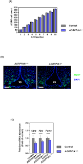
Here researchers study: Yongheng Cao1, Masanori Nakata, Shiki Okamoto, Eisuke Takano, Toshihiko Yada, Yasuhiko Minokoshi, Yukio Hirata, Kazunori Nakajima, Kristy Iskandar, Yoshitake Hayashi, Wataru Ogawa, Gregory S. Barsh, Hiroshi Hosoda, Kenji Kangawa, Hiroshi Itoh, Tetsuo Noda, Masato Kasuga, Jun Nakae.PDK1-Foxo1 in Agouti-Related Peptide Neurons Regulates Energy Homeostasis by Modulating Food Intake and Energy Expenditure. PLoS ONE 6(4): e18324. doi:10.1371/journal.pone.0018324.

IHC Protocol: For immunofluorescence analyses, mice were transcardially perfused with saline followed by 4% paraformaldehyde in 0.1 M phosphate-buffered saline, pH 7.4 (PBS). The brains were dissected and immersed in 4% paraformaldehyde at 4°C overnight and then soaked in 30% sucrose overnight. Frozen, free-floating coronal sections (4 µm thick) were cut through the arcuate nucleus with a microtome (Leica Microsystems). The sections were washed extensively in PBS for 20 min to quench endogenous peroxidase activity. For double staining of PDK1 and AGRP, the sections were stained with a Renaissance Tyramide Signal Amplification kit (#NEL701, Perkin Elmer, Waltham, MA) according to the manufacturer's protocol. The primary antibodies were Ab-241 (Signalway Antibody, Pearland, TX) for PDK1 and GT15023 (Neuromics, Edina, MN) for AGRP; the secondary antibodies were Alexa FluorR 594 chicken anti-rabbit IgG and Alexa FluorR 488 donkey anti-goat IgG (Molecular Probes, Eugene, OR). For double staining of Foxo1 and AGRP, we used an anti-FOXO1A antibody (ab12161, abcamR, Cambridge, UK), respectively. For double staining of FLAG and AGRP, the sections were stained using a Renaissance Tyramide Signal Amplification kit according to the manufacturer's protocol. The primary antibodies were the OctA-Probe (D-8: sc-807, Santa Cruz Biotechnology, Inc, Santa Cruz, CA); the secondary antibodies were Alexa FluorR 594 chicken anti-rabbit IgG and Alexa FluorR 488 donkey anti-goat IgG (Invitrogen, Carlsbad, CA).

Images: Functional defects of AGRP neurons in AGRPPdk1−/− mice. (A) Numbers of AGRP positive cells in the arcuate nuclei of AGRPPdk1+/+ (gray bar) and AGRPPdk1−/− (blue bar) mice. AGRP cell counts indifferent regions of the arcuate nucleus showed different numbers of AGRP neurons in AGRPPdk1+/+ (n = 3) and AGRPPdk1−/− mice (n = 3). (B) Representative Immunofluorescence images of AGRP in the hypothalamic regions of AGRPPdk1+/+ (left panel) and AGRPPdk1−/− mice (right panel). Green, AGRP; blue, DAPI. Scale bars indicate 100 µm. (C) Expression in the fed-state of hypothalamic neuropeptide genes in control (gray bar) and AGRPPdk1−/−(blue bar) mice. Data were normalized to β-actin expression and represent the mean ± SEM of six mice per genotype.

doi:10.1371/journal.pone.0018324.g002

Key Findings:PDK1 and Foxo1 signaling pathways play important roles in the control of energy homeostasis through AGRP-independent mechanisms. data indicated that PDK1 was indispensable for the orexigenic activity of AGRP neurons. Hypothalamic AGRP neurons express AGRP, NPY, the neurotransmitter GABA, and potentially other undiscovered molecules. In AGRPPdk1−/− mice, the expression of Agrp and Npy tended to be lower than that observed in control mice although the difference was not significant. Interestingly, the Δ256Foxo1AGRPPdk1−/− mice exhibited significantly increased food intake compared to AGRPPdk1−/− mice in spite of significantly decreased expression of Agrp and Npy. Therefore, changes in the expression levels of Agrp and Npy may not explain the changes in food intake in AGRPPdk1−/− mice.

Thursday, March 31, 2011

GFAP and Mouse Myenteric Plexus

Our Neuronal-Glial Markers are important tools for our customers investigating expression in the CNS and PNS. I have posted images showing staining of mouse retinal astrocytes and in the ventral horn, funiculus of adult rat spinal cord and mouse medulloblastoma stem cells using our GFAP antibodies.

I wanted to share an excellent image generated by Dr. Kate Ellacott's lab at Vanderbilt University.

GFAP (Chicken-Cat#: CH22102) staining in of enteric glia in the myenteric plexus of the mouse gastrointestinal tract. Staining was performed in methanol/acetone fixed frozen sections using 1:1000 dilution of the antibody followed by 1:500 anti-chicken Alexa 594.

Related Reagents:


Wednesday, March 23, 2011

STEMEZ hNP1 Neural Progenitors and Ion Channels


In my conversation with neuro-drug discover researchers, I am frequently being asked about the potential of using our STEMEZ(TM) hNP1 Human Neural Progenitors Expansion Kits for studying ion channels. How effective are these cells as a source for studying neurodegenerative diseases and for drug screening assays? There is good news.

When differentiated, these  neural progenitors express subunits of glutamatergic,  GABAergic, nicotinic, purinergic and transient receptor potential receptors. In addition, sodium  and calcium channel subunits were also expressed. Functionally, virtually all the NP cells exhibited delayed rectifier potassium channel currents and some differentiated cells exhibited  tetrodotoxin sensitive, voltage-dependent sodium channel current under whole-cell voltage clamp and action potentials could be elicited by current injection under whole-cell current clamp.  These results indicate that removing basic fibroblast growth factor from the neural progenitor cell cultures leads to a post-mitotic state, and also results in the capability to produce excitable cells that can generate action potentials. This is the first data demonstrating capabilitiesof these cells for ionotrophic receptor assays and ultimately for electrically active human neural cell assays for drug discovery.

Images: Glutamate receptor expression in hNP cells and differentiated hNP cells The expression of ionotropic glutamate receptors might also be an indicator of neuronal maturation. These receptors are composed of three distinct families: NMDA, kainate and AMPA receptors. The hNP cells and differentiated hNP cells cultured in the absence of bFGF for 2 weeks were analyzed for mRNA expression of subunits of each glutamate receptor subtype relative to hESCs. Significant increases (p<0.05) in Grin2b were seen in hNP cells (20 fold) and differentiated hNP cells (25 fold) relative to hESCs (Figure 3A). Additionally, Grin1 and Grin2d were significantly increased (p<0.05) only in differentiated hNP cells relative to hESCs, but not in undifferentiated hNP cells (Figure 3A). Of the kainate receptors, Grik4 and Grik5 were significantly (p<0.05) increased only in undifferentiated hNP cells relative to hESCs (Figure 3B); whereas, Grik2 was significantly (p<0.05) increased only in hNP cells where bFGF had been removed (Figure 3B). AMPA receptor subunits were also examined. Gria1 and Gria4 were up regulated in hNP cells relative to hESCs (Figure 3C). Two week differentiated hNP cells showed significant (p<0.05) up regulation of Gria2 and Gira4 relative to hESCs (Figure 3C). To determine if functional glutamate channels exist in differentiated hNP cells, calcium influx in response to AMPA, kainic acid or NMDA application was measured on hNP cells, 14 days after the removal of bFGF. Figure 3G indicates that NMDA could not depolarize differentiated or undifferentiated hNP cells enough to cause significant calcium influx above background. In contrast, AMPA and kainic acid can cause calcium influx which can be potentiated by AMPA receptor specific modulator, cyclothiazide (50 μM, Figure 3G).Calcium influx was detected in the presence of cyclothiazide in calcium activity as measured (Figure 3H).


Images: Sodium channel activity in differentiated hNP cells was measured using whole cell voltage clamp. 81 total hNP cells cultured in the absence of bFGF from 4 to 27 days were analyzed. Of these, 34 exhibited no fast inward currents in response to a step depolarization indicating the 348 absence of functional voltage gated sodium channels (Figure 4G). The remaining cells yielded between 0.04 - 1.5 nA of inward current in response to the step depolarization (Figures 4B and 4G). These currents inactivated rapidly in all cases (Figures 4B and 4C) and could be abolished with the addition of 1 μM TTX (n = 3 cells; Figure 4C). Voltage-dependent steady state inactivation (n = 11 cells; Figure 4D) and recovery from fast inactivation (n = 5 cells; Figure 4E) were also observed on several positive cells. A subset of these cells was subjected to current clamp and action potentials were elicited by current injection (n = 8 cells, Figure 4F). In support of this, increasing concentrations of a sodium channel activator veratridine in a FLIPR assay on differentiated hNP cells show an increasing calcium response (Figure 4H). This probably resulted from voltage-gated sodium channel depolarization of cells that subsequently allowed calcium influx through calcium channels. These data indicate that differentiation of hNP cells by removal of bFGF can lead to a neuronal cell that can generate action potentials and depolarize the cell. The 58% hit rate for voltage-gated sodium channel function (Figure 4G), does not reflect the true proportion of sodium channel positive cells in our differentiated hNP cells, but rather our ability to morphologically distinguish these cells from negative cells by eye. An example of the morphology of a sodium channel positive cell is shown in Figure 4A. The positive cells were phase bright with a few long processes.