Showing posts with label E18 Primary Hippocampal Neurons. Show all posts

Wednesday, January 09, 2019

Rat Hippocampal Neurons

Used to Study Apoptosis Induced by Brain Insults
Although our primary human cells are increasingly being used in drug discovery and toxicology assays versus animal cells, we are always pleased to see the success of the latter in sophisticated studies.

This publication references use of our e18 rat hippocampal neurons. These cells continue to be easy to culture, pure and potent. Chiara Porro, Antonia Cianciulli, Teresa Trotta, Dario Domenico Lofrumento, Rosa Calvello and Maria Antonietta Panaro. (2019). Formyl-methionyl-leucyl-phenylalanine Induces Apoptosis in Murine Neurons: Evidence for NO-Dependent Caspase-9 Activation. Biology 8(1), 4; doi:10.3390/biology8010004.

Highlights
Formyl-methionyl-leucyl-phenylalanine (fMLP) may be present in the brain in the course of some infectious diseases of the central nervous system (CNS), although little is known about its role. This investigation was performed to study the effect of fMLP on neuron apoptosis. The results showed that fMLP treatment of primary cultures of neurons was able to induce morphological features of apoptosis in cell cultures, as well as activation of the intrinsic apoptotic pathway, through the upregulation of caspase-9 and caspase-3.

Figure. (A) Western blot analysis was performed on membrane-enriched cell extracts (25 µg lysate) of primary neurons. The blots were probed with formyl peptide receptor 2 (FPR2) antibody (Ab) and detected by chemiluminescence. A ~41 kDa band corresponding to FPR2 was observed as compared to the positive control. Lane 1: Marker; lane 2: Positive control, lane 3: Primary neuron lysate. (B) Immunofluorescence identification of FPR. Double staining shows the expression of the FPR receptor on the cell membrane and neurofilaments. FPR2 expression (green); skeleton protein staining of neuron-specific neurofilament 68 (red); DAPI nuclear staining (blue); cells stained by both neurofilament 68, FPR2 and DAPI (merged). Scale bar: 100 μm. 1): neurofilaments stain; 2) FPR2 expression; 3) DAPI stain; 4) merged.

The present study emphasizing the potential role of infectious agents, such as N-formyl peptides, in neurodegenerative diseases may help to promote the development of new therapies able to modulate the expression of the N-formyl peptide receptors.

Tuesday, January 31, 2017

Desperately Seeking Data

Answering the Bell
We continue to seek data using our cells. We offer a reward of 25 USD Starbucks' Gift Card.

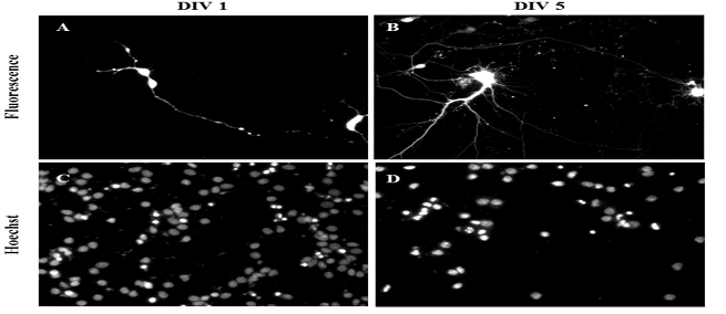
We were pleased to receive a recently published study from Dr. Mahendran Subramanian of Keele University. In this study, researchers showed that oscillating nanomagnetic gene transfection could be used to successfully transfect SH‐SY5Y cells as well as our primary hippocampal and cortical neurons on different days in vitro. This novel technique was used to effectively deliver genetic material into various cell types, resulting in high transfection efficiency and viability. Mahendran Subramanian, Aimee‐Jayne Tyler, Eva Maria Luther, Elena Di Daniel, Jenson Lim and Jon Dobson. Oscillating Magnet Array−Based Nanomagnetic Gene Transfection: A Valuable Tool for Molecular Neurobiology Studies. Nanomaterials 2017, 7, 28; doi:10.3390/nano7020028...Primary rat hippocampal and cortical neurons were obtained from Neuromics (Edina, MN, USA) and disassociated using papain disassociation kit (Worthington, NJ, USA) according to the manufacturer’s instructions. Isolated neurons were maintained using neurobasal medium supplemented with 5% FBS, 0.5 mM Glutamax, 2% B27 supplement, 25 μM L‐glutamine and seeded onto poly‐D‐lysine–coated cells culture plates...
Figure 1. Oscillating magnet array−based nanomagnetic gene transfection experimental setup. (A) Representation of a 96‐well oscillating magnet array–based nanomagnetic transfection setup using NdFeB magnetic array (nanotherics); (B) Dimensions of the permanent magnets and magnetostatic (vectorpotential) algorithm based magnetic field density |B| distribution (T) contour plot for the NdFeB magnetic array.

Figure 2. Gene delivery by oscillating nanomagnetic gene transfection in primary cortical neurons. Images of pmaxGFP plasmid expressed in primary neurons using fluorescence microscopy and its corresponding Hoechst 33,342 stained counterpart of transfected DIV 1 (A,C) and DIV 5 (B,D) mature neurons were taken 48 h post transfection.

If you have data to share email it to me, pshuster@neuromics.com and we'll email you a 25 USD gift card. Thank you. Pete Shuster, CEO & Owner.

Friday, November 04, 2016

We Want Your Data!

50 USD Reward
Our Customer Data program is progressing nicely. I am pleased to present the latest addition from Dr. Vimal Swarup, University of Utah. To qualify for the reward just email Rose Ludescher-rose@neuromics.com.


Hippocampalneuron-based guidance assay. Image of micro-contact patterned chondroitin sulfate proteoglycans (pre-mixed with AlexaFluor 594) purified from astrocyte conditioned media (CM). b) Immuno-stain of the spot in (a) with anti-CSPG antibody CS56. c) Overlay of Embryonic day-18 primary rat hippocampal neurons (Cat# PC35101, Neuromics Inc.) and the underlying surface containing μC patterned CM after 48 h of growth. d) Overlay of neurons and underlying surface containing μC patterned CSPGs purified from CM. e) Average neurite length on pLL and stamped regions after 48 h. f) Average neurite length on plain field surfaces of μC patterned CM, CSPG and pLL after 48 h. Results from this experiment show that neurons avoid CM or CSPGs purified from CM while elongating. Swarup et al. JACS. 2013

Submit your data today and earn you reward.

Tuesday, August 06, 2013

Healthy and Happy Neuron/Astrocytes Cultures

A Track Record of Customer Success

Neuromics is  recognized for the quality of  hNP1™ Human Neural Progenitor,  hN2™ Neuron Discovery Kits, E18 and E20 Rat Primary Neurons and E18 Rat Primary Astroglia. As the company owner, it is important that I keep my finger on the pulse of how well they work for each and every unique application. I personally follow up with each user and if there are any issues, we replace the cells once free of charge. Your success is critical to our growth.

I wanted to share with you recent references. These give and excellent snapshot of the exciting ways our cells can used. Alexzander Asea, Punit Kaur, Alexander Panossian, Karl Georg Wikman, Evaluation of molecular chaperons Hsp72 and neuropeptide Y as characteristic markers of adaptogenic activity of plant extracts. Phytomedicine, Available online 6 August 2013, ISSN 0944-7113, http://dx.doi.org/10.1016/j.phymed.2013.07.001
...using trypan blue exclusion test and routinely found to contain less than ;5% dead cells. Primary human neurons were purchased from Neuromics (Edina, MN)...

Images: Micropictograph of primary culture from micro-dissected hippocampus. (A) Neurons are round and healthy 1 h after plating on poly-d-lysine substrate. (B) Five days in culture, neurons remain healthy and have extended processes. Magnification 60×. http://dx.doi.org/10.1016/j.phymed.2013.07.001

Todd GK, Boosalis CA, Burzycki AA, Steinman MQ, Hester LD, et al. (2013) Towards Neuronal Organoids: A Method for Long-Term Culturing of High-Density Hippocampal Neurons. PLoS ONE 8(4): e58996. doi:10.1371/journal.pone.0058996
... a protocol that allows for culturing of E18 hippocampal neurons at high densities for more than 120 days. These cultured hippocampal neurons are (i) well differentiated with high numbers of synapses, (ii) anchored securely to their substrate, (iii) have high levels of functional connectivity, and (iv) form dense multi-layered cellular networks. We propose that our culture methodology is likely to be effective for multiple neuronal subtypes–particularly those that can be grown in Neurobasal/B27 media. This methodology presents new avenues for long-term functional studies in neurons...
Xiugong Gao, Hsiuling Lin, Radharaman Ray, Prabhati Ray. Toxicogenomic Studies of Human Neural Cells Following Exposure to Organophosphorus Chemical Warfare Nerve Agent VX. Neurochemical Research. February 2013.
...Human hN2 neurons were obtained from Neuromics...
Image: Staining of hN2 Human Neurons with Tuj 1 (Neuron-specific class III beta-tubulin) (red) and Nestin (green). Counter stained with DAPI (blue). hN2 Cells-Electro Phys Data. 

Xiufang Guo, Severo Spradling, Maria Stancescu, Stephen Lambert, James J. Hickman. Derivation of sensory neurons and neural crest stem cells from human neural progenitor hNP1. Biomaterials, In Press, Corrected Proof,Mar 2013.doi:10.1016/j.biomaterials.2013.02.061
...hNP1, were obtained from Neuromics (Edina, Minnesota)...
Wei Zhang , Radhia Benmohamed, Anthony C. Arvanites, Richard I. Morimoto, Robert J. Ferrante, Donald R. Kirsch, Richard B. Silverman. Cyclohexane 1,3-diones and their inhibition of mutant SOD1-dependent protein aggregation and toxicity in PC12 cells. Bioorganic & Medicinal Chemistry. Elsevier Ltd. All rights reserved.doi:10.1016/j.bmc.2011.11.039.
...Primary rat cortical tissue was purchased from Neuromics Inc., Edina, MN and used to initiate primary cortical neuron cultures. The tissue was isolated from micro-surgically dissected E18 embryonic Sprague/Dawley or Fischer 344 rat brain and shipped in a nutrient rich medium under refrigeration. To isolate neurons, the tissue was incubated with papain at a concentration of 2 mg/mL in Hibernate without calcium for 30 min at 37OC. The enzymatic solution was then removed, and 1 mL of culture media (Neurobasal, B27, 0.5 mM glutamine) was added. A sterile Pasteur pipette was used to gently disperse the cells, which were then washed, re-suspended and counted. The cells were plated on poly-D-lysine coated 96-well plates at a density of 20,000 cells/well and incubated at 37OC in a 5% CO2-humidified atmosphere for 5 days prior to use in compound testing. By microscopic inspection, the resulting cultures consisted of app. 90% neurons...
Majumder A, Dhara SK, Swetenburg R, Mithani M, Cao K, Medrzycki M, Fan Y, Stice SL. Inhibition of DNA methyltransferases and histone deacetylases induces astrocytic differentiation of neural progenitors. Stem Cell Res. 2013 Jul;11(1):574-86. doi: 10.1016/j.scr.2013.03.003. Epub 2013 Apr 2.
...Progenitor to Astrocytes Protocol: For astrocytic differentiation of hNP cells, neuronal differentiation media were supplemented with BMP2 (20 ng/mL) and combinations of Aza-C and TSA; Aza-C (500 nM), TSA (100 nM) and BMP2 (20 ng/mL) for 2 days, with one complete media change in between, followed by differentiation media supplemented with BMP2 but not with Aza-C or TSA. Cells were harvested prior to analysis at 5, 15 or 30 days of treatment or for cryopreservation at d6 or d10 of differentiation. For cryopreservation, cells were dissociated with Accutase™ and frozen in differentiation media containing10% DMSO. Viability was assessed at 30 days in Aza-C and TSA treated cultures by trypan blue exclusion, and datawas acquired using a Cellometer Auto T4® (Nexcelom Biosciences)...
Aparna Talekar, Antonello Pessi, and Matteo Porotto. Infection of primary neurons mediated by Nipah virus envelope proteins: Role of host target cells in antiviral action. J. Virol. doi:10.1128/JVI.00452-11.
...Hippocampus, Cortex and Ventricular Cells (Neuromics)...

I will continue posting results here.

Monday, April 29, 2013

Neuron-Glial Cultures-Setting a Higher Bar!

Improved Methods for Long Term, High Denisty Cultures
 
Dr. Randen Patterson and his team at UC Davis have developed new culturing techniques using our e18 Rat Primary Hippocampal Neurons. They have developed a protocol that allows for culturing of E18 hippocampal neurons at high densities for more than 120 days. These cultured hippocampal neurons are (i) well differentiated with high numbers of synapses, (ii) anchored securely to their substrate, (iii) have high levels of functional connectivity, and (iv) form dense multi-layered cellular networks. We propose that our culture methodology is likely to be effective for multiple neuronal subtypes–particularly those that can be grown in Neurobasal/B27 media. This methodology presents new avenues for long-term functional studies in neurons. This is good news indeed: Todd GK, Boosalis CA, Burzycki AA, Steinman MQ, Hester LD, et al. (2013) Towards Neuronal Organoids: A Method for Long-Term Culturing of High-Density Hippocampal Neurons. PLoS ONE 8(4): e58996. doi:10.1371/journal.pone.0058996.
 
Protocol Highlights: 
Substrate Preparation
1. On the day of plating, prepare 25 mm coverslips by removing them from 70% ethanol storage solution and propping them up at an angle in each well of a 6-well culture plate to allow drying. [No more than 5 plates (30 coverslips) should be dried simultaneously for 15–25 minutes in culture hood to avoid over-drying.]
2. Once dry, shake slips down flat into their respective wells and coat with 1 mL 0.1% poly-D-lysine, taking care to form a liquid meniscus on each slip. Carefully transfer coverslips into incubator, taking care to preserve meniscus.
3. Incubate for 1 hr at 37°C. [Keeping poly-D-lysine meniscus on top of coverslip is important; this serves to avoid poly-D-lysine coating under coverslip surface that may lead to problematic flotation of coverslip.] After incubation, remove poly-D-lysine and rinse each coverslip three times with 2 mL sterile deionized water. Take care to ensure coverslips do not completely dry at any point during the rinse. After the third and final rinse, leave coverslips in 2 mL sterile deionized water for at least 1 hr. Remove water just before plating, again, make sure to avoid over-drying. [This critical step requires attention. Take care to aspirate off all sterile water remaining from the final rinse, but also use caution as not to over-dry the coverslips. Ultimately, the coverslip must be mostly dry as to allow for the meniscus formation during plating (Fig. 1, Step 12), whereas over-drying can result in the neurons peeling off the glass coverslips days to weeks after plating.]
Fig 1: Neuron-Glial Culturing Steps
Preparation of Isolated Neurons (Numbers in Fig. 1 correspond to numbers below).
1. Store tissue at 4°C until ready to use. If dissecting your own cultures, upon isolation of the tissue, store in an appropriate storage media.
2. When ready to plate, make 2 mL of enzymatic solution without B27. In our case, we used Hibernate E-Ca, containing 4 mg (2 mg/mL) of papain. If making your own solution, use a commercially available papain dissociation kit. Make sure to sterile filter solution with 0.2 micron filter after adding papain if source of enzyme is not sterile.
3. Remove the storage media from the dissected tissue and transfer into sterile 15 mL screw-cap tube; be careful not to disturb or remove tissue from original tube. Save the storage media, do not discard.
4. Add 2 mL of media made in Step 2 to tissue (in our case, Hibernate E-Ca containing 2 mg/mL of papain). Incubate for 35 min at 37°C. [Be sure to add Hibernate E-Ca containing papain slowly as to avoid disturbing tissue.]
5. Remove enzymatic solution from tissue, again, take care not to disturb or remove tissue. Add back 1 mL of storage media saved in 15 mL tube.
6. Using a 1 mL pipettor with a sterile plastic pipette tip (tissue can adhere to glass pipettes), aspirate the tissue with the medium into the pipette and immediately dispense contents back into same container. Take care not to create bubbles. [This is another critical step that requires attention. Take care to make sure pipette tip remains in a stable position (as shown in Fig. 1, Step 6). Maintain slow, steady speed when both drawing in and re-dispensing media containing tissue.]
7. Repeat this trituration step 10–12 times or until most all the tissue is dissociated and the cells are dispersed. [Under close examination cell dispersion is highly visible. Stop pipetting immediately upon cell dispersion.]
8. Slowly transfer contents of the tissue tube into a new sterile 15 mL screw-cap tube.
9. Use the remainder of storage medium saved in Step 3 and rinse the interior of the tissue tube before adding it to the sterile 15 mL screw cap tube containing dispersed cells from step 7. [This step helps ensure minimal wastage, as any remaining cells should be saved with this extra rinse.]
10. Spin dispersed cells at 1,100 rpm (200Xg) for 1 min.
11. Discard the supernatant while being careful not to remove any of the cells from cell pellet.
12. Flick tube a few times to loosen the cell pellet. Re-suspend pellet in 2.4 mL of pre-warmed B27/Neurobasal/0.5 mM glutamine medium. Re-suspend by gently pipetting up and down. For E18 Hippocampus, medium includes 25 µM glutamate.
13. Plate cells within a meniscus (approx. 10 mm diameter) at a minimum of 40 µL per 25 mm coverslip. Take care not to disturb meniscus. [Periodically pipette up and down throughout plating process (no more than once every plate per 6 coverslips) to help maintain equal cell density. Again, plating with meniscus formation is critical.]
14. Incubate plated cells at 37°C with 5% CO2 and/or 9% or 20% oxygen for 1 hr.
15. Add 1.5 mL per well of pre-warmed 1:1 ACM/NbActiv4. [Slow and steady media addition rate and proper pipette position are necessary for successful plating density consistency. Position pipette tip at 45° angle along middle of 6-well interior sidewalls, dispense 1.5 mL as slowly and steadily as possible (see Steps 1–14).]
16. Incubate cells at 37°C with 5% CO2 and/or 9% or 20% oxygen.
17. Add Cytosine β-D-arabinofuranoside (Ara-C) to a final concentration of 5 µM, 5–6 days after plating to curb glial proliferation. [Remove 1/3 of media from each well and replace with equal volume containing final concentration of Ara-C]
18. After 4 days or longer, neurons are well differentiated. If further culture is desired, change 1/3 of medium with fresh, pre-warmed 1:1 ACM/NbActiv4 every 7–8 days.
Images: 40X Confocal Images of 30 DIV Hippocampal Cultures. Immunofluorescence detection of MAP-2 (green) and GFAP (red) in 30 DIV (A–C) cultured E18 hippocampal cells using a 40X objective. These images (A and B) clearly depict the intimate physical contact between glia processes and dendritic arbors. Under closer examination (CI and CII), it is clear that the dendrites have grown both bellow (blue arrows) and above (white arrows) glial processes, forming a highly interconnected three-dimensional network by 30 DIV. doi:10.1371/journal.pone.0058996.g004.
All Primary Neuron Assay Customer Publications
 
Related Content: If you have any questions on optimizing your cell cultures, do not hesitate to contact me @ pshuster@neuromics.com or 612-801-1007

Saturday, March 02, 2013

Increasing Returns in Drug Discovery

"Harnessing the Power of Cells"
 
Cell based assays presentation v1_03_2012 from Pete Shuster

This presentation provides an executive overview of how our Solutions can improve Drug Discovery processes.

Why Consider our Solutions?
Research proven solutions used by large Pharmas, Biotechs, Academic and Government Labs. 2500+ customers-Publications/Testimonials
Lower per well costs-we can aggressively discount cells and media
Customer success focused                                  
Expert technical support
Detailed protocols and methods
Quick replacements and reorders
Large expert resource network
Published expert input
Non confidential; customer feedback and data sharing through social media outlets and blogs

Thursday, April 26, 2012

Primary Hippocampal Neurons Performing!

I advertise our Primary Neurons and Astrocytes as being easy to culture, grow and maintain. We confirm this via the data/images our customers generously share and the many publications referencing use of the cells.

I would like to thank George Kenneth Todd (Patterson Lab at UC Davis) for these wonderful images of our e18 Primary Rat Hippocampal Neurons. These were taken at day 67!


Prior to staining, the cells were treated with Wnt5 for 2 min, then Wnt5 + Wnt3 for an additional 2 min during Calcium Imaging experiments. The cells were then fixed and stained for IP3R (green), Frizzled2 (blue), and B-Catenin (red), and the confocal images were captured at 10x. Notice the parallel neurites formation in image 3.


For staining culture options, check out our neuron-glial-astrocyte markers.

Thursday, October 20, 2011

Immunostaining Neurons and Glia

I would like to thank Dr. Gerry Shaw, University of Florida for his excellent work with our Primary Neurons and Astrocytes and Neuronal-Glial Markers. Here's an example image with many more to follow:

Image: E18 hippocampal neurons stained with MAPT (red) and Doublecortin (green). The two proteins overlap in the proximal dendrites, but doublecortin is more abundant in the growth cones and periphery. As a result, the periphery appears green while the more proximal regions of the cells are yellow. The single longer process of this cell, presumably an axon, has a low doublecortin content and so appears red. Blue staining is the nuclear DNA. Protocol on datasheet.