Showing posts with label neuron cultures. Show all posts

Monday, July 13, 2015

Apoptosis and Neurodegeneration

Towards the Development of Disease Specific Assays
Neurodegenerative diseases are becoming increasingly prevalent, especially in the Western societies, with larger percentage of members living to an older age. They have to be seen not only as a health problem, but since they are care-intensive, they also carry a significant economic burden.

Apoptotic pathways are induced in many of these diseases and are key culprits in disease progression.
Figure: Schematic representation of apoptotic pathways. Apoptosis triggered by internal (intrinsic) or external (extrinsic) stress signals that is activated by binding of ligands (e.g. FasL, APO-2L, TRAIL, TNF) to cell surface receptors (e.g. Fas, DR4, DR5, TNF-R1). The intrinsic apoptosis pathway might be triggered by p53 upon DNA damage following exposure to cellular stress. In the intrinsic pathway, death signal reaches mitochondria, leading to release of cytochrome c, which can binds to Apaf1. The cytochrome c/Apaf1 make a complex with pro-caspase-9 (in the presence of dATP), activates caspase-9, which promotes caspase-3 activation, eventually leading to cell death. The extrinsic pathway is initiated through the stimulation of the members of tumor necrosis factor receptor (TNF-R) family (transmembrane death receptors) by their respective ligands. These receptors activate pro-caspases-8, -10 by recruiting the endogenous adaptor protein FADD. Procaspase-8, -10 cleave themselves to form activated caspase-8 or -10. Ultimately, effector enzymes such as caspase-3, -6, -7 are activated in this cascade to mediate apoptosis. Likewise, there can be cross-talk between the intrinsic and extrinsic pathways. For example caspase-8 may cleave Bid to form tBid that is a strong activator of the intrinsic/mitochondrial apoptotic pathway. The intrinsic pathway is usually activated by the recruitment of BAX and BAK to outer mitochondrial membrane, causing cytochrome c release formation of apoptosome and subsequent activation of caspase-9. Activated caspase-9 proteolytically activates caspases-3, -6, and -7. Moreover, some of the effector caspases also can activate caspase-8, forming a positive amplification loop. doi:10.1016/j.pneurobio.2013.10.004.

Working with the Apoptosis Experts at Immunochemistry Technologies and Human Astroglial-Neuron Biosensors Experts at ArunA Biomedical, we plan on  developing disease specific assays. Here's a map of the general process.
We welcome feedback and input on your interests. You can email rose@neuromics.com. We will continue to post updates on exciting new developments.


Monday, January 05, 2015

Racing into 2015

Thank you for a Great 2014!
EU PCWow. I would like to thank our customers, collaborators and friends for helping us achieve unprecedented success in 2014.

We plan on racing hard into 2015. I would like to highlight our focus areas for the New Year.

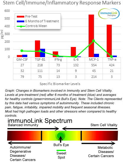
Stem Cell Activating Solutions-We have been pilot testing solutions foir balanced immunity and stem cell vitality. Our clients have been mostly peopole with autoimmune related issues. After 6 months of therapy, the improvement in symptoms and test results, for many, has been stunning (see below).
We have developed migration and other assays to test these solutions in vitro using our UCB Derived hMesenchymal Stem Cells. We also use medical testing to prove and improve their therapeutic value. These assays and arrays are available for labs and clinics interested in testing stem cell related solutions. Here're representative data from our Quantibody Array testing program.
IT

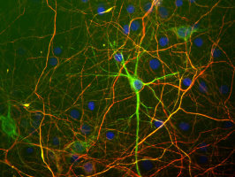
Stem and Primary Cell Based Assays-We will continue to add new solutions for your assays. These will include mixed culture that more closely mimic in vivo environments like mixed astroglial-neuron cultures. Below is our hN2TM Human Neurons stained with one of our neuron markers.
hN2
Image: hN2 cells grown in culture for 4 days and stained with our chicken polyclonal to Neurofilament light or low molecular weight chain NF-L, a marker of neurons. Many of the differentiating cells show strong cytoplasmic and clearly fibrillar staining for NF-L. Blue stain is DAPI and reveals cell nuclei of some non neuronal cells in this culture.

Check out and join my Stem Cell Clinical Trails Group on Linkedin (4500+members strong).
We wish you a rewarding 2015.

Pete Shuster-
CEO & Owner--pshuster@neuromics.com or 612-801-1007

Saturday, November 01, 2014

GFP Labeled Mouse Motor Neurons-Buy Now Save 100 USD

Designed for Motor Neuron Disease Research

I am pleased to add (finally) to our catalog potent, pure and ready to culture motor neurons. I know from the many request I have gotten for these that demand could be out stripping supply.

These are designed foruse in High-throughput fluorescent screening applications. Derived from transgenic mice expressing eGFP using Hb9 motorneuron promoter enables easy tracking and vi sualization of spinal motor neurons without the need for additional fluorescent markers and extended cultures.

Image: GFP+ mMN Mouse Motor Neurons at 2 days post thaw 20X

If you want to learn more about our any of our Neuron-Glial-Astrocyte Based Assay Solutions, do not hesitate to contact me (612-801-1007) or pshuster@neuromics.com. Pete Shuster, Owner and CEO, Neuromics.

Thursday, April 26, 2012

Primary Hippocampal Neurons Performing!

I advertise our Primary Neurons and Astrocytes as being easy to culture, grow and maintain. We confirm this via the data/images our customers generously share and the many publications referencing use of the cells.

I would like to thank George Kenneth Todd (Patterson Lab at UC Davis) for these wonderful images of our e18 Primary Rat Hippocampal Neurons. These were taken at day 67!


Prior to staining, the cells were treated with Wnt5 for 2 min, then Wnt5 + Wnt3 for an additional 2 min during Calcium Imaging experiments. The cells were then fixed and stained for IP3R (green), Frizzled2 (blue), and B-Catenin (red), and the confocal images were captured at 10x. Notice the parallel neurites formation in image 3.


For staining culture options, check out our neuron-glial-astrocyte markers.
