Showing posts with label hMSCs. Show all posts

Thursday, May 04, 2017

Culturing Stem Cells in 3-D

Requires Potent Media + Supplements

Neuromics is responding to the many challenges our clients face in building 3-D, in-vivo like, cell- based assays. We do this by offering the most potent Media plus Supplements like FGFS.

Here's a protocol for single cell 3-D assays using hMSCs and Hydrogels. It features use of our ISOKineTM FGF


Images: Cell-centering in cytocompatible microgels enables long-term single-cell 3D culture by preventing cell escape. a) Qualification of Dex-TA microgel crosslinking as a function of the microemulsion flow rate (Qemulsion) and concentration of the H2O2 feed ([H2O2]feed). Blue, green, and red indicate incomplete crosslinking, complete crosslinking, and H2O2 excess, respectively. b,c) Amplex Red assay to quantify the concentration of residual H2O2 ([H2O2]emulsion) in Dex-TA microgel precursor droplets and crosslinked microgels after their retrieval from the diffusion-based crosslinking platform. d) The microencapsulation procedure had no detrimental effect on short-term cell survival. e) Delayed crosslinking resulted in 4 ± 1% cell escape after 7 d of in vitro culture, as compared to 27 ± 5% cell escape when using coupled emulsification and gelation. f) The number of encapsulated cells per microgel tightly followed the Poisson distribution and remained similar throughout long-term (28 d) of in vitro culture, which confirmed that cell centering prevents cell escape. g–i) MSCs encapsulated in delayed enzymatically crosslinked microgels remained viable and metabolically active throughout 28 d of in vitro culture. j) Positive Oil Red O and k) Alizarin Red staining confirmed that l) more than 60% of the microencapsulated MSCs could differentiate into the adipogenic and osteogenic lineage, respectively. Black scale bars: 50 µm, white scale bars: 5 µm. DOI: 10.1002/smll.20160371.

Protocol for Cell Isolation and Expansion: Human MSCs were isolated from fresh bone marrow samples and cultured as previously described. The use of patient material was approved by the local ethical committee of the Medisch Spectrum Twente and informed written consent was obtained for all samples. In short, nucleated cells in the bone marrow aspirates were counted, seeded in tissue culture flasks at a density of 500 000 cells cm−2, and cultured in MSC proliferation medium, consisting of 10% FBS, 100 U mL−1 penicillin, 100 mg mL−1 streptomycin, 1% GlutaMAX, 0.2 × 10⁻³ m ascorbic acid, and 1 ng mL−1 bFGF (added fresh) in αMEM. Mouse insulinoma MIN6-B1 cells (provided by Dr. P. Halban, University Medical Center, Geneva, Switzerland) were cultured in MIN6 proliferation medium, consisting of 10% (v/v) FBS, 100 U mL−1 penicillin, and 100 mg mL−1 streptomycin, and 71 × 10−6 m 2-mercaptoethanol (added fresh) in DMEM. When cells reached near confluence, the cells were detached using 0.25% Trypsin-EDTA at 37 °C and subsequently subcultured or used for experimentation.

I am at your beck and call to answer questions on our Cell Based Assay Solution. Pete Shuster-CEO and Owner, direct phone: (612) 801-1007 or pshuster@neuromics.com.

Tuesday, November 15, 2016

Infection of Mesenchymal Stem Cells

UCB derived huMSCs infected with RSVs

Here's cool customer data of our Human Mesenchymal Stem Cells.

Figure. RSV infects and replicates in human MSCs. Human MSCs grown on coverslips, infected with 1 MOI of RSV, and immunostained at 48 or 72 hours post-infection with a monoclonal antibody to RSV tagged with FITC (green), Evan’s blue dye (red) and DAPI (blue). Total magnification of images is 200X (inset 1000X) and scale bar is 50μm (A). RSVN transcripts detected in MSCs at 6, 12, 24 and 72 hours post infection normalized to mock (B). RSV titers (PFU/ml) isolated from the culture medium of infected and mock-infected MSCs is shown (C). Results representative of at least duplicate experiments. doi.org/10.1371/journal.pone.0163709.

Your data is wanted. Just email rose@neuromics.com and claim your 50 USD reward. #datawanted!

Tuesday, August 23, 2016

Human Mesenchymal Stem Cells Conjugates

Targeted Delivery of Stem Cells
Here's a recent publication referencing use of our Human Mesenchymal Stem Cells to form conjugates that could have application for drug delivery and cell therapies: Yun Seop Kim, Won Ho Kong, Hyemin Kim, Sei Kwang Hahn. Targeted Systemic Mesenchymal Stem Cell Delivery Using Hyaluronate - Wheat Germ Agglutinin Conjugate. http://dx.doi.org/10.1016/j.biomaterials.2016.08.027


Images: Fluorescence microscopic images of dissected livers and lungs from normal rats 4 h after intravenous injection of PBS, WGA-FITC/hMSC, and HA-WGA-FITC/hMSC (Filter for FITC). (b) Quantification of hMSCs in dissected organs estimated from ROI (n = 3). **P versus the WGA/hMSC group. (c) Representative fluorescence microscopic images of WGA-FITC/hMSC and HA-WGA-FITC/hMSC in the liver and lung (green = FITC, blue = DAPI for nucleus). Arrows indicate the location of hMSCs (scale bar = 200 μm). http://dx.doi.org/10.1016/j.biomaterials.2016.08.027
Protocol: Cultured MSCs were trypsinized, washed, and resuspended at 1 × 10⁶ cells/mL in PBS. HA-WGA conjugate at 10 μg/mL of WGA was added to MSCs in suspension, and incubated in an ice bath for 10 min with mild mixing. To remove the unbound HA-WGA conjugate to MSCs, cell suspension was washed with PBS and collected by centrifugation (1000 × g, 3 min at 4 °C). After surface modification with HA-WGA conjugate, HA-WGA/hMSC were resuspended in the cell culture medium on 96-well plates (1 × 10⁴ cells per well). At the predetermined time, cell viability of HA-WGA/hMSC was measured using an EZ-cytox cell viability assay kit according to the manufacturer’s instructions. Zeta potentials were analyzed using a Zetasizer Nano (Malvern Instruments, UK) to assess the change of surface charge after the surface modification.
Conclusions: Researchers successfully developed a target-specific systemic delivery system of MSCs to the liver using HA-WGA conjugate. HA-WGA conjugate was synthesized by the coupling reaction of HA-aldehyde with amine group of WGA. GPC analysis revealed the successful synthesis of HA-WGA conjugate. CD analysis corroborated that the secondary structure of WGA remained stable even after conjugation to HA. HA-WGA conjugate appeared to bind to the surface of MSCs and remain stably for up to 1 h. After cell surface modification, most of HA-WGA/MSC complex could be systemically delivered to the liver 4 h after intravenous injection, whereas MSCs were trapped mainly in the lung. This new strategy to target-specifically deliver MSCs to the liver using HA-WGA conjugate might be successfully exploited for treatment of various liver diseases.

We will continue to post new applications developed by users of our Stem Cells.

Friday, January 30, 2015

Sensitive, Specific and Cost Effective Protein Expression Analysis

Reaching for Cell Based Assay Solutions Excellence

We have been receiving positive feedback from clients using our Quantibody® and RayBio® C-Series Membrane-Based Antibody Arrays. Our manufacturing partner, Raybiotech, is ISO 9000 certified insuring all arrays are made to exacting specification. The cost for measuring a single cytokine runs as low as 16 USD/Cytokine!

We use these arrays to test the blood serum of clients with autoimmune diseases to determine levels of cytokine and growth factor expression vs healthy controls. We also use them to determine proteins secreted by our  USB Derived hMSCs cultured in our hMSC Media. This gives us insight as to the protein expression patterns in our hMSC, Chondrogenesis and Osteogenesis Assays.

Figure: The graph shows cytokine secretion of hMSCs after a continuous 24 day culture. These results were obtained using our select markers from our cytokine and bone metabolism arrays. Data courtesy of Dr. Jim Musick and Tiana Tonrey, Vitro Biopharma.

We are pleased to announce the addition of these new human and mouse arrays:

Quantibody® Human Growth Factor+Neurotrophin+Cytokine Array Q1-10. Precisely measures: Amphiregulin, BDNF, bFGF, BMP-4, BMP-5, BMP-7, beta-NGF, EGF, EGFR, EG-VEGF (PK1), FGF-4,FGF-7 (KGF), GDF-15, GDNF, Growth Hormone, HB-EGF, HGF, IGFBP-1, IGFBP-2, IGFBP-3, IGFBP-4, IGFBP-6, IGF-1, Insulin, M-CSF R, NGFR (TNFRSF16), NT-3, NT-4, Osteoprotegerin (TNFRSF11B), PDGF-AA, PLGF, SCF, SCF R (CD117/c-kit), TGF alpha, TGF beta 1, TGF beta 3, VEGF-A, VEGFR2, VEGFR3 and VEGF-D.
Mouse C-Series Cytokine Array. C1-2-Semi-quantitavely measures: GCSF,GM-CSF, IL-2, IL-3, IL-4, IL-5, IL-6, IL-6, IL-9, IL-10, IL-12 p40/p70, IL-12 p70, IL-13, IL-17A, IFN-gamma, MCP-1 (CCL2), MCP-5, RANTES (CCL5), SCF, TNF RI (TNFRSF1A), TNF alpha, Thrombopoietin, (TPO) and VEGF-A.


Images: Representative images obtained with RayBio® C-Series Antibody Arrays. These membranes were probed with conditioned media from two different cell lines. Membranes were exposed with Kodak X-Omat® film at room temperature for 1 minute. Note the strong signals of the Positive Control spots in the upper left and lower right corners.

Questions? Do not hesitate to contact me: pshuster@neuromics.com or direct phone-612-801-1007. Thank you. Pete Shuster, CEO and Owner, Neuromics

Monday, October 27, 2014

100% Serum-Xeno Free Media for Expanding hMSCs

Works as well as our Serum Containing Media

 Selection of Growth and Differentaition media for Mesenechymal Stem Cell Assays is important for the ultimate performance of your cell based assays. The better the media the better the cutures & the lower your costs.


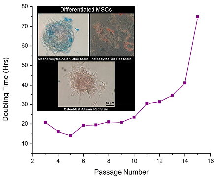
Images: Human mesenchymal stem cells (Catalog no. SC00A1) were plated at 5,000/cm² in a Falcon BD TC-coated T-25 flasks and maintained in serum containing media (Cat. No. SC00B1) and serum-free/xeno-free media (Cat. No. SC00B3) in reduced O2 environment (1% O2, 5% CO2, 90% N2) at 37°C in a humidified chamber. When cells became 80-90% confluent, they were subcultured, counted on a Beckermen Z2 particle counter (range 10-30uM), and passaged. Doubling time of 20-25hrs were calculated in each pass using ln(2*dT)/ln(Cf/Ci), where dT is the time, in hours, from inoculation to detachment; Ci is the initial number of cells plated and Cf is the final number of cells recovered from subculture. There were no apparent differences in doubling time with TC-coated flasks and Laminin/Fibronectin treated flasks.

Testimonial: “We tested the effects of MSCGroTM defined medium using several different lots of human adult primary stem cells and found that MSCGro supports a more robust proliferation rate than normal undefined media. This provided shorter doubling times and increased cellular yield, and maintained the cells in an undifferentiated state. We also found that MSCGro medium is stable under normal laboratory conditions for an extended time period compared to other defined media.” Ben Buehrer, VP and CSO, Zen-Bio

Sunday, March 23, 2014

Autism, Inflammation and Stem Cell Enhancers

Proving the Therapeutic Value

We have been running Quantibody® Antibody Arrays on blood serum of children diagnosed with Autism Spectrum Disorder (ASD). All reside in areas of heavy industry in Central Europe. All have elevated levels of one or several heavy metals.

These assays are being run as part of our strategy of treating these children with natural stem cell enhancing supplements. Here are the average serum levels of 2 cytokines (IL-6 and TNF-alpha) and 1 related chemokine (CCL3). All of the children had elevated levels vs healthy controls:

Figure: Serum ASD levels vs Healthy Controls (pg/ml)

TNF-alpha and IL-6 promotes the immune/inflammatory response. These two cytokines are guided to sites (including the CNS) of infection or tissue damage by the chemokine CCL-3 and others. In the normal process, the site(s) of immune response are cleaned (the response) of infection and/or damaged tissue and then repaired. The key with autoimmune diseases and disorders, is that that this process becomes a continuous loop; hence, these cytokines and chemokines are elevated. If the loop is broken or down modulated, the the levels of these should decrease.

Mesenchymal stem cells (MSCs) are immunomodulating and anti-inflammatory. We plan on testing candidate substances (all our currently available as natural supplements) on kinetic assays using our umbilical cord blood human mesenchymal stem cells. These will enable us to quantify  cell growth and expansion. We also plan on testing the best candidates on our human neurons to see the effects on cell behavior.

We will then determine safe dosing working with experts in the U.S. and Europe. During treatment, we will again be testing serum to see if the levels of these and other key Cytokines, Chemokines and Growth Factors. This will give proof as to to whether or not the treatments are working. If so, we should see the serum levels of these move toward to those of healthy controls.

I plan on making more data as it becomes available.

Tuesday, February 25, 2014

Kinetic Assays and Stem Cell Toxicology Studies

UCB Derived hMSCs and Cobalt

Here're results from a recent Kinetic Assay study we conducted using our Umbilical Cord Blood Derived Stem Cells:

Images and Figure: Images: Dose-Response curve for Co++ toxicity to Hoechst-stained hMSCs (UCB Derived catalog number SC00A1-HC). The bar graph on the lower right shows cell counts verses [Co++] at 24, 48 & 72 hours exposure to Co++. Results at 144 hours showed massive cell death. The initial increase in cell counts at low concentration may reflect the well-known activation of HIFs by Co++. Counts were determined by Hoechst-stained MSCs and simultaneous propidium iodide staining shows increasing numbers of permeable (presumably dead) cells at 24, 48 & 72 hours post-Co++ during exposure to 10% DMSO. Images acquired on Biotek Cytation 3 imaging system.

We are currently designing  assays for testing small molecules and compounds. These are customized for Musculoskeletel Diseases Drug Discovery. They will be released in Q2 2014.

We also offer CRO Services. We have the ability to test different analytes and their impact on: Cell Migration, Differentiation and Expansion. This could include the study of toxic analytes on these behaviors. We are also assaying various supplements that claim to endogenously boost stem cells or other natural substances like Li-VPA. We can do these studies using your stem cells or ours.

Questions or interest? I can be reached at pshuster@neuromics.com or cell: 612-801-1007.

Tuesday, December 17, 2013

hMSCs and Mesen-X Media in Actions

Customer Feedback on Neuromics' Human Mesenchymal Stem Cells

The demand for these UCB derived hMSCs continues to grow. We do everything we can to insure users' success. This includes replacing cells if there are any issues.

It is always great to get documented feedback. Dr. Rodney Nash, CEO of Javeen Biosciences used their new Mesen-X media to grow the cells. This GMP manufactured media is TOTALLY serum and animal free. It is shipped at room temperature and requires no attachment agents.

Here's Dr. Nash's feedback: We recently used the hMSCs derived from Umbilical Cord Blood. Their performance was nothing less than excellent. We were highly impressed with their morphology and their doubling rate. In addition the cells respond very well to accutase and maintain their performance after passaging. We highly recommend this cell line."

Neuromics' hMSCs grown using Mesen-X Media

The poperties of this Media makes it an excellent solution for drug discovery. Neuromics will be distributing in early 2014 so there will be much more data to follow.

Sunday, November 24, 2013

Microelectrodes for On-Chip Manipulation of Human Mesenchymal Stem Cells

Amping up Osteogenesis Assays for Drug Discovery

Neuromics and Vitrobiopharma are developing kinetic, differentiation asssays for improving the Drug Discovery process for Osteo-related diseases like Osteoporosis and Osteoarthritis. We have the ability to develop in vivo like assays with quantitative endpoints. The foundation of these assays are potent, easy to culture and cost effective Human Mesenchymal Stem Cells (hMCSCs).

I wanted to share an excellent study using our hMSCs for Osteogenesis Assays: Hsiao, Y.-S., Kuo, C.-W. and Chen, P. (2013), Multifunctional Graphene–PEDOT Microelectrodes for On-Chip Manipulation of Human Mesenchymal Stem Cells. Adv. Funct. Mater., 23: 4649–4656. doi: 10.1002/adfm.201203631. This represents a novel approach for Osteogenesis Assays and illustrates the capabilities of our hMSCs.
Absract: All-solution-processed multifunctional organic bioelectronics composed of reduced graphene oxide (rGO) and dexamethasone 21-phosphate disodium salt (DEX)-loaded poly(3,4-ethylenedioxythiophene) (PEDOT) microelectrode arrays on indium tin oxide glass are reported. They can be used to manipulate the differentiation of human mesenchymal stem cells (hMSCs). In the devices, the rGO material functions as an adhesive coating to promote the adhesion and alignment of hMSC cells and to accelerate their osteogenic differentiation. The poly(L-lysine-graft-ethylene glycol) (PLL-g-PEG)-coated PEDOT electrodes serve as electroactive drug-releasing electrodes. In addition, the corresponding three-zone parallel devices operate as efficient drug-releasing components through spatial-temporal control of the release of the drug DEX from the PEDOT matrix. Such devices can be used for long-term cell culturing and controlled differentiation of hMSCs through electrical stimulation.

Protocols: The hMSCs (Neuromics, Edina, MN) used in experiments were at passage 3-9. Each passage of hMSCs was maintained on the TCPS dishes with a pre-coating of Geltrex reduced growth factor basement membrane matrix (Invitrogen, CIBCO, NY). All hMSCs were maintained in the growth medium, Dulbecco's modified Eagle's medium-low glucose (DMEM-LG) supplemented with mesenchymal cell growth supplement (MSCGM, Lonza) containing L-glutamine, penicillin, and streptomycin, and incubated in an atmosphere containing 5% CO2 at 37 °C. The medium was replenished every 3 to 4 days. For osteogenic differentiation, the hMSCs were cultured in the osteogenesis induction medium, DMEM-LG supplemented with mesenchymal stem cell osteogenesis kit (Chemicon, Cat. No. SCR028), and incubated on the TCPS dishes (control) and test devices. To study the drug release from the devices, hMSCs were cultured in the osteogenesis induction medium in the absence of DEX. The fresh medium was replaced every 2 to 3 days.


Figure: a–d) Osteocalcin expression in hMSCs cultured on rGO–PEDOT microelectrode arrays of various sizes (rGO–PEDOT-20, rGO–PEDOT-50, rGO–PEDOT-100), revealed through immunofluorescence staining. Cells were cultured for 9 days in osteogenesis induction medium in the absence of DEX; drug release was modulated electrically through three cycles of ES (three-day interval); osteocalcin (green) revealed the bone matrix and DAPI (blue) revealed the nucleus. e) Enlarged view of immunofluorescence of osteocalcin expression in hMSCs cultured on rGO–PEDOT-100. Arrows indicated the formation of bone matrix nodules. Scale bar: 100 μm. f) Schematic representation of some bioelectronic features integrated into our rGO–PEDOT devices. doi: 10.1002/adfm.201203631

Conclusion: Authors explored the effect of the dimensions of the rGO–PEDOT microelectrode arrays on the cell spreading and expression of differentiation; whereas the smaller rGO–PEDOT-20 array exhibited better cell alignment ability, the larger rGO–PEDOT-100 exhibited better performance at inducing osteocalcin expression in hMSCs. They also found that using three-zone parallel devices made it possible to release drugs at three points in time. This concept could presumably be extended to release different types of drugs at different lineages on defined patterns. Eventually, such devices might be applicable in tissue engineering and regenerative medicine.

I plan on frequent updates on ours and others development of Osteogenesis Assays for Drug Discovery.

Friday, February 22, 2013

Osteoblasts Off

I am pleased to announce we now have unlabeled and FITC-labeled Osteoblasts. These are differentiated from our Umbilical Cord Blood derived Human Mesenchymal Stem Cells.

They are designed for:
  • Osteogenesis/Bone Formation Studies
  • Compound/Small Molecule Testing
  • Gene Expression Analysis

Images: (A) Human cord-blood MSCs were expanded in low-serum MSC-GroTM to confluence as shown here. (B) were differentiated in osteogenic MSC-GroTM. Early stage osteoblasts are shown here; the arrow shows early formation of mineralized matrix. (C)&(D) Mature osteoblasts stained positive for Alizarin red. Phase contrast image at 200 x, scale bar is 50 mmeters.

I will continue to update you on new Cell Based Assay Solutions.

Thursday, January 24, 2013

QCing our hMSC Derived Chondrocytes

We routinely internally test our Human Mesenchymal Stem Cells (hMSCs) and terminally differentiated cells. I would like to share the latest on our hMCS Derived Human Chondrocytes.
Msc derived human chondrocytes in culture-01-2013 from Pete Shuster
We will soon be adding QC and related images for our Osteoblasts and Endothelial Cells.

Thursday, January 17, 2013

Shiny, New GF nanoparticle labeled hMSCs

Human Mesenchymal Stem Cells-Green Fluorescent Nanoparticle-labeled-These cells are generated by chemical transfection of CdSe/ZnS nanoparticles, 100 nm in diameter and show a strong fluorescent signal (Excitation maximum= 515 nm; Emission maximum= 520-550 nm) detectable by standard FITC filters.


Image: FITC analysis of cells during fourth passage following transfection. Image obtaining using FITC filter on Olympus CKX41 microscope at 200 X equipped with a Retiga 2000 digital camera and Q-Capture software program. Scale bar is 25 micrometers. Fluorescent nanoparticles are less prevalent within the cytoplasm than earlier passages. Technical Information & Data.

They retain GFNP labeling through 3+ passages and can be differentiation into chondrogenic, adipogenic or osteogenic lineages. Excellent for regeneration, toxicity and related studies. Technical Information & Data.


Image: Osteogenic differentiation of GF nanoparticle-labeled MSCs. Image obtaining using phase contrast Olympus CKX41 microscope at 100 X equipped with a Retiga 2000 digital camera and Q-Capture Pro software program. Scale bar is 25 micrometers. Note the presence of mineral deposits indicating differentiation into osteoblasts. Chondrogenic and adipogenic differentiation was also apparent.


Thursday, July 05, 2012

eSC Derived human Mesenchymal Progenitor Pubs

We have added hMProTM Mesenchymal Progenitors to our Stem Cell Research Reagents category. These progenitors coupled with our UBC derived Human Mesenchymal Stem Cells give more options to basic and drug discovery researchers. Since they serve as potent research tools, it is a strategy of ours to continue to broaden our offerings in this products category.

Publications matter when assessing the capabilities of bio-reagents. I would like to share several that prove the utility of these cells:  David L. Simpson, Nolan L. Boyd, Sunjay Kaushal, Steve L. Stice, Samuel C. Dudley Jr. Use of human embryonic stem cell derived-mesenchymal cells for cardiac repair. Biotechnology and Bioengineering Volume 109, Issue 1, pages 274–283, January 2012. DOI: 10.1002/bit.23301

Nolan L. Boyd, Ph.D., Kelly R. Robbins, Ph.D., Sujoy K. Dhara, D.V.M., Ph.D., Franklin D. West, Ph.D., and Steven L. Stice, Ph.D. Human Embryonic Stem Cell–Derived Mesoderm-like Epithelium Transitions to Mesenchymal Progenitor Cells. Tissue Eng Part A. 2009 August; 15(8): 1897–1907. Published online 2009 January 15. doi: 10.1089/ten.tea.2008.0351.

Images: (A) Phase contrast image of hMPro™ mesenchymal progenitor cells in culture. (B) Confocal image of hMPro™ cells stained for the early smooth muscle marker (αSMA;green), F-actin (red) and nuclei (blue). Exposure to 10ng/mL of transforming growth factor beta 1 (TGF-β1) for 12 days induces expression of alpha smooth muscle actin (αSMA) in WA09-derived mesenchymal progenitor cells, suggesting their ability to differentiate along the smooth muscle lineage.

These cells have the ability to:
•Form adherent monolayers – ideal for 96- and 384-well formats for high throughput and high content cell-based assays.
•Differentiate into a wide variety of mesenchymal subtypes, including osteogenic and chondrogenic lineages (but not adipogenic)– ideal for drug discovery for a wide variety of biological targets and basic research in bone, cartilage, metabolic and immunological diseases.
•Passage up to 10X.

I will keep you posted on new applications.

Friday, April 13, 2012

Potent Mesenchymal Stem Cells and Media

Researchers require a dependable and cost-effective source for Mesenchymal Stem Cells (hMSCs). These cells can be differentiated into cartilage, bone, fat, muscle and even neural cells using our MSCGro™ Media for growth and differentiation. Better cells and media means satisfaction with your Cell Based Assays
This makes them an ideal solution for research studies that include developmental biology, regenerative medicine, cell therapy, and tissue engineering.

These cells have the capabilities of passaging a minimum of 10-fold.

Cells and Media
Images: MSCs (Catalog #: SC00A1) Growth in MSCGroTM, Low Serum Medium (Catalog#: SC00B1). Inset: MSCs differentiatiated using MSCGro Differentiation Media.

I will continue to update you on customer data and pubs using these cells and media. I wish you exciting and rewarding discoveries.