Showing posts with label Glial Markers. Show all posts

Wednesday, June 21, 2017

Gap Junctions (GJs) and Glaucoma

GJs Can Offer Neuroprotection
This study references use of our GFAP Antibody,

Gap junctions (GJs), intercellular channels composed of subunit connexins, can play a major role in secondary cell death by forming conduits through which toxic molecules from dying cells pass to and injure coupled neighbors. Secondary cells like glia and astrocytes are involved in this process though the precise mechanisms have yet to be defined: Abram Akopian, Sandeep Kumar, Hariharasubramanian Ramakrishnan, Kaushambi Roy, Suresh Viswanathan, and Stewart A. Bloomfield. Targeting neuronal gap junctions in mouse retina offers neuroprotection in glaucoma. J Clin Invest. doi:10.1172/JCI91948. Copyright © 2017, The American Society for Clinical Investigation. ...anti-GFAP (1:1,000, RA22101; Neuromics)...

Figure: Reactive gliosis in retinas of microbead-injected mice is significantly reduced by GJ blockade/ablation. (A) Confocal images of retinal layers stained for GFAP, SMI32, and DAPI in control and glaucomatous retinas. Scale bar: 50 μm in all panels. Z-stack: 7 sections, 3-μm steps. (B) GFAP expression in the retinal layers of CxWT and Cx36–/– mouse retinas under different conditions (n = 6 retinas per group). (C) GFAP labeling in retinal sections from control and microbead-injected CxWT (n = 5 retinas), Cx36–/– (n = 5 retinas), and Cx36–/– Cx45–/– mice (n = 3 retinas). GFAP expression is presented as percentage of immunolabeling per area.
Our Neuron/Synapse, Astrocytes, Glia, Microglia, Oligodendrocytes, Progenitors and Schwann Cell Markers are frequently referenced and I will continue to post new developments.

Sunday, June 12, 2016

Neuroscience Cell Based Markers Applications

Progenitors, Neuronal, Astroglia and PNS Markers A to Z
Our Neuron/Synapse, Astrocytes, Glia, Microglia, Olidogodendrocytes, Progenitors and Schwann Cell Markers continue to widely used and frequently published.
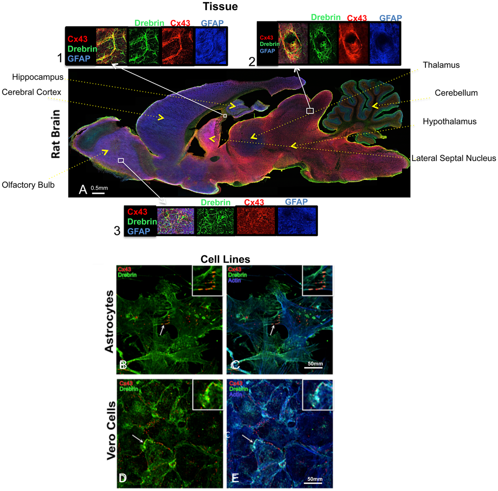
Here're some recent examples: Cinzia Ambrosi , Cynthia Ren, Gaelle Spagnol, Gabriel Cavin, Angela Cone, Elena E. Grintsevich, Gina E. Sosinsky, Paul L. Sorgen. Connexin43 Forms Supramolecular Complexes through Non-Overlapping Binding Sites for Drebrin, Tubulin, and ZO-1. Published: June 9, 2016. http://dx.doi.org/10.1371/journal.pone.0157073...chicken anti-GFAP in blue (Neuromics, Catalog # CH22102)...
Figure 1. Cx43 and drebrin colocalization analysis in brain and cellular models. (A) Rat brain transversal slice mosaic shown after multiple immunolabeling with antibodies anti-Cx43 (red), anti-drebrin (green), and anti-GFAP (blue) as astrocytes marker. White boxes localize the area enlarged in insets 1, 2, and 3 (six fold enlargement). Colocalization of drebrin and Cx43 (yellow) is especially noticeable around the blood vessels (inset 2) and in regions rich of astrocytes (insets 1 and 3). The different regions of the brain were labeled. Cultured astrocytes (B and C) and Vero cells (D and E) were immunolabeled with anti-Cx43 (red), anti-drebrin (green), and anti-actin (blue). White arrows indicate zones of colocalization of Cx43, drebrin and actin that were enlarged in the insets (white boxes, three fold enlargement).

Xiangchen Li, Yu Guo, Yaxin Yao, Jinlian Hua, Yuehui Ma, Changqing Liu, Weijun Guan.Reversine Increases the Plasticity of Long-Term Cryopreserved Fibroblasts to Multipotent Progenitor Cells through Activation of Oct4. International Journal of Biological Sciences 2016; 12(1): 53-62. doi: 10.7150/ijbs.12199...anti-GFAP and NSE (1:200,Neuromics, MN, USA)...
Save 70 USD on High Titer Neuron/Synapse Markers-Only 225/100 ul (Through June 30, 2016).
We will continue to post news on our Neuroscience Research Solutions!

Monday, May 02, 2016

New Neuronal Astroglial PNS Markers

Save 70 USD on High Titer Antibodies
The foundation of our company is built on our catalog of proven, published and high titer Neuron-Astroglia and PNS Markers. We are pleased to offer 75 USD off our latest additions.
Antibody
Type
Species Reactivity
Applications
Mouse IgG
Chicken IgY
Chicken IgY
Mouse IgG
Mouse IgG
Mouse IgG
Mouse IgG
Mouse IgG
Chicken IgY
Rabbit IgG
Mouse IgG
B; H; M; R
B; H; M; R
H; M; R
B; H; M; R
B; H; M; R
H; R
Ch; H; M; Pr; R
Ch; H; M; R
H; M; R
H; M; R
B; H; M; R
IF; WB
ICC; IF; IHC; WB
ICC; IF; IHC; WB
IF; WB
IF; WB
IF; WB
ICC;
IHC; WB
ICC; IHC; WB
ICC; IF; IHC; WB
ICC; IF; IHC; WB

Images: Left: View of mixed neuron/glial cultures stained with Aldolase-C (green) and our rabbit antibody to NeuN/FOX3 (red). MCA-4A9 antibody reveals strong cytoplasmic staining in astrocytes, while Rabbit Fox3/NeuN antibody shows nuclear and distal cytoplasmic staining in neuron cells and is complete absence of astrocytes. Blue is a DNA stain. Middle and Right: Mouse brain sections (fixed by transcardial perfusion with 4% paraformaldehyde) stained with Aldolase-C (red) and our chicken Vimentin antibody (green). In the striatum (Middle), Aldolase-C positive astrocytes are highly co-stained Vimentin, which results in yellow to gold colors. In the cerebellum (Right), however, Aldolase-C positive Purkinje cells do not express vimentin, which results in red color. Insets show a higher magnification picture of MCA-4A9 single labeling in red. Nuclei are labeled with DAPI (blue).
Our clients often use these antibodies for double and triple labeling like the above example.


We plan on aggressively be adding more and more of these type of antibodies. Stay tuned.

Thursday, September 03, 2015

Connective-Tissue Growth Factor (CTGF/CCN2) Induces Astrogenesis

SOX-2 Proves a Marker For Astrogenesis

This proved  surprising to the authors of a recent publication referencing use of our Monoclonal SOX-2 Antibody. We have added our SOX-2 abs to our Glial-Astrocyte Markers. See: Fabio A. Mendes , Juliana M. Coelho Aguiar , Suzana A. Kahn, Alice H. Reis, Luiz Gustavo Dubois, Luciana Ferreira Romão, Lais S. S. Ferreira, Hervé Chneiweiss, Vivaldo Moura Neto, José G. Abreu. Connective-Tissue Growth Factor (CTGF/CCN2) Induces Astrogenesis and Fibronectin Expression of Embryonic Neural Cells In Vitro. Published: August 4, 2015DOI: 10.1371/journal.pone.0133689...monoclonal anti-Sox2 (Neuromics, Acris, Germany) in blocking solution were incubated with the membranes overnight at 4°C, followed by incubation with Peroxidase-conjugated anti-rabbit IgG and peroxidase-conjugated anti-mouse IgG secondary antibodies...


Figure: Exogenous CTGF protein increases the number of Sox2-positive cells of a neural progenitor culture. Immunostaining showing Sox2 (A and D) expression of untreated and CTGF-treated cells. C and F show merged pictures of Sox2 immunostaining together with nuclei-DAPI staining. Scale bars 10 μm. G shows the percentage of cells that were positive for Sox2. doi:10.1371/journal.pone.0133689.g002

Neuromics have a significant catalog of potent and proven Asytoglial, Neuronal, Schwann Cell and Oligodendrocyte Markers.


Monday, January 19, 2015

Solutions for Studying Neuro-degeneration

Data Rich and Frequently Published

The Neuromics' brand is built, in part, by our proven ability to provide solutions for the study of neuro-degeneration. These include:
A recent example shows the use of one of  MAP-2 markers to study hearing decline with age: Radtke-schuller S, Seeler S and Grothe B(2015) Restricted loss of olivocochlear but not vestibular efferent neurons in the senescent gerbil (Meriones unguiculatus). Front. Aging Neurosci. 7:4. doi:10.3389/fnagi.2015.00004.

Figure: Lipofuscin granules in MSO neurons of an aged gerbil. MSO neurons are MAP2 immunostained (Alexa Fluor 647, red). Lipofuscin granules have been excited with the DAPI excitation wavelengths and appear blue. Confocal images show a maximum projection of image stacks in A and a single optical image of 0.3 µm thickness in the enlargement in B. Scale bar in A: 50 µm and 20 µm in B.

We stand ready to serve you. Should you have interest or questions, do not hesitate to contact me directly: Pete Shuster-Owner/CEO-pshuster@neuromics.com or direct phone: 612-801-1007. Thank you.

Wednesday, July 16, 2014

Potents Tools for Neuroscience Based Toxicology Assays

Neuromics' Offers Best in Class Cell and Markers

I am always on the hunt for proof that are tools work in the many different applications required by Researchers Studying Neurotoxicology. Success is confirmed to us through Customer Data/Pubs and Testimonials.

I would like to feature here some examples:


Figures: Neurons stained with Neuromics' MAP2 antibody to determine Neurite Damage.

We guarantee results. If you would like to learn more, please contact me directly at pshuster@neuromics.com or direct phone line: 612-801-1007. Thank you.

Thursday, September 19, 2013

New Astrocyte-Glial Markers

More Options!

We get a lot of request for markers that will more specifically stain astrocytes, glia and microglia. Let's say, for example, you want to pinpoint these cell types in a mixed neuron-glial culture. You can now do a dual label and generate these results.

Image: Neuron-glial cell mixture cultures stained with  ALDH1L1 (red) and our monoclonal antibody against GFAP (green). Blue is a DNA stain. ALDH1L1 stains astrocytes and excludes from neuron cells. ALDH1L1 stains the astrocytes cell body and processes, whereas GFAP labels the intermediate filament of the cytoskeleton in subset of astrocytes. Astrocytes are positive for both ALDH1L1 and GFAP appear yellow. ALDH1L1 also labels many astrocytes not labeled by GFAP, which appear as red. Inset:Blot of rat liver tissure homogenates blotted with ALDH1L1. The antibody binds strongly a band at ~100 kDa.

Or how about these results:
Image: Mixed neuron-glial cultures stained with Mouse Monoclonal GFAP, and Chicken Polyclonal Neurofilament-NF-L (green). The GFAP antibody stains the network of astrocytes in these cultures, while the NF-L antibody stains neurons and their processes. The blue channel shows the localization of DNA. This antibody also works on formalin fixed paraffin embedded brain tissues. Inset: Strip blot of rat spinal cord protein extract stained with GFAP. A prominent band at about 55 kDa corresponds to the major isoform of GFAP.

We will continue to post new additions to our Neuronal-Glial Markers.

Monday, July 15, 2013

MAP-2-A Versatile Neuron Marker

Neuromics is a leader in providing Neuron-Glial Markers for Neuroscientists.

We are constantly on the search for publications that reference use of these markers in unique applications. In this posting I would like to share a publication where researchers used on of our MAP-2 antibodies to stain medial superior olive (MSO) neurons. Baumann Veronika, Lehnert Simon, Leibold Christian, Koch Ursula. Tonotopic Organization of the Hyperpolarization-activated Current (Ih) in the Mammalian Medial Superior Olive. Front. Neural Circuits 7:117. doi: 10.3389/fncir.2013.00117.
 ...Following recording, slices were fixed in 4% paraformaldehyde for 30 min. After extensive washing in phosphate-buffered saline (PBS) slices were exposed to blocking buffer (0.5% trition X-100/0.1% saponin/1% BSA in PBS) followed by incubation with the primary antibody (chicken anti-microtubule-associated protein 2, MAP2, 1:1000, Neuromics) in blocking buffer. Slices were then rinsed in washing buffer (0.5% Trition X-100/0.1% saponin in PBS) and immunoreactivity was visualized by incubating the slices with the Cy3-conjugated secondary antibody raised in donkey (1:300; Dianova). Finally, slices were washed and mounted on slides with vectashield mounting reagent (Vector Laboratories, USA)...

Here the MAP-2 antibody is used to help identify the dorsal, medial and ventral portion of the MSO of p18 and p22 gerbils.

Figure . Ih varies systematically along the dorsoventral axis. (A) A brain slice containing the MSO with Alexa-488-filled neurons (green) verifies the distribution of the patched neurons along the dorsoventral axis (red: MAP-2). (B) Pharmacologically isolated Ih current traces were elicited by depolarizing and hyperpolarizing voltage steps from −60.5 mV to potentials between −40.5 mV and −120.5 mV for 1 s in 5 mV step increment and then to −100.5 mV for 0.5 s to elicit the tail current to determine the voltage dependence of Ih activation. Current traces are representative for the dorsal, the intermediate and the ventral part of the MSO. (C) I-V relationships of steady-state (red arrow in B) Ih density for ventral (n = 15), intermediate (n = 12) and dorsal (n = 18) neurons emphasize that Ih density amplitudes are smallest in dorsal neurons and largest in ventral neurons (C1). Ih density amplitudes for a voltage step to −110.5 mV (C2). (D) Weighted activation time constants at −110.5 mV (D1). The weighted activation time constants are voltage dependent and largest in the dorsal part of the MSO (D2). (E) The voltage-dependence of Ih activation was measured from the tail current 20 ms after the end of the voltage steps (red arrow) (E1). Values were fitted with a Boltzmann function to obtain the half-maximal activation voltage. In dorsal neurons the Ih activation curve is shifted to more negative voltages (E2). Half-maximal activation voltage was measured in each experiment and averaged (E3). Black symbols: dorsal neurons; gray symbols: intermediate neurons; white symbols: ventral neurons. **P < 0.01, ***P < 0.001, single-factor ANOVA test followed by a Scheffe's post-hoc test.

I will continue to post interesting applications using our Neuron-Glial Markers.

Tuesday, July 31, 2012

Neuron-Glial Antibodies Hit the Mark!

Neuromics' Neuron-Glial Markers offerings include:
Glial-Astrocyte MarkersNeural Progenitor Markers
Neuron/Synapse MarkersOligodendrocyte, Oligodendroglial Oligodendrocyte Lineage Markers
Schwann Cell or PNS Neuronal Markers -Peripheral Nervous System (PNS) Related
They are widely used and frequently published. Customers have also shared data which is included in product descriptions. Here is a listing of the most recent pubs:
Wiebke Kallenborn-Gerhardt, Katrin Schröder, Domenico Del Turco, Ruirui Lu, Katharina Kynast, Judith Kosowski, Ellen Niederberger, Ajay M. Shah, Ralf P. Brandes, Gerd Geisslinger, and Achim Schmidtko. NADPH Oxidase-4 Maintains Neuropathic Pain after Peripheral Nerve Injury. The Journal of Neuroscience, 25 July 2012, 32(30): 10136-10145; doi: 10.1523/​JNEUROSCI.6227-11.2012.


Images: MPZ and PMP22 expression in the sciatic nerve of WT and Nox4−/− mice after SNI. A, Western blot analysis of the myelin-specific proteins MPZ and PMP22 in the day 14 SNI sciatic nerve (proximal nerve stump) and the uninjured control sciatic nerve. GAPDH was used as loading control. Note that MPZ and PMP22 protein expression is significantly decreased after SNI in WT mice but not in Nox4−/− mice. n = 3 mice per group. Data are presented as mean ± SEM (*p < 0.05). B, Immunostaining of the day 14 SNI sciatic nerve shows increased MPZ immunoreactivity in Nox4−/− mice compared with WT mice, whereas immunoreactivity of the neuronal marker NF200 is similar in both genotypes. Scale bar, 10 μm.

Maria Maddalena Valente, Valeria Bortolotto, Bruna Cuccurazzu, Federica Ubezio, Vasco Meneghini, Maria Teresa Francese, Pier Luigi Canonico, Mariagrazia Grilli.Alpha2delta ligands act as positive modulators of adult hippocampal neurogenesis andprevent depressive-like behavior induced by chronic restraint stress. Molecular Pharmacology Fast Forward Published on May 9, 2012 as doi:10.1124/mol.112.077636...chicken anti-nestin polyclonal (1:4,000, Neuromics, Edina, Minnesota)...

Frances Y. Cheng, Xi Huang, Anuraag Sarangi, Tatiana Ketova, Michael K. Cooper, Ying Litingtung, Chin Chiang. Widespread Contribution of Gdf7 Lineage to Cerebellar Cell Types and Implications for Hedgehog-Driven Medulloblastoma Formation. PLoS ONE 7(4): e35541. doi:10.1371/journal.pone.0035541...rabbit anti-GFAP (Neuromics, 1:500), mouse anti-GFAP (Neuromics, 1:200)...

Juan M Jimenez-Andrade and Patrick W Mantyh. Sensory and sympathetic nerve fibers undergo sprouting and neuroma formation in the painful arthritic joint of geriatric mice. Arthritis Research & Therapy 2012, 14:R101...to label primary afferent sensory nerve fibers, an antibody against neurofilament 200 kDa (NF200, chicken anti neurofilament 200 kDa; NF200, 1:5000; Neuromics; catalog #CH22104)...

I will be posting new developments.

Thursday, April 26, 2012

GFAP Antibodies-Daily Double

I consider publications important for confirming our Neuronal-Glial Markers are working as advertised. These tools often used by our customers for lineages of differentiating Neural Progenitors. This publication references use of our Mouse Monoclonal GFAP and Rabbit Polyclonal GFAP Antibodies: Frances Y. Cheng, Xi Huang, Anuraag Sarangi, Tatiana Ketova, Michael K. Cooper, Ying Litingtung, Chin Chiang. Widespread Contribution of Gdf7 Lineage to Cerebellar Cell Types and Implications for Hedgehog-Driven Medulloblastoma Formation. PLoS ONE 7(4): e35541. doi:10.1371/journal.pone.0035541.
Abstract:The roof plate is a specialized embryonic midline tissue of the central nervous system that functions as a signaling center regulating dorsal neural patterning. In the developing hindbrain, roof plate cells express Gdf7 and previous genetic fate mapping studies showed that these cells contribute mostly to non-neural choroid plexus epithelium. We demonstrate here that constitutive activation of the Sonic hedgehog signaling pathway in the Gdf7 lineage invariably leads to medulloblastoma. Lineage tracing analysis reveals that Gdf7-lineage cells not only are a source of choroid plexus epithelial cells, but are also present in the cerebellar rhombic lip and contribute to a subset of cerebellar granule neuron precursors, the presumed cell-of-origin for Sonic hedgehog-driven medulloblastoma. We further show that Gdf7-lineage cells also contribute to multiple neuronal and glial cell types in the cerebellum, including glutamatergic granule neurons, unipolar brush cells, Purkinje neurons, GABAergic interneurons, Bergmann glial cells, and white matter astrocytes. These findings establish hindbrain roof plate as a novel source of diverse neural cell types in the cerebellum that is also susceptible to oncogenic transformation by deregulated Sonic hedgehog signaling.

Images: A subset of Gdf7-lineage cells express neural stem cell markers.(A–B″′) Many cells within the tumor tissue of Gdf7Cre/+;SmoM2 mice coexpress neural stem cell marker Nestin (red) and glial marker GFAP (green). Arrows indicate co-localization.


I will continue to post publication and customer data showing results with our Neuron-Glial Markers.

Monday, January 16, 2012

Primary Neuron Assays for Studying Neurodegeneration

Our goal is to provide our customers and collaborators the tools they need to insure success. This is defined by having the specific Primary Neurons, Growth Factor plus the Markers to meet unique research needs.

The proof is in the results. Here are some highlights.
Images/Data: FIGURE 5. Microglial p38α MAPK-dependent TNFα is involved in LPS-induced neurite degeneration. (A) Photomicrographs of MAP-2 immunocytochemistry show the morphology of neurons after 72h of co-culture with microglia. The arrow points to the appearance of neurites that have been damaged by LPS-activated WT microglia. In contrast, the arrowhead points to the morphological appearance of healthy, undamaged neurites. (B) Diagram of the Sholl method for quantifying the total number of healthy neurites that intersect the concentric circles. (C) Quantification of healthy neurites by the Sholl analysis demonstrates that LPS stimulation of p38α WT microglia in co-culture causes neurite degeneration as seen by a significant reduction in the number of intersections by healthy neurites in the LPS-stimulated group compared to the unstimulated group (white bars). This degeneration can be attenuated by the addition of a blocking antibody to TNFα (5μg/ml), while the non-immune IgG control was not protective (gray bars). Microglia from p38α KO mice stimulated with LPS (black bar) also have significantly less neurite degeneration than the LPS-stimulated p38α WT microglia (white bar). However, by adding TNFα back to the p38α KO microglia co-culture, there is a significant decrease in the healthy neurite arborization compared to the p38α KO microglia stimulated with LPS alone (black bars). (***p<0.005; Bonferroni’s multiple comparison test). Data represents 2 independent experiments. Scale bar equals 25μm. Molecular Neurodegeneration 2011, 6:84 doi:10.1186/1750-1326-6-84
hN2 cells grown in culture for 4 days and stained with our chicken polyclonal to Neurofilament light or low molecular weight chain NF-L, a marker of neurons. Many of the differentiating cells show strong cytoplasmic and clearly fibrillar staining for NF-L. Blue stain is DAPI and reveals cell nuclei of some non neuronal cells in this culture.

We will continue to post relevant images and data that demonstrate our capabilities.

Thursday, October 20, 2011

Immunostaining Neurons and Glia

I would like to thank Dr. Gerry Shaw, University of Florida for his excellent work with our Primary Neurons and Astrocytes and Neuronal-Glial Markers. Here's an example image with many more to follow:

Image: E18 hippocampal neurons stained with MAPT (red) and Doublecortin (green). The two proteins overlap in the proximal dendrites, but doublecortin is more abundant in the growth cones and periphery. As a result, the periphery appears green while the more proximal regions of the cells are yellow. The single longer process of this cell, presumably an axon, has a low doublecortin content and so appears red. Blue staining is the nuclear DNA. Protocol on datasheet.

Wednesday, December 09, 2009

New Mouse Monoclonal GFAP Antibody

Check it out!

GFAP Antibody

Excelent marker for human astrocyte intermediate filaments in the central nervous system. It has also been detected in the glial cells of the enteric nervous system and some Schwann cells in the peripheral nervous systems.

Posted using ShareThis

Saturday, August 08, 2009

ENS Development Markers

This is an excellent study on the developing Enteric Nervous Stem (ENS). This system is often referred to as the "second brain".

Despite this, neurogenesis has been much less studied in the ENS than in the brain. Understanding how neurons are formed in the gut is the foundation for finding cures for ENS disorders. The key finding here is that the ability of 5-HT4 receptors to unmask a regulation of enteric neurogenesis in adult animals suggests that the mature ENS is capable of an unexpected degree of plasticity (potentially good new for discovering therapies for ENS related disorders).

Min-Tsai Liu, Yung-Hui Kuan, Jingwen Wang, René Hen, and Michael D. Gershon. 5-HT4 Receptor-Mediated Neuroprotection and Neurogenesis in the Enteric Nervous System of Adult Mice. The Journal of Neuroscience, August 5, 2009, 29(31):9683-9699; doi:10.1523/JNEUROSCI.1145-09.2009.

More details @ http://www.anxietyinsights.info/serotonin_the_gut_and_neurogenesis.htm

On a side note, this publication is rich with excellent ICC and Western Blot images of a variety of Neurogenesis Markers. This include referencing of 4 of our markers:

GFAP-CH22102-ICC Dilution 1:2000
Musashi-1-RA14128-ICC Dilution 1:100 WB Dilution 1:1000
Neurofilament NF-H-CH22104-ICC Dilution 1:2000
S100B-RA25022-ICC Dilution 1;1000

Thursday, October 09, 2008

Neuron/Neuron Glial Markers

We have a comprehensive and growing catalog of Neuron/Glial Marker Antibodies and Proteins.

These reagents must work everytime in our customers' applications. Quality is confirmed by pro-active customers follow up and publication referencing the reagents.

Here we have several recent publications.

We are pleased to first feature Dr. Dr. Juana Maria Pasquini, University of and colleagues from University of Buenos Aires. She and her team use our Olig1,2,3 as a marker to study de-myelinating disease.

P.G. Franco, L. Silvestroff, E.F. Soto and J.M. Pasquini. Thyroid hormones promote differentiation of oligodendrocyte progenitor cells and improve remyelination after cuprizone-induced demyelination. doi:10.1016/j.expneurol.2008.04.039
...Olig 1-2-3 antibodies were from Neuromics Antibodies (Edina, MN); ...

Featured Product:

Related Products:
Antibodies:
Olig2
Neuron-Glial Markers
Neurotrophins and Growth Factors
Neurodegenerative Disease
Proteins:
Neurotrophins-Neuron/Glial Markers
Neurodegenerative Disease
SC Reagents

The second references one of our GFAP antibodies.

Sun Jin-qiao, Sha Bin, Zhou Wen-hao and Yang Yi. Basic fibroblast growth factor stimulates the proliferation and differentiation of neural stem cells in neonatal rats after ischemic brain injury. doi:10.1016/j.braindev.2008.06.005.
For the immunofluorescence assays, sections from the SVZ were washed (0.1 M Tris, pH 7.6, 15 min), denatured (2 N HCl, 37oC, 30 min), rinsed (0.1 M P10 min), incubated with 1% H2O2 in 0.1 M Tris for 30 min, rinsed, blocked (10% normal goat serum, 37oC, 30 min).
...GFAP (1:100, Neuromics)...