Showing posts with label cancer markers. Show all posts

Sunday, September 15, 2013

Antibody Array-ELISA Kits for Cancer Researchers

We continue to build our catalog of Antibody Arrays & ELISA Kits
for Cancer Researchers. Our RayBio® C-Series Ctyokine Arrays
enable you to analyze the expression of up to 120 Cytokines for about
the price of a single ELISA (585 USD).
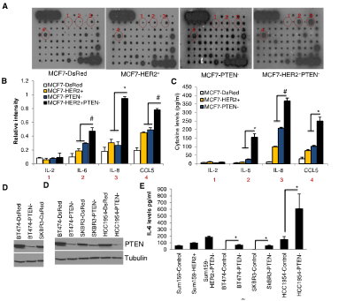
 Antiboy Array 5
Figure 3. PTEN Downregulation in HER2-Overexpressing Cells Activates an IL6/NF-kB-Mediated Inflammatory Feedback Loop (A–E) MCF7-HER2+PTEN cells secreted 3- to 5-fold higher levels of IL6, IL8, and CCL5 compared to MCF7-HER2+ or MCF7-PTEN cells as determined by RayBio human cytokine antibody Array 5 (A). The intensity of each blot compared to control was determined by Kodak image analyzer (B) and confirmed by ELISA (C). Downregulation of PTEN in HER2-amplified breast cancer cells, BT474, SKBR3, HCC1954, and Sum159-HER2+ (D) results in increased levels of these cytokines in vitro (E). http://dx.doi.org/10.1016/j.molcel.2012.06.014

Array Capabilities:
  • Detects expression of 96 secreted proteins in a single sample, in a single day. 
  • If you can do a Western, you can use a C-Series Antibody Array. And get excellent results the first time!  
  • Sandwich ELISA pairs give C-Series arrays high sensitivity and specificity.  
  • C-Series arrays are compatible with practically ANY liquid sample. 
References have cited using membrane-based C-Series Cytokine Antibody Arrays with nearly every liquid sample type imaginable, including: cell-cultured and co cell cultured media, cell and tissue lysates, tissue/organ perfusates, serum, plasma, urine, and many other body fluids, including cyst, interstitial, synovial, blister, cerebrospinal, prostatic and amniotic fluids, as well as abscesses, broncho-alveolar lavage, sputum, saliva, tears, breath condensates, and even human milk and colostrum. Publications referencing use of our Antibody Arrays.

I will be posting more new developments here.

Saturday, February 04, 2012

Neurotrophins-Growth Factors Update

Our Neurotrophins and Growth Factor Antibodies and Recombinant Proteins help support a wide span of research areas. These areas include: neuroscience, immunology, cardiac disease research and cancer.

I would like to update you on recent publications highlighting use of some of these reagents: Aiko Sada, Kazuteru Hasegawa, Pui Han Pin, Yumiko Saga. NANOS2 Acts Downstream of Glial Cell Line-Derived Neurotrophic Factor Signaling to Suppress Differentiation of Spermatogonial Stem Cells. DOI: 10.1002/stem.790. Copyright © 2011 AlphaMed Press...anti-GFRA1 (1:200, Neuromics, Edina, MN)...


Images: Ngn3-Cre targets GFRA1-negative cells. (A-F): At P7, Rosa-YFP; Ngn3-Cre double transgenic testes were immunostained with the indicated markers. Most of YFP-positive spermatogonia (Ngn3-lineage cells) were not stained with GFRA1. Scale bar, 100 μm. (G): Quantification of YFP-positive cells per GFRA1-positive cells at P7 and 6 weeks (N=3).

Karine Bédard, Stéphanie Segura, Stéphanie Mahaut, Catherine Tardivel, Guylaine Ferland, Bruno Lebrun, Pierrette Gaudreau. Effects of aging and caloric restriction on brainstem satiety center signals in rats. Mechanisms of Ageing and Development. dx.doi.org/10.1016/j.mad.2012.01.004...goat serum and 0.3% Triton-X-100) (Sigma–Aldrich), overnight at 4 °C with chicken anti-rat BDNF antibody (Neuromics, Edina, MN; diluted 1/200 in blocking buffer), rinsed 3× 10 min in fresh PBS and incubated for 2 h, in the dark...

Tali Kobilo, Qing-Rong Liu, Kriti Gandhi, Mohammed Mughal, Yavin Shaham and Henriette van Praag. Running is the neurogenic and neurotrophic stimulus in environmental enrichment. doi: 10.1101/lm.2283011. Learn. Mem. 2011. 18: 605-609... human recombinant BDNF (0.1 µg) monomer (Neuromics)...

Masamichi Shinoda, Masatake Asano, Daisuke Omagari, Kuniya Honda, Suzuro Hitomi, Ayano Katagiri, and Koichi Iwata. Nerve Growth Factor Contribution via Transient Receptor Potential Vanilloid 1 to Ectopic Orofacial Pain. The Journal of Neuroscience, 11 May 2011, 31(19):7145-7155; doi:10.1523/JNEUROSCI.0481-11.2011....expression immunohistochemically in TG- anti-p75 goat antibody (1:200; Neuromics)...


Images: Mandibular nerve fibers were labeled by β-NGF which was administrated into the lower lip with CFA (A) or saline (F). On day 1 after β-NGF administration into the lower lip with CFA or saline, β-NGF-positive whisker pad or lower lip TG neurons defined by FG or DiI, respectively. B, G, β-NGF-positive TG neurons. C, H, DiI-labeled TG neurons. D, I, FG-labeled TG neurons. E, J, DiI- and FG-labeled β-NGF-positive TG neurons. Mandibular nerve fibers (K) and TG neurons (L) on day 1 after labeled BSA administration into the lower lip with CFA. Open arrow, β-NGF-positive nerve fibers. Arrow, DiI-labeled β-NGF-positive TG neurons. Arrowhead, FG-labeled β-NGF-positive TG neurons. Scale bar, 50 μm. M, Frequency of β-NGF-positive neurons in FG- or DiI-labeled TG neurons after CFA or saline injection into the lower lip (n = 5 in CFA-injected group; n = 4 in saline-injected group; Student's t test).

F. R. Carreño, J. D. Walch, M. Dutta, T. P. Nedungadi, J. T. Cunningham. Brain-Derived Neurotrophic Factor-Tyrosine Kinase B Pathway Mediates NMDA Receptor NR2B Subunit Phosphorylation in the Supraoptic Nuclei Following Progressive Dehydration. Journal of Neuroendocrinology. Volume 23, Issue 10, pages 894–905, October 2011. DOI: 10.1111/j.1365-2826.2011.02209.x...TrkB receptor (Neuromics, Edina, MN, USA)...

Jie Shen, Yoko Ishii, Guihua Xu, Thanh Chung Dang, Takeru Hamashima, Takako Matsushima, Seiji Yamamoto, Yuichi Hattori, Yusuke Takatsuru, Junichi Nabekura and Masakiyo Sasahara. PDGFR-β as a positive regulator of tissue repair in a mouse model of focal cerebral ischemia. Journal of Cerebral Blood Flow & Metabolism , (28 September 2011). doi:10.1038/jcbfm.2011.136...goat polyclonal anti-PDGFR-β antibody (1:100; Neuromics, Edina)...

Thomas Karsten Kilvaer, Andrej Valkov, Sveinung W. Sorbye, Tom Donnem, Eivind Smeland, Roy Martin Bremnes,and Lill-Tove Busund. Platelet-Derived Growth Factors in Non-GIST Soft-Tissue Sarcomas Identify a Subgroup of Patients with Wide Resection Margins and Poor Disease-Specific Survival. Volume 2010 (2010), Article ID 751304, 10 pages doi:10.1155/2010/751304...PDGF-CC (goat polyclonal; GT15151; Neuromics; 1 : 80)...

I will keep you posted on new developments.

Thursday, August 18, 2011

Plasma netrin-1 is a diagnostic biomarker of human cancers

I am pleased to report broadening application for our Stem Cell Markers as a diagnostic for cancers. This publication references use of our Netrin-1 antibody.

Ganesan Ramesh, Arthur Berg, and Calpurnia Jayakumar. Plasma netrin-1 is a diagnostic biomarker of human cancers. Biomarkers. Author manuscript; available in PMC 2011 July 26. Published in final edited form as: Biomarkers. 2011 March; 16(2): 172–180. Published online 2011 February 8. doi: 10.3109/1354750X.2010.541564.
Objectives: To determine whether plasma netrin-1 can be used as a diagnostic biomarker of human cancer.

Materials and Methods: A total of 300 cancer plasma samples from breast, renal, prostate, liver, meningioma, pituitary adenoma, glioblastoma, lung, pancreatic and colon cancer patients were compared against 138 control plasma samples. Netrin-1 levels were quantified by ELISA and immunohistochemistry.

Results: Plasma netrin-1 levels were significantly increased in breast, renal, prostate, liver, meningioma, pituitary adenoma, and glioblastoma cancers as compared to control samples.

Discussion and Conclusion: Our results suggest that plasma netrin-1 can be used as a diagnostic biomarker for many human cancers.

Image: Immunohistochemical localization of netrin-1 in renal cell carcinoma (RCC) tissues. A. Secondary antibody control showing no staining. B. Normal adjacent tissues do not show any staining for netrin-1. C. Stage I RCC shows staining for netrin-1. D. Stage II RCC shows staining for netrin-1. E-F.


Netrin-1 Immunohistochemistry: Stage I–III renal cell carcinoma and normal tissue section (Tissue Array) was obtained from Biomax to immunolocalize netrin-1, as described previously (29). Briefly, tissue sections were dewaxed and rehydrated with graded ethanol (100%, 90%, 70% and 30%) and then washed with PBS. Antigen retrieval was carried out using citrate buffer and steamer. The tissue section was permeabilized with 0.2% Triton X-100 in PBS, and washed and blocked with PBS containing 5% donkey serum and 1% BSA. Primary antibodies included a chicken anti-netrin-1 polyclonal antibody (Neuromics cat # CH23002). Primary antibodies were detected using secondary antibodies conjugated with biotin, which was followed by incubation with streptavidin-horseradish peroxidase (Pierce). Slides were mounted in permount and photographed using an Olympus microscope attached to a CCD camera.
Potent diagnostic tools for cancers save lives. This is especially true of markers that can diagnose them in early stages. I will post these important studies as the cross my radar scope.
