Showing posts with label Glial Fibrillary Acidic Protein Antibody. Show all posts

Wednesday, April 08, 2020

Schwann Cell Data

Quality Counts
Though rare, we occasionally have clients challenge the phenotype of our 21-CFR compliant human primary cells. This challenge is usually a result of  certain markers not being expressed when they run immuno-fluorescence assays.

When this happens, we too test the cells in question using the antibodies against the markers in question. We have a world class team doing immuno-fluorescence assays.

Recently a customer claimed our schwann cells were negative for GFAP and P0. Here're our testing results.

For P0 (CH23009) we used green fluorescence +DAPI.
For GFAP (RA22101) we used red fluorescence + DAPI (blue) counterstain
We take these challenges for seriously and will always test the same lot of cells internally to make sure they are "walking and talking" as advertised.

Wednesday, June 21, 2017

Gap Junctions (GJs) and Glaucoma

GJs Can Offer Neuroprotection
This study references use of our GFAP Antibody,

Gap junctions (GJs), intercellular channels composed of subunit connexins, can play a major role in secondary cell death by forming conduits through which toxic molecules from dying cells pass to and injure coupled neighbors. Secondary cells like glia and astrocytes are involved in this process though the precise mechanisms have yet to be defined: Abram Akopian, Sandeep Kumar, Hariharasubramanian Ramakrishnan, Kaushambi Roy, Suresh Viswanathan, and Stewart A. Bloomfield. Targeting neuronal gap junctions in mouse retina offers neuroprotection in glaucoma. J Clin Invest. doi:10.1172/JCI91948. Copyright © 2017, The American Society for Clinical Investigation. ...anti-GFAP (1:1,000, RA22101; Neuromics)...

Figure: Reactive gliosis in retinas of microbead-injected mice is significantly reduced by GJ blockade/ablation. (A) Confocal images of retinal layers stained for GFAP, SMI32, and DAPI in control and glaucomatous retinas. Scale bar: 50 μm in all panels. Z-stack: 7 sections, 3-μm steps. (B) GFAP expression in the retinal layers of CxWT and Cx36–/– mouse retinas under different conditions (n = 6 retinas per group). (C) GFAP labeling in retinal sections from control and microbead-injected CxWT (n = 5 retinas), Cx36–/– (n = 5 retinas), and Cx36–/– Cx45–/– mice (n = 3 retinas). GFAP expression is presented as percentage of immunolabeling per area.
Our Neuron/Synapse, Astrocytes, Glia, Microglia, Oligodendrocytes, Progenitors and Schwann Cell Markers are frequently referenced and I will continue to post new developments.

Sunday, June 12, 2016

Neuroscience Cell Based Markers Applications

Progenitors, Neuronal, Astroglia and PNS Markers A to Z
Our Neuron/Synapse, Astrocytes, Glia, Microglia, Olidogodendrocytes, Progenitors and Schwann Cell Markers continue to widely used and frequently published.
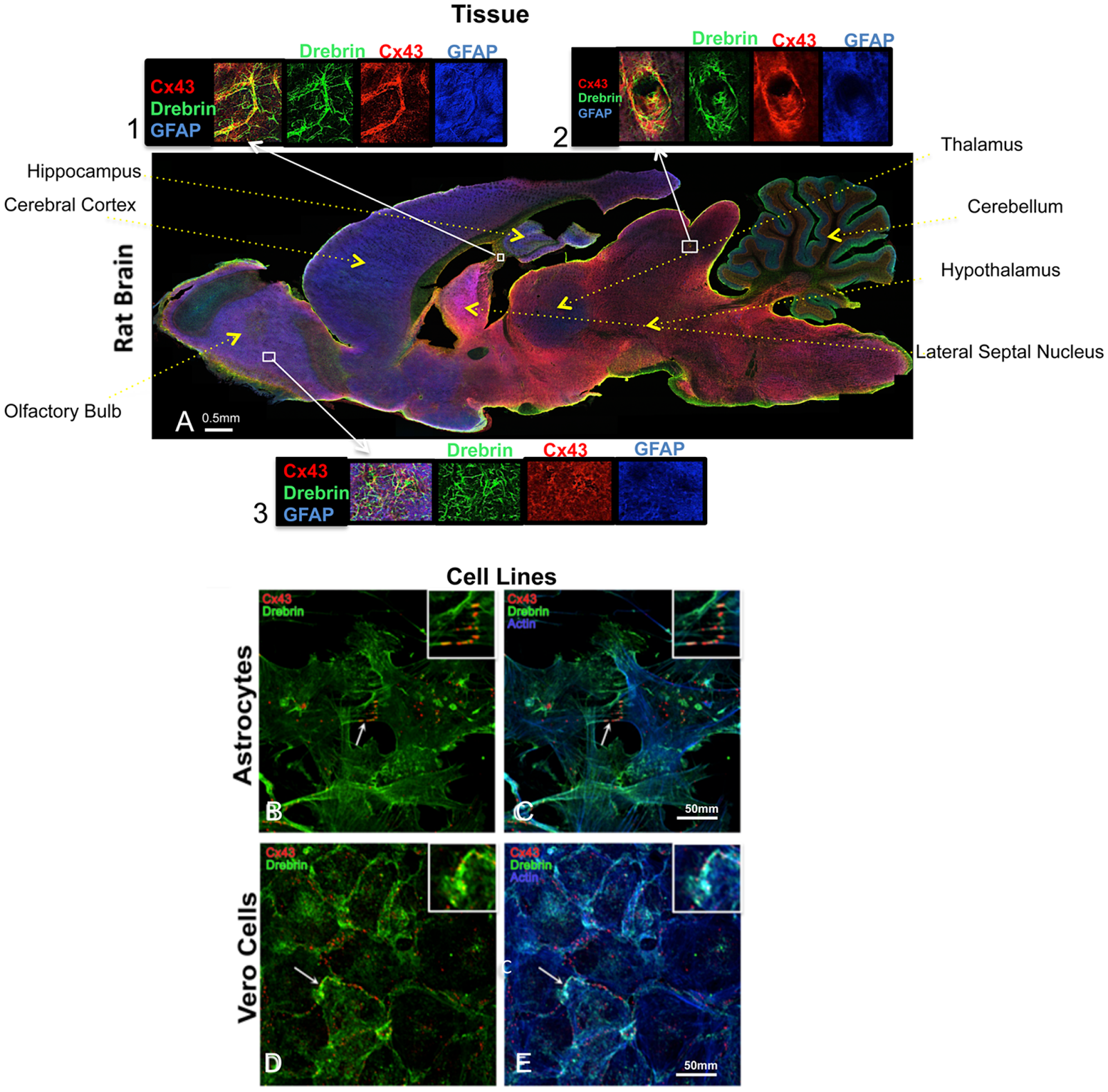
Here're some recent examples: Cinzia Ambrosi , Cynthia Ren, Gaelle Spagnol, Gabriel Cavin, Angela Cone, Elena E. Grintsevich, Gina E. Sosinsky, Paul L. Sorgen. Connexin43 Forms Supramolecular Complexes through Non-Overlapping Binding Sites for Drebrin, Tubulin, and ZO-1. Published: June 9, 2016. http://dx.doi.org/10.1371/journal.pone.0157073...chicken anti-GFAP in blue (Neuromics, Catalog # CH22102)...
Figure 1. Cx43 and drebrin colocalization analysis in brain and cellular models. (A) Rat brain transversal slice mosaic shown after multiple immunolabeling with antibodies anti-Cx43 (red), anti-drebrin (green), and anti-GFAP (blue) as astrocytes marker. White boxes localize the area enlarged in insets 1, 2, and 3 (six fold enlargement). Colocalization of drebrin and Cx43 (yellow) is especially noticeable around the blood vessels (inset 2) and in regions rich of astrocytes (insets 1 and 3). The different regions of the brain were labeled. Cultured astrocytes (B and C) and Vero cells (D and E) were immunolabeled with anti-Cx43 (red), anti-drebrin (green), and anti-actin (blue). White arrows indicate zones of colocalization of Cx43, drebrin and actin that were enlarged in the insets (white boxes, three fold enlargement).

Xiangchen Li, Yu Guo, Yaxin Yao, Jinlian Hua, Yuehui Ma, Changqing Liu, Weijun Guan.Reversine Increases the Plasticity of Long-Term Cryopreserved Fibroblasts to Multipotent Progenitor Cells through Activation of Oct4. International Journal of Biological Sciences 2016; 12(1): 53-62. doi: 10.7150/ijbs.12199...anti-GFAP and NSE (1:200,Neuromics, MN, USA)...
Save 70 USD on High Titer Neuron/Synapse Markers-Only 225/100 ul (Through June 30, 2016).
We will continue to post news on our Neuroscience Research Solutions!

Thursday, December 17, 2015

Fibroblasts into Neurons

Via Activation of Oct4

We have a variety of stem cell, progenitor and neuron markers that are often referenced in publication. There represent a qualitative way to determine the state of stem cells as they differentiate.

Here're researchers take Fibroblasts and differentiate them into various progenitors. They were able to further differentiate these progenitors into astrocytes and neurons by: For neuronal differentiation, after 2 days of incubation with 20 nM reversine, cells were then treated with 0.5 μM all-trans-retinoic acid (RA) in serum-free DMEM/F-12 medium supplemented with the ITS for 2 days and switched into serum-free medium in the absence of RA with replacement of the medium every 2-3 days. 

Our NSE and GFAP were used to confirm this differentiation.
To learn more see: Xiangchen Li, Yu Guo, Yaxin Yao, Jinlian Hua, Yuehui Ma, Changqing Liu, Weijun Guan.Reversine Increases the Plasticity of Long-Term Cryopreserved Fibroblasts to Multipotent Progenitor Cells through Activation of Oct4. International Journal of Biological Sciences 2016; 12(1): 53-62. doi: 10.7150/ijbs.12199

Thursday, September 19, 2013

New Astrocyte-Glial Markers

More Options!

We get a lot of request for markers that will more specifically stain astrocytes, glia and microglia. Let's say, for example, you want to pinpoint these cell types in a mixed neuron-glial culture. You can now do a dual label and generate these results.

Image: Neuron-glial cell mixture cultures stained with  ALDH1L1 (red) and our monoclonal antibody against GFAP (green). Blue is a DNA stain. ALDH1L1 stains astrocytes and excludes from neuron cells. ALDH1L1 stains the astrocytes cell body and processes, whereas GFAP labels the intermediate filament of the cytoskeleton in subset of astrocytes. Astrocytes are positive for both ALDH1L1 and GFAP appear yellow. ALDH1L1 also labels many astrocytes not labeled by GFAP, which appear as red. Inset:Blot of rat liver tissure homogenates blotted with ALDH1L1. The antibody binds strongly a band at ~100 kDa.

Or how about these results:
Image: Mixed neuron-glial cultures stained with Mouse Monoclonal GFAP, and Chicken Polyclonal Neurofilament-NF-L (green). The GFAP antibody stains the network of astrocytes in these cultures, while the NF-L antibody stains neurons and their processes. The blue channel shows the localization of DNA. This antibody also works on formalin fixed paraffin embedded brain tissues. Inset: Strip blot of rat spinal cord protein extract stained with GFAP. A prominent band at about 55 kDa corresponds to the major isoform of GFAP.

We will continue to post new additions to our Neuronal-Glial Markers.

Thursday, March 31, 2011

GFAP and Mouse Myenteric Plexus

Our Neuronal-Glial Markers are important tools for our customers investigating expression in the CNS and PNS. I have posted images showing staining of mouse retinal astrocytes and in the ventral horn, funiculus of adult rat spinal cord and mouse medulloblastoma stem cells using our GFAP antibodies.

I wanted to share an excellent image generated by Dr. Kate Ellacott's lab at Vanderbilt University.

GFAP (Chicken-Cat#: CH22102) staining in of enteric glia in the myenteric plexus of the mouse gastrointestinal tract. Staining was performed in methanol/acetone fixed frozen sections using 1:1000 dilution of the antibody followed by 1:500 anti-chicken Alexa 594.

Related Reagents:


Wednesday, December 09, 2009

New Mouse Monoclonal GFAP Antibody

Check it out!

GFAP Antibody

Excelent marker for human astrocyte intermediate filaments in the central nervous system. It has also been detected in the glial cells of the enteric nervous system and some Schwann cells in the peripheral nervous systems.

Posted using ShareThis