Friday, August 01, 2008

Stressed Induced Analgesia (SIA) and OFQ

This is to highlight excellent work done by our friends at SRI and AfaSci.

Our Orphanin FQ ab was used in experiments by the team. Here's the latest publication highlighting their work:

Xinmin Xie, Jonathan P. Wisor, Junko Hara, Tara L. Crowder1, Robin LeWinter, Taline V. Khroyan, Akihiro Yamanaka, Sabrina Diano, Tamas L. Horvath, Takeshi Sakurai, Lawrence Toll and Thomas S. Kilduff. Hypocretin/orexin and nociceptin/orphanin FQ coordinately regulate analgesia in a mouse model of stress-induced analgesia. J. Clin. Invest. 118(7): 2471-2481 (2008). doi:10.1172/JCI35115. Copyright © 2008, The American Society for Clinical Investigation.

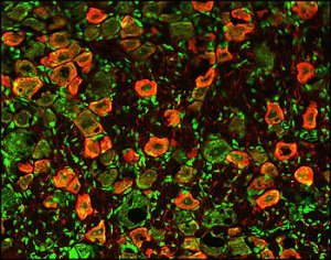
...Brain sections (10 μm) were treated with 0.3% H2O2 to quench endogenous peroxidases and then incubated overnight in primary anti-N/OFQ (1:5,000; RA10106, anti-FGGFTGARKSARKLANQ, Neuromics) and anti-orexin-B (1:5,000; sc-8071, Santa Cruz Biotechnology Inc.) antisera at 4°C with agitation. Sections were incubated in blocking buffer for 1 hour, followed by a 2-hour incubation at room temperature in secondary antisera (Alexa Fluor 546 donkey anti-goat [1:750] and 488 donkey anti-rabbit [1:500]; Molecular Probes, Invitrogen). As a negative control, additional sections were treated similarly, but the primary antibody was omitted. Preadsorption with the N/OFQ peptide FGGFTGARKSARKLANQ was used as a positive control and blocked all specific staining as also found by others...

N/OFQ-containing fibers innervate Hcrt neurons, and N/OFQ inhibits Hcrt neuronal activity. (A) Left panel shows confocal image of N/OFQ-immunoreactive fibers in the vicinity of, and in putative contact with, Hcrt-immunoreactive neurons in the PLH of WT mice. N/OFQ (green) fibers are in close proximity to Hcrt-immunoreactive (red) cells. The arrow indicates the N/OFQ innervation of an Hcrt cell body. Middle panel shows light micrograph of a light brown immunolabeled Hcrt neuron contacted by a dark black bouton (arrow) representing immunolabeling for N/OFQ. Right panel shows electron micrograph taken from ultrathin sections of the same labeled terminal and dendrite shown in the light micrograph in the middle panel. Black arrow indicates the N/OFQ-immunolabeled axon terminal in synaptic contact (red arrowhead) with the dendrite of the Hcrt cell. Scale bars: 10 μm (left and middle panels); 1 μm (right panel). (B) Under current clamp, bath application of N/OFQ (1 μM) hyperpolarized Hcrt neurons, decreased input resistance, and blocked spontaneous firing of action potentials. The resting potential of this cell was –54 mV and was manually adjusted to –60 mV with DC current. Membrane resistance was monitored using hyperpolarizing current pulses (–0.3 nA, 800 ms) delivered every 5 seconds throughout the experiment. (C) Under voltage-clamp mode at a Vh of –60 mV, N/OFQ (1 μM) induced an outward current (–53 pA) in an Hcrt neuron. Notice that the frequency but not the amplitude of the miniature synaptic currents (inward currents) recorded in the presence of TTX (0.5 μM) was also reduced.

Wednesday, July 30, 2008

TH-Marker for Diabetic Neuropathy

New Pub Referencing our Tyrosine Hydroxylase.

Blount, Andrew L. B.A.; Peled, Ziv M. M.D.; Dexter, Erica L. B.S.; Nagle, Raymond B. M.D., Ph.D.; Maloney, Christopher T. M.D.; Dellon, A Lee M.D., Ph.D. Sympathetic Nerves in the Tarsal Tunnel: Implications for Blood Flow in the Diabetic Foot. Plastic & Reconstructive Surgery. 122(1):188-191, July 2008.

...Specimens were fixed in 10% neutral buffered formalin for 3 to 4 hours and then embedded in paraffin. Three-micron sections were obtained from the block and used for immunohistochemistry, which was performed using a mouse monoclonal antibody against tyrosine hydroxylase (MO20001; Neuromics, Edina, Minn.) diluted 1:50 in Discovery Diluent (Ventana Medical Systems, Inc., Tucson, Ariz.) on a Discovery XT Automated Immunostainer (Ventana Medical Systems)...
Images: (A) Tyrosine hydroxylase immunohistochemical staining of tibial epineurium from the tarsal tunnel. Positively staining sympathetic fibers (arrows) are seen within a larger nerve bundle. (B) Tyrosine hydroxylase immunohistochemical staining of connective tissue from the tarsal tunnel. Several positively staining sympathetic fibers (arrows) are seen innervating the media of a local venule.

Thursday, July 24, 2008

SP is Back and Better than Ever

We have successfully re-made our widely used Guinea Pig Substance P antibody. It has been tested both internally and by an interested customer. The results exceeded expectations.

This antibody has proven useful for Pain/Inflammation and Diabete/Obesity Research.

Image: Immuofluorescent detection of Substance P in rat spinal cord dorsal horn (red fluorescence). DAPI (blue) was used as counter stain.

Here're are related publications:

P . Tsai , A . Alonso , M . Pinto , D . Leigh , E . Weiler , J . Fricton , M . Erickson , L . Stone , L . Kehl. Substance P is co-localized with protein gene product 9.5-immunoreactive nerve fibers in intervertebral discs from patients with painful degenerative disc disease. doi:10.1016/j.jpain.2006.01.096
...guinea pig anti-substance P (1:500; Neuromics, Edina, MN) ...
Mei Bigliardi-Qi, Claire Gaveriaux-Ruff, Katrin Pfaltz, Pierre Bady, Tommy Bauman, Theo Rufli, Brigitte L Kieffer and Paul L Bigliardi. Deletion of - and -Opioid Receptors in Mice Changes Epidermal Hypertrophy, Density of Peripheral Nerve Endings, and Itch Behavior. Journal of Investigative Dermatology (2007) 127, 1479–1488. doi:10.1038/sj.jid.5700661; published online 21 December 2006.
.. substance P (Neuromics 1:200) staining, the sections were incubated at 4°C over night in Zamboni buffer. ...

Related Reagents:
Neurokinin-1 (NK 1) Receptor
Neurokinin-1 (NK 1) Human Receptor
Neurokinin-3 (NK 3) Receptor
proNeurokinin B (proNKB or P2)
All Neuropeptides
Pain and Inflammation Antibodies
Diabetes and Obesity Antibodies
Substance P Customer Data

Wednesday, July 23, 2008

ELISPOT

We will be releasing soon a website dedicated to ELISA and ELISpot.

Here are excellent "how to" videos.


Sunday, July 13, 2008

Molecular Pathways and Heart Development

Owen WJ Prall, Mary K Menon, Mark J Solloway, Yusuke Watanabe, Stéphane Zaffran, Fanny Bajolle, Christine Biben, Jim J McBride, Bronwyn R Robertson, Hervé Chaulet, Fiona A Stennard, Natalie Wise, Daniel Schaft, Orit Wolstein, Milena B Furtado, Hidetaka Shiratori,6 Kenneth R Chien, Hiroshi Hamada,6 Brian L Black, Yumiko Saga, Elizabeth J Robertson, Margaret E Buckingham, and Richard P Harvey. An Nkx2-5/Bmp2/Smad1 negative feedback loop controls second heart field progenitor specification and proliferation. Cell. 2007 March 9; 128(5): 947–959. doi: 10.1016/j.cell.2007.01.042.

Summary: During heart development the second heart field (SHF) provides progenitor cells for most cardiomyocytes and expresses the homeodomain factor Nkx2-5. We now show that feedback repression of Bmp2/Smad1 signaling by Nkx2-5 critically regulates SHF proliferation and outflow tract (OFT) morphology. In the cardiac fields of Nkx2-5 mutants, genes controlling cardiac specification (including Bmp2) and maintenance of the progenitor state were up-regulated, leading initially to progenitor over-specification, but subsequently to failed SHF proliferation and OFT truncation. In Smad1 mutants, SHF proliferation and deployment to the OFT were increased, while Smad1 deletion in Nkx2-5 mutants rescued SHF proliferation and OFT development. In Nkx2-5 hypomorphic mice, which recapitulate human congenital heart disease (CHD), OFT anomalies were also rescued by Smad1 deletion. Our findings demonstrate that Nkx2-5 orchestrates the transition between periods of cardiac induction, progenitor proliferation and OFT morphogenesis via a Smad1-dependent negative feedback loop, which may be a frequent molecular target in CHD.

...goat anti-Isl1, raised against full length human Isl1, GT15051, Neuromics)...

Monday, June 30, 2008

Combined Neuron Cultures Assays

We would like to thank Dr. Matteo Porotto, Weill Cornell Medical College

Larger Image

Primary rat neurons: Transfection of functional HeV glycoproteins and infection with HeV pseudotyped virions.In order to establish the feasibility of carrying out the proposed experiments in primary neurons, we show (figure ) that our assays are amenable to use in primary neurons. In the experiment, Combined Hippocampus, Cortex, and Ventricular -E18 (Neuromics) were plated, and at 3 days were transfected with plasmids encoding HeV G/F as well as YFP. On the following day, these cells were infected with HeV or VSV pseudotyped viruses bearing RFP. In the figure, (A) the phase contrast photos show the differentiated neurons; (B) upon excitation for RFP, the red fluorescence indicates neurons infected by HeV pseudotyped virions; (C) upon excitation for YFP, and the green fluorescence shows the efficiency of transfection in neurons. This experiment indicates that the proposed experiments can be carried out in primary neurons, which are transfectable and infectable in our systems, and thus supports all the proposed aims. Data Courtesy of Dr. Matteo Porotto, Weill Cornell Medical College

Primary Neuron Cultures Featured
Combined Hippocampus, Cortex, and Ventricular -E18
Other Reagents to Consider
All Primary Neurons/Astrocytes, Stem Cells and Media
Transfection Reagents
Neuron-Glial Markers
Stem Cell Markers

Thursday, June 19, 2008

Amped Up ORL-1

We have been collaborating with a customer to amp up our ORL-1. Here's an example of a resulting IHC images.

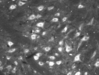
Image: ORL 1 staining in rat brain (singulate cortex). ORL1 Detection was done using anti-rabbit Cy3 conjugated antibodies (red color). DAPI was used to counterstain cell nuclei (blue color). Working dilution is 1:40-1:100.

Wednesday, June 04, 2008

TRPV1 Pub

Katrin Schnizler, Leonid P. Shutov, Michael J. Van Kanegan, Michelle A. Merrill, Blake Nichols, G. Stanley McKnight, Stefan Strack, Johannes W. Hell, and Yuriy M. Usachev. Protein Kinase A Anchoring via AKAP150 Is Essential for TRPV1 Modulation by Forskolin and Prostaglandin E2 in Mouse Sensory Neurons. J. Neurosci., May 2008; 28: 4904 - 4917 ; doi:10.1523/JNEUROSCI.0233-08.2008

......anti-TRPV1 antibody (catalog #RA14113; Neuromics, Edina, MN) and 30 mul of prewashed protein-A...mm NaCl). Probing was done using the Neuromics anti-TRPV1 antibody (1:500 dilution...with other TRPV1 antibodies either from Neuromics (N-terminus rabbit anti-TRPV1) or from......

Thursday, April 03, 2008

Recent Pubs Referencing Neuromic's Products

Neurotensin Receptor 1 (NTS1)-GP14020


Geneviève Roussy, Marc-André Dansereau, Louis Doré-Savard, Karine Belleville, Nicolas Beaudet, Elliott Richelson and Philippe Sarret. Spinal NTS1 receptors regulate nociceptive signaling in a rat formalin tonic pain model. doi:10.1111/j.1471-4159.2007.05205.x


Images: Expression of NTS1 receptors in sensory neurons and spinal cord. (a) Immunoperoxidase staining reveals the presence of NTS1-like immunoreactivity throughout the lumbar dorsal horn of the spinal cord. Immunolabeling is most prominent in the superficial layers of the dorsal horn. The lateral spinal nucleus (lsn) is also moderately labeled (arrow). (b) High magnification of (a). A dense NTS1 immunostaining is observed over laminae I and II of the dorsal horn. Numerous immunopositive nerve cell bodies are visible in the superficial laminae and in the nucleus proprius of the dorsal horn (arrows). (c) Light microscopic analysis of NTS1 expression in primary afferent neurons. NTS1 is expressed in subpopulations of small- and medium-ganglion cells. No apparent labeling is detected in large DRG neurons (arrows). At higher magnification, NTS1 neurons exhibit a cytoplasmic pattern of immunoreactivity (d, arrowheads). (e) Identification of endogenously expressed NTS1 receptors, by western blotting, in homogenates from DRGs, lumbar spinal cord and brain. The 47 kDa protein band corresponds to the molecular weight deduced from the cDNA sequence of NTS1. Each lane represents the transfer of 25 lg of protein. Scale bars: (a, b, c, and d) 300, 150, 70 and 20 lm, respectively. doi:10.1111/j.1471-4159.2007.05205.x

Primary Neurons and Astrocytes


Cortex
Hippocampus
Striatum
E18 and E20 Rat Primary Neuronal Tissue -NEURON CULTURES
E18 Rat Primary Neuronal Tissue - ASTROCYTE CULTURES
E18 Mouse Neuronal Tissue -NEURON CULTURES
E18 Mouse Neuronal Tissue -ASTROCYTE CULTURES
Frozen Primary Rat Neurons-NEW
Frozen Primary Mouse Neurons and Astrocytes-New
FluoGreen Tracer-Stains Cell Nuclei and Cytoplasm

Primary Neurons and Astrocytes

Shouqing Luo, Haruo Mizuta and David C. Rubinsztein. p21-activated kinase 1 promotes soluble mutant huntingtin self-interaction and enhances toxicity. Human Molecular Genetics 2008 17(6):895-905; doi:10.1093/hmg/ddm362

...Rat E18 cortical and striatal neurons were purchased from Neuromics (MN) or prepared from E18 rat brain tissues according to instructions from Neuromics, cultured in neurobasal (Invitrogen) medium supplemented with B27 (Invitrogen) and 0.5 mM glutamine and transfected with Lipofectamine 2000 according to standard methods...

Friday, March 07, 2008

New Data and Pubs referencing Neuromics

Cullen L. Schmid, Kirsten M. Raehal and Laura M. Bohn. Agonist-directed signaling of the serotonin 2A receptor depends on ?;-arrestin-2 interactions in vivo. Published online on January 14, 2008, 10.1073/pnas.0708862105
...Neuromics, Edina, MN; rabbit anti-5HT ..... 5A-5D...


Thomas W. Gould, Shigenobu Yonemura, Ronald W. Oppenheim, Shiho Ohmori, and Hideki Enomoto. The Neurotrophic Effects of Glial Cell Line-Derived Neurotrophic Factor on Spinal Motoneurons Are Restricted to Fusimotor Subtypes. The Journal of Neuroscience, February 27, 2008, 28(9):2131! -2146
...mouse Ret (Neuromics, Edina, MN), human Ret (Neuromics), and activated caspase-3 (Cell Signaling, Danvers, MA). Muscles or sections were rinsed and then...


Image: Nuclei of D3 or IGnT-KO ES cells stained with goat anti-Oct3/4 antibody. doi: 10.1093/glycob/cwm138

Thursday, January 17, 2008

Nov-Dec Publication Referencing Neuromics

Diversity in the Neural Circuitry of Cold Sensing Revealed by Genetic Axonal Labeling of Transient Receptor Potential Melastatin 8 Neurons
CELLULAR/MOLECULAR:Yoshio Takashima, Richard L. Daniels, Wendy Knowlton, James Teng, Emily R. Liman, and David D. McKemyDiversity in the Neural Circuitry of Cold Sensing Revealed by Genetic Axonal Labeling of Transient Receptor Potential Melastatin 8 NeuronsJ. Neurosci., Dec 2007; 27: 14147 - 14157 ; doi:10.1523/JNEUROSCI.4578-07.2007 ......211; Santa Cruz Biotechnology, Santa Cruz, CA), 1:500 rabbit anti-transient receptor potential vanilloid 1 (TRPV1; RA14113; Neuromics, Edina, MN), and 1:500 rabbit anti-TRPM8 (a gift from M. Tominaga, Okazaki Institute for Integrative Bioscience, Okazaki......
Abstract
Full Text
PDF
Supplemental Data
G 5 Is Required for Normal Light Responses and Morphology of Retinal ON-Bipolar Cells
BRIEF COMMUNICATIONS:Anjali Rao, Rebecca Dallman, Scott Henderson, and Ching-Kang ChenGβ5 Is Required for Normal Light Responses and Morphology of Retinal ON-Bipolar CellsJ. Neurosci., Dec 2007; 27: 14199 - 14204 ; doi:10.1523/JNEUROSCI.4934-07.2007 ......2003) 1:100 RGS6 Imgenex (San Diego, CA) 1:100 Goalpha Millipore (Billerica, MA) 1:100 mGluR6 Neuromics (Northfield, MN) 1:100 PKCalpha Sigma 1:100 FLAG Sigma 1:100 Transmission electron microscopy......
Abstract
Full Text
PDF
Exogenous Delivery of Heat Shock Protein 70 Increases Lifespan in a Mouse Model of Amyotrophic Lateral Sclerosis
NEUROBIOLOGY OF DISEASE:David J. Gifondorwa, Mac B. Robinson, Crystal D. Hayes, Anna R. Taylor, David M. Prevette, Ronald W. Oppenheim, James Caress, and Carolanne E. MilliganExogenous Delivery of Heat Shock Protein 70 Increases Lifespan in a Mouse Model of Amyotrophic Lateral SclerosisJ. Neurosci., Nov 2007; 27: 13173 - 13180 ; doi:10.1523/JNEUROSCI.4057-07.2007 ......overnight, at 4C. The primary antibodies used for glial cell activation were glial fibrillary acidic protein (GFAP; RA22101; Neuromics, Minneapolis, MN) and ionized calcium binding adaptor molecule 1 (Iba1; 016-20001; Wako, Neuss, Germany). The primary antibody......
Abstract
Full Text
PDF
Modulation of Semaphorin3A Activity by p75 Neurotrophin Receptor Influences Peripheral Axon Patterning
DEVELOPMENT/PLASTICITY/REPAIR:Ayal Ben-Zvi, Liat Ben-Gigi, Hagit Klein, and Oded BeharModulation of Semaphorin3A Activity by p75 Neurotrophin Receptor Influences Peripheral Axon PatterningJ. Neurosci., Nov 2007; 27: 13000 - 13011 ; doi:10.1523/JNEUROSCI.3373-07.2007 ......domain) used in Western blot analyses was purchased from Alamone Labs (Jerusalem, Israel). Anti-Neuropilin1 was purchased from Neuromics (Edina, MN). Secondary antibodies were obtained from Jackson Immunoresearch Laboratories (Jackson, PA). Protease inhibitor......
Abstract
Full Text
PDF
Supplemental Data
Somatostatin Peptides Produce Multiple Effects on Gating Properties of Native Cone Photoreceptor cGMP-Gated Channels That Depend on Circadian Phase and Previous Illumination
CELLULAR/MOLECULAR:Shih-Kuo Chen, Gladys Y.-P. Ko, and Stuart E. DryerSomatostatin Peptides Produce Multiple Effects on Gating Properties of Native Cone Photoreceptor cGMP-Gated Channels That Depend on Circadian Phase and Previous IlluminationJ. Neurosci., Nov 2007; 27: 12168 - 12175 ; doi:10.1523/JNEUROSCI.3541-07.2007 ......into the plasma membrane, and this can be monitored to infer activation of PLC. Transfection was performed using pn-Fect from Neuromics (Edina, MN). Briefly, retinal cells were grown for 4 d in LD cycles, until 24 h before analysis. At that time, coverslips......

Wednesday, November 07, 2007

Striatal Neurons Image


Thank you to Dr. Henry Guo, Intracellular Therapies
E18 and E20 Rat Primary Neuronal Tissue -NEURON CULTURES
Fresh Tissue and media are provided to initiate a culture of primary neurons.
E18 Rat Primary Neuronal Tissue - ASTROCYTE CULTURES
Fresh Tissue and media are provided to initiate a culture of primary astrocytes.
Human Brain Tissue (Blocks, Lysates and Slides)-New
Healthy adult human brain tissue
Frozen Primary Rat Neurons-NEW
From E18 Rat Cortex
Frozen Primary Mouse Neurons-New
Growth Media and Poly-D-Lysine Coverslips

Tuesday, October 30, 2007

SfN Presentation-i-Fect and siRNA in vivo

We are excited about this upcoming presentation at Society for Neuroscience comference presesentation. Neuromics' i-Fect ™ reagent was used to deliver siRNA in vivo to silence the NTS2 gene.

Program#/Poster#:509.6/PP9
Title:Small interfering RNA-mediated selective knockdown of NTS2 receptors reverses neurotensin-induced analgesia in rats
Location:San Diego Convention Center: Halls B-H
Presentation Start/End Time:Monday, Nov 05, 2007, 2:00 PM - 3:00 PM
Authors:*L. DORE-SAVARD1,2, G. ROUSSY1, M.-A. DANSEREAU1, K. BELLEVILLE1, N. BEAUDET1, M. BEHLKE2, P. SARRET1; 1
Physiology and Biophysics, Univ. Sherbrooke, Sherbrooke, PQ, Canada; 2Integrated DNA Technologies Inc., Coralville, IA

2007 Copyright by the Society for Neuroscience all rights reserved. Permission to republish any abstract or part of any abstract in any form must be obtained in writing by SfN office prior to publication.

Thursday, September 13, 2007

Musashi Customer Data


Image: Musashi-positive cells in the ventricular zone of E14.5 mouse medulla. Picture was taken at 200x magnification. The sections are 4% PFA fixed, paraffin-embedded and cut at 5 micron. Courtesy of Xi Huang, Chiang Lab, VUMC

Sunday, August 19, 2007

Hot off the Presses

Blackwell Synergy - Headache, Volume 47 Issue 7 Page 1008-1023 ...07 Jun 2007 Issue online: 12 Jul 2007. Accepted for publication April 19, .... Neuromics, Edina, MN, USA), active p38 MAP kinase (1:200; Cell Signaling ...
www.blackwell-synergy.com/doi/abs/10.1111/j.1526-4610.2007.00854.x - Similar pages

Thursday, July 19, 2007

New GF Proteins

We are pleased to inform you of new Growth Factor Proteins. These reagents are well characterized, research proven and data rich. Even better, they are priced at a level that makes the intial buy decision easy. These are the initial proteins we have added to our catalog. We will be adding 30-50 per month and keep you updated.

BMP-7 CHO
CNTF
EGF
EGF
EGF Pichia
EGF-21 Leu
FGF-1, acidic
FGF-1, acidic, Sf9
FGF-19
FGF-2
FGF-2, basic
FGF-2, basic, Sf9
FGF-21
FGF-22
FGF-9
FGF-9
GDNF
NGF-b
NGF-b CHO
ProNGF
Image: GDNF Structure and Sequence

Wednesday, April 25, 2007

Nesting Staining of Grafted Human Neural Progenitor Cells

We would like to thank Dr. Rike Zietlow of Cardiff University for generously providing us this image.

Image: Nestin (Catalog# GT15012) staining of human neural progenitor cells grafted into the rat brain, 24 weeks post-op. One is of processes running along the host corpus callosum, one of cells in the hippocampus, weakly counterstained with human nuclear antigen (brown). Nestin concentration was 1:1000, overnight at room temp, visualised using the DAKO vector SG kit.


Wednesday, March 21, 2007

APJ (Apelin Receptor) in the Gut


Great Image of our APJ-Catalog#:MO15011
Image: Confocal microscopy of rabbit gastric gland distinguishing the distribution of HDC and the apelin receptor. Green pseudocolor: FITC-coupled polyclonal anti-HDC ab. Red pseudocolor: TRITC-coupled monoclonal anti-APJ receptor ab. Two cells in this gland show specific anti-AJP receptor and anti-HDC staining. Right: a higher magnification scan of the cell in the top right corner of the left panel showing peripheral APJ receptor localization and central perinuclear HDC staining. Physiol. Genomics 25: 153-165, 2006


APJ (Apelin Receptor) in the Gut


Great Image of our APJ-Catalog#:MO15011
Image: Confocal microscopy of rabbit gastric gland distinguishing the distribution of HDC and the apelin receptor. Green pseudocolor: FITC-coupled polyclonal anti-HDC ab. Red pseudocolor: TRITC-coupled monoclonal anti-APJ receptor ab. Two cells in this gland show specific anti-AJP receptor and anti-HDC staining. Right: a higher magnification scan of the cell in the top right corner of the left panel showing peripheral APJ receptor localization and central perinuclear HDC staining. Physiol. Genomics 25: 153-165, 2006


Tuesday, March 06, 2007

Ret in Recent Pub

Our Ret-Catolog#: GT15002 is referenced in the publication:
Bhupinder P.S. Vohra , William Planer , Jennifer Armon , Ming Fu , Sanjay Jain , Robert O. Heuckeroth Reduced endothelin converting enzyme-1 and endothelin-3 mRNA in the developing bowel of male mice may increase expressivity and penetrance of Hirschsprung disease-like distal intestinal aganglionosis
Developmental Dynamics 236:106–117, 2007... goat anti-Ret antibody (Neuromics,. Inc., Minneapolis, MN, 1:200)...


Figure 2. Testosterone and Mullerian inhibiting substance (MIS) do not have deleterious effects on enteric nervous system (ENS) precursor proliferation, differentiation, or survival. A-D: Cultured immunoselected embryonic day (E) 12.5 C57BL/6 ENS precursors were grown for 24 hr with or without glial cell line-derived neurotrophic factor (GDNF), testosterone, or MIS. A-C: Triple-label immunohistochemistry was performed with antibodies to Ret (A), neuron-specific beta III tubulin (TuJ1; B), or histone 3 phosphate (H3P; C). D: Shows the merged image. Arrowheads show a Ret+ TuJ1+ H3P- cell. Arrows identify a cell with a punctate nuclear staining pattern characteristic of H3P immunohistochemistry. Both TuJ1 and H3P antibodies were generated in rabbits, but easily were distinguished based on characteristic staining patterns. E,H: Quantitative analysis of TuJ1 immunoreactivity in Ret+ cells shows that treatment with testosterone or MIS does not alter expression of the neuronal differentiation marker recognized by TuJ1 in the presence or absence of GDNF. F,I: Quantitative analysis of proliferation (H3P+ cells) shows that neither testosterone nor MIS influences proliferation of Ret+TuJ1+ cells in the presence or absence of GDNF. G,J: Quantitative analysis of proliferating Ret+TuJ1- cell demonstrates increased proliferation in response to testosterone, but not MIS in the absence of GDNF. There was no additive effect of testosterone plus GDNF under these conditions. Asterisks indicate P < n =" 3" bar =" 50">


Wednesday, February 14, 2007

Customer Data on New Alpha 2c


Alpha 2c staining of rat ventral spinal cord (dilution 1:100). para-picric fixative. Please note perinuclear labeling. This appears around most neurons in spinal cord.

Thank you to Dr. Patrick Carr, UND

Sunday, February 04, 2007

GDF-3 and Cancer, Diabetes

We have just added a new GDF-3 to our BMPs/GDFs antibodies category.

Image: GDF Staining of mouse thymus. Tissue was stained with goat HRP-DAB (brown) and counterstained with Hematoxylin (blue

Description: The TGFp superfamily of ligands plays key functions in development and disease. In both human and mouse embryonic stem cells, a member of this family, GDF3, is specifically expressed in the pluripotent state. We show thatGDF3 is an inhibitor of its own subfamily, blocks classic BMP signaling in multiple contexts, interacts with BMP proteins and is expressed specifically in the node during gastrulation in a pattern consistent with BMP inhibition. Furthermore, we use gain- and reduction-of-function to show that in a species-specific manner, GDF3 regulates both of the two major characteristics of embryonic stem cells: the ability to maintain the undifferentiated state and the ability to differentiate into the full spectrum of cell.

GDF-3 is suspected to be expressed in certain forms of cancer. These could include Breast and Testicular Cancer (Link).


Friday, January 05, 2007

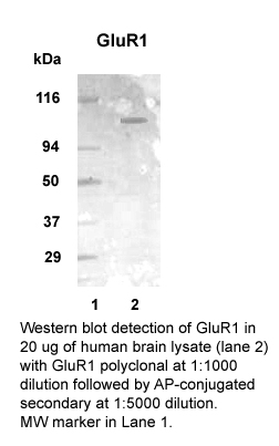
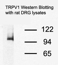
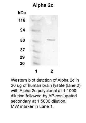
mGluR1 Alpha, Alpha 2c and TRPV1 Westerns


mGluR1 alpha (Catalog#: RA19065)

Species: Human, Mouse an Rat

Applications: ICC; IHC and WB

Immunogen: 1159-1171 SSVPSSPVSESVL GAEGGAGGADGQGAGP

VR1 (TRPV1) (Catalog#: GT15129)

Species: Rat

Applications: IHC and WB

KLH-coupled synthetic peptide corresponding to amino acid residues 4 - 21 of Vanilloid Receptor 1


2c Adrenergic Receptor (Catalog#: RA19064)

Species: Human and Rat

Applications: ICC; IHC and WB

Immunogen: 309-324

Thursday, December 21, 2006

Customer Data-GFAP IHC

We would like to thank Dr. Qi-Lin Cao, University of Louisville Medical School for this excellent image.

Image: GFAP (Catalog#: CH22102) Staining of of Astrocytes in the Ventral horn and Funiculus of Adult Rat Spinal Cord.

Dilution 1:500

Wednesday, December 13, 2006

Wednesday, November 29, 2006

P2Y2-It is baaack!

We have finally managed to make a new P2Y2 (Catalog#: RA14102) that passed our IHC certification process.


Image: P2Y2 Staining of Rat DRG.

Related Antibodies to Consider:
P2X1
P2X2-Guinea Pig
P2X2-Rabbit
P2X3-Rabbit
P2X3-Guinea Pig
Neuropeptides
Opioids
Opioid Neuropeptides
TRPV1s
Vision and Retina
Cancer

Friday, October 27, 2006

Excellent TUJ-1 IHC

We would like to thank Christine Graham and Doug Vetter, Tufts Univ. School of Medicine, Boston for the excellent image.


Tuj-1 Catalog#: MO15013 staining of
staining of mouse cochlea (whole mount, ~50um).

Tuj-1 Rabbit Polyclonal
All Neuron-Glial Markers
All Stem Cell Reagents

Monday, October 16, 2006

P2X3 Western Blot

There have recently been customer question on the expected band for our P2X3 Antibody (Catalog#: RA10109)
Here's an excellent publication showing both WB and IHC for this antibody.

P2X2 and P2X3 receptor expression in postnatal and adult rat urinary bladder and lumbosacral spinal cord Simon Studeny, Ali Torabi and Margaret A. Vizzard.


Figure: Western blot of whole urinary bladder (40 mg) for P2X3 receptor expression in postnatal (P0-P36) and adult (A) rats. Erk staining was used as a loading control. B. Preabsorption of P2X3 antibody with immunogen (1 mg ml−1) eliminated the band at 75kD. C. Histogram of relative P2X3 band density in all groups examined normalized to erk-1 staining. P2X3 receptor expression in urinary bladder is significantly increased from P14-P21 compared to adult rat bladder. *, p ≤ 0.005, #, p ≤ 0.01.

Friday, September 29, 2006

ESC to Neural Progenitors-Role of MOR-1C

Features use of Neuromics' MOR-1C

Eunhae Kim, Amy L. Clark , Alexi Kiss, Jason W. Hahn, Robin Wesselschmidt. MU AND KAPPA OPIOIDS INDUCE THE DIFFERENTIATION OF EMBRYONIC STEM CELLS TO NEURAL PROGENITORS
JBC Papers in Press. Published on September 1, 2006 as Manuscript M603862200


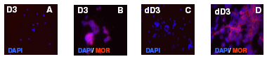
Image: A-D-Staining of Differentiated Stem Cells. D3 ES cells were maintained in serum/LIF/ME induced self-renewal conditions (undifferentiated cells, D3). Upon treatment with 1 µM RA they undergo differentiation (dD3). Cells were fixed and permeabilized. Treatments were followed by overnight incubation with polyclonal MOR-1 (C-terminus, 1:2500)

Tuesday, September 19, 2006

New TRPV1-C Publication

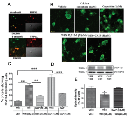
Amol M. Patwardhan, Nathaniel A. Jeske, Theodore J. Price, Nikita Gamper, Armen N. Akopian, and Kenneth M. Hargreaves. The cannabinoid WIN 55,212-2 inhibits transient receptor potential vanilloid 1 (TRPV1) and evokes peripheral antihyperalgesia via calcineurin. PNAS 103 (July 2006): 11393-11398.


Figure: WIN activates calcineurin in TG neurons and dephosphorylates TRPV1. (A) Calcineurin subunits are coexpressed with TRPV1. The colocalization of calcineurin A subunit (Upper) and B subunit (Lower) with TRPV1isdemonstratedin the respective panels.(B) The effect of various treatments on the nuclear translocation of NFATc4 (calcineurin activation). Cultured TG neuronswere exposed to vehicle, ionomycin (1 M), capsaicin (1 M), WIN (25 M), or WINCAIP(25 M50 M), and immunohistochemistry was performed by using an antibody againstNFATc4. (C) Graphical representation of the percent of NFATc4 positive neurons showing nuclear translocation of NFAFc4 after treatment with VEHVEH, VEHWIN (25 M), CAIP (50 M)WIN, VEHCAP (1 M), or CAIP(50 M)CAP (n 4 independent cultures assessed by blinded observer; n 152–180 cells per condition; **, P<0.01,anovawith n="3">

Saturday, September 09, 2006

4 New Interleukins

These Interleukins have been tested for IHC and WB apps and recommended for use by Diabetes/Obesity, Cancer, Neurodegenerative Disease and Pain/Inflammation Researchers

IL-1Alpha/IL-F1


Image: IL-1α staining of blood lymphocytes. Cells were stained using anti-gat-Cy3 (red) and counterstained with Fluoro Nissl Green


IL-1Beta


Image: IL-1 beta staining (red color) in mouse splenocytes stimulated with Con A. Cell nuclei counterstained green.


IL-1 R1


Image: IL-1R staining of mouse thymus. Staining was done using HRP-DAB (brown) detection. Tissue was counterstained with haematoxylin (blue).


IL-6


Image: IL-6 staining of mouse T cells. Cells were stained with goat anti-rat IL-6 (red) and nuclei counterstained with Fluoro Nissl Green.

Thursday, August 31, 2006

Monoclonal Ubiquitin Antibodies-Great IHC

Ubiquitin is a protein that plays a role in the targeting of proteins for proteolytic degradation. Ubiquitin becomes covalently bonded to many types of pathological inclusions seen in neurodegenerative diseases that appear to be resistant to normal degradation. The neurofibrillary tangles and paired helical filaments diagnostic of Alzheimer's disease, the Lewy bodies seen in Parkinson's disease, and Pick bodies found in Pick's disease are all heavily ubiquitinated and can all be readily visualized with this antibody.


Ubiquitin-Catolog#:19005


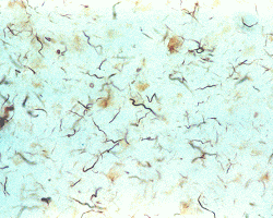
Image: ABC-HRP staining with Ubiquitin monoclonal antibody (diluted 1:1,000) of Alzheimer’s disease cerebral cortex showing dystrophic neuritis, which contain ubiquitinated tau. The inclusions are detected by the Ubiquitin antibody. Normal cortex does not contain inclusions and would be white in color.

Ubiquitin-Catalog#: MO18001


Confocal images of hippocampal CA1 sections from rats, (A) mock-treated or (B) subjected to 15 minutes ischemia followed by 24 hours of reperfusion, using ubiquitin mouse antibody (green) and propidium iodide (red). Data courtesy of Cell Signaling Technology, Inc.

Saturday, July 29, 2006

New P2X3 Data

P2X3 Catalog#: RA10109

Data Courtesy of Dr. Yan Sun University of Maryland School of Medicine Baltimore


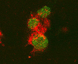
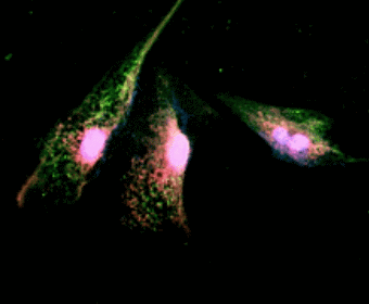
Image: Double immunofluorescence staining in cultured human bladder urothelial cells (BUCs). BUCs show cytokeratin (red areas) and P2X3 (green areas) expression.

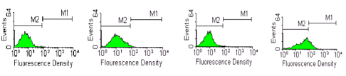
Figure: Flow cytometry and FACS analysis of P2X3 expression in human BUCs. P2X3 expression in BUCs were compared among in vitro stretched samples and unstretched samples in both normal and interstitial cystitis (IC) patients. A, unstretched normal BUCs. B, stretched normal BUCs. C, unstretched IC BUCs. D, stretched IC BUCs. M1 region represents P2X3 localization detected.

Monday, July 24, 2006

New AD Research Antibodies

We continue to add to our catalog of Neurodegenerative Disease Reagents:

BACE-1
Cleavage of amyloid precursor protein (APP) into 40 or 42 amino acid amyloid β (aβ) peptides is a result of the action of two proteases β- and γ-secretase. The beta-site APP cleaving enzyme, or BACE-1, is a transmembrane aspartic protease that is localized in the golgi and endosomes. Low levels of BACE-1 mRNA are found in most adult tissues with a higher level of expression in pancreas.


Image: Detection of BACE-1 in human cerebellum. Stained with HRP-DAP (brown) and counter-stained with hemtoxylin (blue).


Abeta 40 (beta amyloid 1-40) and Abeta 42 (beta amyloid 1-42)
Amyloid beta-protein (Abeta) is associated with neuronal injury and death in Alzheimer's disease. These proteins are the main components of amyloid plaques. Abeta 42 plays a bigger role in deneurogeneration and is more strongly associated with Alzheimer's disease than Abeta 40.


Image: Detection of Abeta 40 on 5 ng of peptide per lane using NB 300-225. Lane 1: Abeta-40, lane 2: Abeta-42, lane 3: Abeta-40 and -42 mix

Saturday, July 22, 2006

TRPV1-N and C Data

Some interesting results from:

Sharif Naeini R, Witty MF, Seguela P, Bourque CW (2006) An N-terminal variant of Trpv1 channel is required for osmosensory transduction. Nature Neuroscience. (1):93-8.


Figure: SON Neurons express an N-terminal variant(s) of TRPV1 (a) Simplified structure of mRNA (numbered boxes show exons of TRPV1 gene) encoding TRPV1 (above). Gray arrow tails and lines illustrate mRNA regions detected by primer sets A and B. RT-PCR analysis of mRNAs expressed in DRG and SON (below). Note the lack of signals for the N terminus of TRPV1 (lane 2, exons 2-6, primer set A) in SON. (b) Immunocytochemical staining confirmed the presence of full length TRPV1 in DRG neurons. In contrast, only the C-terminus of the TRPV1 protein (C-TRPV1) was detected in the AVP positive SON neurons. (c) Whole-cell recordings reveal that DRG neurons are sensitive to capsaicin ('Cap,' 10 uM, 5s) but that SON neurons are insensitive.

Sunday, June 25, 2006

New Publication-Neuromic's E-18 Primary Hippocampal Neurons

Brian Hoffpauir , Emily McMains, and Evanna Gleason Nitric Oxide Transiently Converts Synaptic Inhibition to Excitation in Retinal Amacrine Cells J Neurophysiol 95: 2866-2877, 2006

Hippocampal cultures were made from 18-day-old rat (Fischer 3440) embryos. Micro-dissected hippocampal tissue was obtained from Neuromics (Bloomington, MN) and prepared and maintained according to the supplier’s protocols. Recordings from single hippocampal neurons were made after 4–7 days in culture. No physiological assessments of the relative maturity of these cells have been made. According to their developmental timeline alone, they would be considered embryonic.

Thursday, June 15, 2006

New Goat VR1 (TRPV1) and Rabbit NK1 Antibody


Catalog #: GT15129

Image: VR1 staining in Rat DRG: red color - TRPV1 labeling, green-counterstain.

Description: VR1, also known as TRPV1 (Transient Receptor Potential Vanilloid 1) and capsaicin receptor, is a 115 kDa integral membrane ion channel that has six transmembrane domains and contains intracellular N-and C-termini. It contributes to normal pain and temperature sensation and also has a "sensory-effector" function. By alternative splicing, at least three VR1 isoforms are known. The sequence of residues 4 - 21 of rat VR1 is 78% and 28% identical to that of mouse and human VR1, respectively.


Catalog #: RA25001

Image: NK 1 staining of lamina III neuron in the spinal cord of a rat.

Description: The tachykinins belong to an evolutionary conserved family of peptide neurotransmitters that share the c-terminal sequence Phe-X-Gly-Leu-Met-NH2 and have an established role in neurotransmission. The mammalian tachykinins include substance P, neurokinin A (NKA) and neurokinin B (NKB) which exert their effects by binding to specific receptors. Tachykinin peptides are important in the mediation of many physiological and pathological processes including inflammation, pain, migraine headache and allergy induced asthma. Three tachykinin receptor types have been characterized, NK-1, NK-2 and NK-3 which have preferential affinities for SP, NKA and NKB respectively. All three receptors share a high degree of sequence homology, have seven transmembrane spanning domains and similar signal transduction mechanisms (e.g. G-protein coupled activation of phospholipase C).

Wednesday, June 07, 2006

New GFAP IHC Image


GFAP (Cat# 22102)-Staining in mouse retina. Courtesy od Dr. Sal Stella.

IHC Protocol

Wednesday, May 24, 2006

New BDNF and NT-3 IHC Images

Images from Dr. Matt Ramer's Lab, University of British Columbia, Courtesy of Dr. Lowell McPhail.


NT-3 Staining in Rat Dorsal Horn


BDNF Staining in Rat Cerebellum

Monday, May 08, 2006

You've Been Patient-Now We Have It!

We have finally produced a new and robust P2X3 Guinea Pig Antibody. It tested out beautifully in IHC.

Friday, April 14, 2006

The Neuromics-AlphaGenix alliance

We are thrilled to be working with Neuromics in the area of RNAi and believe it has enormous potential for genetic-based therapies for neurological disorders, neuro-oncology, viral infections and inherited genetic disorders.

Neuromics and AlphaGenix work closely with Dr. Mark Behlke, Vice President of Molecular Genetics at Integrated DNA Technologies to develop optimum strategies for the design of RNAi molecules.

This is followed by developing in vitro studies on primary cells, which are required in order to design in vivo strategies to inhibit targeted gene expression. While transfection studies enhance our understanding of RNAi technology, we believe that it is vital to demonstrate validation by including sufficent controls as well as demonstrating knockdown of endogenous message.

We will be routinely publishing new publications to this blog. In this way, researchers are able to get leading papers and reviews in these fields of research as well as having the ability to bring their own expertise to the table. Indeed, science is a collective effort and we encourage you as colleagues to help all of us keep on the cutting edge.

Best regards,

Stephen Hall, Ph.D.

Thursday, April 13, 2006

RNAi Transfection Alliance


Neuromics and Alphagenix would like to announce our RNAi Transfection Alliance. We will be partnering with IDT to develop and commercialize better ways to deliver RNAi to Glia, Neurons and the Nervous System.

Here are some related publications:

Progress Towards in vivo Use of siRNAs
siRNAi: Applications in Functional Genomics and Potential as Therapeutics
Assembly and Function of RNAi Silencing Complexes
Synthetic dsRNA Substrates Enhance RNAi Potency and Efficacy

In vivo Publications:

Luo MC, Zhang DQ, Ma SW, Huang YY, Shuster SJ, Porreca F, Lai J (2005). An efficient intrathecal delivery of small interfering RNA to the spinal cord and peripheral neurons. Molecular Pain 2005, 1:29.

Priti Kumar, Sang Kyung Lee, Premlata Shankar, N. Manjunath (2006) A Single siRNA Suppresses Fatal Encephalitis Induced by Two Different Flaviviruses. PLOS Medicine, Volume 3 Issue 4 APRIL 2006

Monday, April 03, 2006

New Article with RNAi and i-Fect in vivo Results

A Single siRNA Suppresses Fatal Encephalitis Induced by Two Different Flaviviruses

Priti Kumar1, Sang Kyung Lee2, Premlata Shankar1*, N. Manjunath1*
1 The CBR Institute for Biomedical Research and Department of Pediatrics, Harvard Medical School, Boston, Massachusetts, United States of America, 2 Department of Bioengineering, Hanyang University, Seoul, Korea

ABSTRACT
Background
Japanese encephalitis virus (JEV) and West Nile virus (WNV) are neurotropic flaviviruses that can cause acute encephalitis with a high fatality rate. Currently there is no effective treatment for these infections.
Methods and Findings
We tested RNA interference (RNAi)-based intervention to suppress lethal JE and WN encephalitis in mice. To induce RNAi, we used either lentivirally expressed short hairpin RNA (shRNA) or synthetic short interfering RNA (siRNA). As target, we selected the cd loop-coding sequence in domain II of the viral Envelope protein, which is highly conserved among all flaviviruses because of its essential role in membrane fusion. Using as a target a species-specific sequence in the cd loop that is conserved only among the different strains of either JEV or WNV, we could achieve specific protection against the corresponding virus. However, by targeting a cross-species conserved sequence within the cd loop, we were able to protect mice against encephalitis induced by both viruses. A single intracranial administration of lentivirally delivered shRNA or lipid-complexed siRNA before viral challenge or siRNA treatment after viral challenge was sufficient for protection against lethal encephalitis.
Conclusions
RNAi-based intervention affords near complete protection from both JEV- and WNV- induced encephalitis in mice. Our results show, to our knowledge for the first time, that siRNA can be used as a broad-spectrum antiviral agent for treating encephalitis caused by multiple related viruses.
Funding: This work was supported by NIH grant U19 AI 056900 to NM and PS.
Competing Interests: The authors have declared that no competing interests exist.
Academic Editor: Clifford Lane, National Institutes of Health, United States of America
Received: September 20, 2005; Accepted: December 16, 2005; Published: February 14, 2006
DOI: 10.1371/journal.pmed.0030096
Copyright: © 2006 Kumar et al. This is an open-access article distributed under the terms of the Creative Commons Attribution License, which permits unrestricted use, distribution, and reproduction in any medium, provided the original author and source are credited.


Relevant to i-Fect (TM)

"Although there is a report of the successful use of naked siRNA targeting the pain-related cation channel P2X3 to treat chronic neuropathic pain in a rat model [28], other studies suggest that naked siRNA is poorly taken up by brain parenchymal cells [29,30]. Recently, a cationic lipid formulation, i-Fect (Neuromics) has been found to deliver siRNA into neuronal cells without toxicity (Dr. Josephine Lai, University of Arizona Health Sciences Center, personal communication). We tested if synthetic FvEJ siRNA (siFvEJ) complexed with i-Fect can protect mice against viral encephalitis. After confirming that i-Fect can transduse siFvEJ siRNA as efficiently as lipofectamine to inhibit JEV infection in Neuro 2a cells (Figure 3A), we infected mice by IC injection with JEV and, after allowing 30 min for viral adsorption, injected the synthetic siFvEJ or control luciferase siRNA (siLuc) complexed with i-Fect at the same site. All mice injected with siLuc died by day 5, whereas all of the siFvEJ-injected mice survived indefinitely (Figure 3B), suggesting that i-Fect can deliver siRNA into neuronal cells and result in protection that is similar to the lentivirally delivered shRNA."

Saturday, April 01, 2006

More Customer Data-Ret and SHANK 1a-C-terminus

Ret-(Cat#GT15002)

Thank you Kim Carnes, UIUC, for the image and protocol for the Ret staining of spermatagonial stem cells.

Protocol

Depraffinize and hydration
Peroxidase blocking - 30 minutes in 0.06% H2O2 in methanol.
Antigen retrieval - 0.01M Citrate buffer (pH6.0) in microwave (10 minutes, power level 7)
Serum blocking - horse serum (unfiltered normal, diluted1:100) for 30 minutes.
Primary antibody - Neuromics Ret antibody (1:200), overnight at 4º C in a moisture chamber.
Secondary Ab - this was a horse universal biotinylated ab that came in a kit (with the above horse serum) diluted 1:50 with PBS and horse serum (also 1:50) for 60 minutes at room temp.
ABC incubation (Vector ABC Elite kit) - 30 minutes.
Colorization with DAB, counterstain with hematoxylin followed by dehydration.

We use PBS rinses between steps.

SHANK 1a C-terminus

Thank you Alex Vila, UCLA, for your image and protocol for the SHANK 1a C-terminus staining of retinol tissue

Protocol

Species: Mouse C57B6
Fixation: 15 minute 4% paraformaldehyde fixation
Antibody dilution: 1:800 overnight.
Secondary Alexa 568 1:500 for 45 minutes
Tissue preparation: Blocked for 2 hrs at room temp in 10% normal goat serum and
Triton X-100 (0.5%) in 0.1 M Phosphate buffered saline (PBS)
Washed: 3X 0.1 M PBS between primary and secondary step.
Mounted tissue with Paramount.
Confocal: Images are 2048 X 2048, 12 bit, collected with a 40X water objective, 10 stacks 0.55 um per stack

Wednesday, March 01, 2006

New Park7 Data-Thank you Amanda Ashley

We have received extremely positive feedback on the quality of our PARK7 (DJ-1) antibody. In addition to feedback, Amanda Ashley, in Dr. Legare's Laboratory, Colorado State University, resently sent us a very clean Western Blot using the PARK7 (DJ-1) antibody comparing wildtype to PARK7/DJ-1 knockout animals


Click to see full sized image.


WB: Wild type and PARK7 (DJ-1) KO animals
Data courtesy of Amanda Ashley and Dr. Marie Legare, Colorado State University.

Western conditions:
– 25mg protein from mouse brain lysate
– 1:2000 Park7 (DJ-1) antibody (Cat # RA19006)
– 12% gel
– Electrotransferred onto PVDF
– Blocked with 5% BSA in TBS-Tween
– Detected with ImmunStar HRP (BioRad)
– Dually probed for beta-actin (1:1000, Sigma)