
IL-1Alpha/IL-F1

Image: IL-1α staining of blood lymphocytes. Cells were stained using anti-gat-Cy3 (red) and counterstained with Fluoro Nissl Green
IL-1Beta

Image: IL-1 beta staining (red color) in mouse splenocytes stimulated with Con A. Cell nuclei counterstained green.
IL-1 R1
.jpg)
Image: IL-1R staining of mouse thymus. Staining was done using HRP-DAB (brown) detection. Tissue was counterstained with haematoxylin (blue).
IL-6
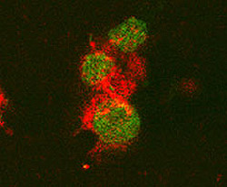
Image: IL-6 staining of mouse T cells. Cells were stained with goat anti-rat IL-6 (red) and nuclei counterstained with Fluoro Nissl Green.


No comments:
Post a Comment