Saturday, January 31, 2015

Neural Progenitors and Cool Science

Titania Nanotubes and Neural Prostheses

I am always on the hunt for the use of our Neural Progenitor Markers in cool and important human health applications. Here our Nestin Antibody is use to access the growth and maintenance of C17.2 neural stem cell line cultured in these nanotubes: Jonathan A. Sorkin, Stephen Hughes, Paulo Soares, Ketul C. Popat. Titania nanotube arrays as interfaces for neural prostheses. Materials Science and Engineering: C Volume 49, 1 April 2015, Pages 735–745. doi:10.1016/j.msec.2015.01.077

Image: Nestin staining eSC Derived hNP1 Human Neural Progenitors. Cells were stained using goat anti-mouse Alexa Fluor 488 secondary antibody (green) (Molecular Probe, A-11001) and counterstained with DAPI (blue).

Abstract: Neural prostheses have become ever more acceptable treatments for many different types of neurological damage and disease. Here we investigate the use of two different morphologies of titania nanotube arrays as interfaces to advance the longevity and effectiveness of these prostheses. The nanotube arrays were characterized for their nanotopography, crystallinity, conductivity, wettability, surface mechanical properties and adsorption of key proteins: fibrinogen, albumin and laminin. The loosely packed nanotube arrays fabricated using a diethylene glycol based electrolyte, contained a higher presence of the anatase crystal phase and were subsequently more conductive. These arrays yielded surfaces with higher wettability and lower modulus than the densely packed nanotube arrays fabricated using water based electrolyte. Further the adhesion, proliferation and differentiation of the C17.2 neural stem cell line was investigated on the nanotube arrays. The proliferation ratio of the cells as well as the level of neuronal differentiation was seen to increase on the loosely packed arrays. The results indicate that loosely packed nanotube arrays similar to the ones produced here with a DEG based electrolyte, may provide a favorable template for growth and maintenance of C17.2 neural stem cell line.

I will continue to how our stem cell solutions are used in cool, new discoveries.

Friday, January 30, 2015

Sensitive, Specific and Cost Effective Protein Expression Analysis

Reaching for Cell Based Assay Solutions Excellence

We have been receiving positive feedback from clients using our Quantibody® and RayBio® C-Series Membrane-Based Antibody Arrays. Our manufacturing partner, Raybiotech, is ISO 9000 certified insuring all arrays are made to exacting specification. The cost for measuring a single cytokine runs as low as 16 USD/Cytokine!

We use these arrays to test the blood serum of clients with autoimmune diseases to determine levels of cytokine and growth factor expression vs healthy controls. We also use them to determine proteins secreted by our  USB Derived hMSCs cultured in our hMSC Media. This gives us insight as to the protein expression patterns in our hMSC, Chondrogenesis and Osteogenesis Assays.

Figure: The graph shows cytokine secretion of hMSCs after a continuous 24 day culture. These results were obtained using our select markers from our cytokine and bone metabolism arrays. Data courtesy of Dr. Jim Musick and Tiana Tonrey, Vitro Biopharma.

We are pleased to announce the addition of these new human and mouse arrays:

Quantibody® Human Growth Factor+Neurotrophin+Cytokine Array Q1-10. Precisely measures: Amphiregulin, BDNF, bFGF, BMP-4, BMP-5, BMP-7, beta-NGF, EGF, EGFR, EG-VEGF (PK1), FGF-4,FGF-7 (KGF), GDF-15, GDNF, Growth Hormone, HB-EGF, HGF, IGFBP-1, IGFBP-2, IGFBP-3, IGFBP-4, IGFBP-6, IGF-1, Insulin, M-CSF R, NGFR (TNFRSF16), NT-3, NT-4, Osteoprotegerin (TNFRSF11B), PDGF-AA, PLGF, SCF, SCF R (CD117/c-kit), TGF alpha, TGF beta 1, TGF beta 3, VEGF-A, VEGFR2, VEGFR3 and VEGF-D.
Mouse C-Series Cytokine Array. C1-2-Semi-quantitavely measures: GCSF,GM-CSF, IL-2, IL-3, IL-4, IL-5, IL-6, IL-6, IL-9, IL-10, IL-12 p40/p70, IL-12 p70, IL-13, IL-17A, IFN-gamma, MCP-1 (CCL2), MCP-5, RANTES (CCL5), SCF, TNF RI (TNFRSF1A), TNF alpha, Thrombopoietin, (TPO) and VEGF-A.


Images: Representative images obtained with RayBio® C-Series Antibody Arrays. These membranes were probed with conditioned media from two different cell lines. Membranes were exposed with Kodak X-Omat® film at room temperature for 1 minute. Note the strong signals of the Positive Control spots in the upper left and lower right corners.

Questions? Do not hesitate to contact me: pshuster@neuromics.com or direct phone-612-801-1007. Thank you. Pete Shuster, CEO and Owner, Neuromics

Tuesday, January 27, 2015

Human Growth Factor+Neurotrophin+Cytokine Array-Test 40 Markers in on Assay

Sensitive, Specific and Cost Effective

Neuro-immuno and degenerative diseases and disorders commonly show dysregulation of Growth Factors and Cytokines. Using our Quantibody Neuroscience Arrays, we have measured the blood serum of clients with Neuro-inflammatory/immune response diseases/disorders including Autism Spectrum Disorder (ASD). Most showed lowered levels of Growth Factors/Neurotrophins and elevated levels of Inflammatory Response Cytokines.

In order to expand the number of biomarkers measured, we are pleased to announce the addition of our New Growth Factor+Neurotrophin+Cytokine Array. This will enable us to further determine the "finger prints" of these diseases/disorders at the protein level. This array includes: Immunogen: Amphiregulin, BDNF, bFGF, BMP-4, BMP-5, BMP-7, beta-NGF, EGF, EGFR, EG-VEGF (PK1), FGF-4,FGF-7 (KGF), GDF-15, GDNF, Growth Hormone, HB-EGF, HGF, IGFBP-1, IGFBP-2, IGFBP-3, IGFBP-4, IGFBP-6, IGF-1, Insulin, M-CSF R, NGFR (TNFRSF16), NT-3, NT-4, Osteoprotegerin (TNFRSF11B), PDGF-AA, PLGF, SCF, SCF R (CD117/c-kit), TGF alpha, TGF beta 1, TGF beta 3, VEGF-A, VEGFR2, VEGFR3 and VEGF-D.

I will continue to post testing results here.

Monday, January 19, 2015

Solutions for Studying Neuro-degeneration

Data Rich and Frequently Published

The Neuromics' brand is built, in part, by our proven ability to provide solutions for the study of neuro-degeneration. These include:
A recent example shows the use of one of  MAP-2 markers to study hearing decline with age: Radtke-schuller S, Seeler S and Grothe B(2015) Restricted loss of olivocochlear but not vestibular efferent neurons in the senescent gerbil (Meriones unguiculatus). Front. Aging Neurosci. 7:4. doi:10.3389/fnagi.2015.00004.

Figure: Lipofuscin granules in MSO neurons of an aged gerbil. MSO neurons are MAP2 immunostained (Alexa Fluor 647, red). Lipofuscin granules have been excited with the DAPI excitation wavelengths and appear blue. Confocal images show a maximum projection of image stacks in A and a single optical image of 0.3 µm thickness in the enlargement in B. Scale bar in A: 50 µm and 20 µm in B.

We stand ready to serve you. Should you have interest or questions, do not hesitate to contact me directly: Pete Shuster-Owner/CEO-pshuster@neuromics.com or direct phone: 612-801-1007. Thank you.

Monday, January 12, 2015

Cancer Associated Fibroblasts (CAFs)-Flow Cytometry and Immunostaining Results

If they Walk and Talk Like CAFs, They Are!

We are experiencing growing demand for are CAFs. They are closely associated with primary tumor cells and participate in the neoplastic process. There is reciprocal communication between CAFs and tumor cells through paracrine effects of secreted growth factors, cytokines & chemokines from both fibroblasts, tumor cells and other tumor-associated cells.

Potential users are asking how well the cells are characterized. The purpose of this posting is to share key gene expression data:
CELL LINES
CD105
CD90
CD44
CD326
CD133
FAP
+
+
+
-
-
-
+
+
+
-
-
-
+
+
+
-
-
+
+
+
+
-
-
+
+
+
+
-
-
+
Table: expression of key proteins
Images: Human Lung Adenocarcinoma CAFs stained with Vimentin, aSMA, b-Catenin and DAPI (blue),


Cancer Associated Fibroblasts Gene Expression Data(pdf - 784Kb)-Flow Cytometry and Immunostaining Results.
Questions? Please contact me directly-Pete Shuster- CEO & Owner-pshuster@neuromics.com or 612-801-1007.

Monday, January 05, 2015

Racing into 2015

Thank you for a Great 2014!
EU PCWow. I would like to thank our customers, collaborators and friends for helping us achieve unprecedented success in 2014.

We plan on racing hard into 2015. I would like to highlight our focus areas for the New Year.

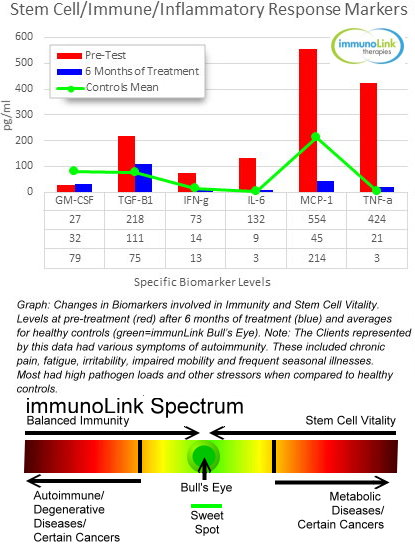
Stem Cell Activating Solutions-We have been pilot testing solutions foir balanced immunity and stem cell vitality. Our clients have been mostly peopole with autoimmune related issues. After 6 months of therapy, the improvement in symptoms and test results, for many, has been stunning (see below).
We have developed migration and other assays to test these solutions in vitro using our UCB Derived hMesenchymal Stem Cells. We also use medical testing to prove and improve their therapeutic value. These assays and arrays are available for labs and clinics interested in testing stem cell related solutions. Here're representative data from our Quantibody Array testing program.
IT

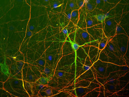
Stem and Primary Cell Based Assays-We will continue to add new solutions for your assays. These will include mixed culture that more closely mimic in vivo environments like mixed astroglial-neuron cultures. Below is our hN2TM Human Neurons stained with one of our neuron markers.
hN2
Image: hN2 cells grown in culture for 4 days and stained with our chicken polyclonal to Neurofilament light or low molecular weight chain NF-L, a marker of neurons. Many of the differentiating cells show strong cytoplasmic and clearly fibrillar staining for NF-L. Blue stain is DAPI and reveals cell nuclei of some non neuronal cells in this culture.

Check out and join my Stem Cell Clinical Trails Group on Linkedin (4500+members strong).
We wish you a rewarding 2015.

Pete Shuster-
CEO & Owner--pshuster@neuromics.com or 612-801-1007

Wednesday, December 24, 2014

immunoLink Therapies and Treating ASD Children

6-Months of Therapy and Testing Results

I have previously posted our testing and treatment regime for ASD Children. For some of the children, we are seeing marked improvement after 6 months of our treatment regime using solutions aimed at balance immunity and stem cell vitality.

We find that for the children we treat all have varying dysregulated levels of immune/inflammatory response, oxidative stress and growth factor/neurotrophin biomarkers.  We customize treatment plans based on pre-treatment testing results. We then re-test after 6 months to see the level of moderation in these biomarkers/factors. The re-tests are used to show the parents how well these children are responding to treatment and to further adjust plans.

Here I would like to share the results from our more traditional medical testing. These are representative results from one of the Children we are treating.
Graph: ASD Child Pre-treament and 6 month treatment analyte levels vs healthy sweet spots

If you are interested in learning more, please contact me directly (612-801-1007) or pshuster@neuromics.com. Pete Shuster, CEO and Owner of Neuromics and ImmunoLinkTMTherapies.

Thursday, December 18, 2014

immunoLink Therapies-Initial Products

Feeling and performing your best depends on balanced immunity and stem cell vitality

Your current investment in exercise, healthy diet and supplements are aiming you towards what we call the immunoLink sweet spot. If you have hit the bull’s eye, you would be seeking ways to stay there, because this is where you feel awesomely great and are most productive. If you have never been there, now is the time.

Illness, injury and medical procedures push you from the spot, but if you are near or on it, your recovery will be faster and more complete. The far reaches of the immunoLink Spectrum represent a cold wasteland of misery and despair.

How do we Know These Solutions Work?-Our Clients have autoimmune and degenerative diseases or are seeking peak performance. Prior to customizing their treatment strategies, we measure the levels of immune/inflammatory response, oxidative stress, allergens and stem cell health factors in our Clients' blood serum. The levels of these factors are compared with healthy controls. We then re-test after 6 months of treatments. For many the improvement in health and performance are startling. This is confirmed in the re-tests.
Stem-Kine and StemTrophin are central to our treatment strategies. Stem-Kine boosts the levels of circulating blood stem cells. These cells differentiate into immune factors and red blood cells. This boosts oxygen delivery and immunity resulting in greater endurance, mental acuity and cardiovascular health. StemTrophin activates mesenchymal stem cell migration. These cells are essential for immune suppression and then initiating the process of repair and regeneration at the sites of injury or degeneration. To learn more or order by phone, please call me directly at 612-801-1007 or you can also inquire via my e-mail: pshuster@neuromics.com. Pete Shuster, CEO and Owner, Neuromics and immunoLink Therapies.

Monday, December 15, 2014

Stem Cell Based Therapy For Tendinosis

Using Collagen I Producing Cells from Hair Follicles

Learn how these cells can be these cells can be harvested, purified and expanded to treat a chronic, degeneration of tendons.


Saturday, December 06, 2014

Treating Autism-Immune System Balancing and Stem Cell Activating Therapies

Linking the Immune System with Stem Cell Health to Treat ASD Children

We have been testing immune/inflammatory response, oxidative stress and growth factor/neurotrophin biomarkers in the blood sera of Children with Autism Spectrum Disorder (ASD). We did this testing using our Quantibody Arrays.

The purpose of these tests were to establish a baseline for the state of these children's immune system and stem cell health. In this baseline testing, we found that all had high immune/inflammatory response, oxidative stress and low levels of the growth factors/neurotrophins needed for repairing and regenerated damaged cells/tissue (stem cell health).

We then treated these children with customized combinations of immune balancing and stem cell activating agents with the goal of showing that any improvement in ASD related symproms would be confirmed by positive modulations of biomarkers that are dysregulated in this disorder. We re-test at 6 months after initiation of the treatment plans.

The goal of our treatment strategies is to balance the immune system and activate the natural cell/tissue repair and regeneration (stem cell) processes. We call this the immunoLinkTM.

We are seeing improvement in the symptoms of the first 2 children recently retested and a marked improvement in the levels of their biomarkers. Here's data from one of the children. Note that key immune/inflammatory response markers have now moderated to the levels of healthy controls.

Graphs: Comparison of initial testing (red) and 6 month re-test (green) vs healthy controls (blue) for one of our ASD Children

We are developing a website that will have a wealth of information to help parents determine treatment strategies for ASD Children. We are also making our treatment plans and strategies available to interested parents. The website ecosystem will be named immunoLinkTM Therapies.
In the meantime, should you want to investigate if we could help your child, do not hesitate to contact me e-mail: pshuster@neuromics.com or cell: 612-801-1007. Pete Shuster, CEO and Owner.

Monday, November 24, 2014

Good Axon; Bad Axon: Regeneration in the CNS and Age

"The incapacity of the central nervous pathways to regenerate is a dogma accepted by science..." - Ramon y Cajal


Harvard University has opened access to Michio Wendell Painter's Dissertation: Regeneration in the aging peripheral nervous system. Will Spinal Cord Injury (SCI) and Neurodegenerative Diseases of the PNS become treatable with regenerative therapies? This work provides important insights.

Age plays a central role in regenerative capacities.

Monday, November 17, 2014

Neuropathy Research Solutions

New Publications and Past Postings

Here're are several key postings on Neuropathy and Neuropathic Pain:
Here're some "hot of the press" publications referencing use of Neuron-Glial Markers
Bethany L. Johnson-Kerner, Faizzan S. Ahmad, Alejandro Garcia Diaz, J. Palmer Greene, Steven J. Gray, R. Jude Samulski, Wendy K. Chung, Rudy Van Coster, Paul Maertens, Scott A. Noggle, Christopher E. Henderson and Hynek Wichterle. Intermediate filament protein accumulation in motor neurons derived from giant axonal neuropathy iPSCs rescued by restoration of gigaxonin. Hum. Mol. Genet. (2014) doi: 10.1093/hmg/ddu556. First published online: November 4, 2014.
...MAP2 (1:2,000, Neuromics, CH22103), NF-H (1:2000, Neuromics, CH22104), NF-L (1:200, Neuromics, MO22104)...

Images: Cells grown from adult rat brain. Large cell in middle is stained with mouse monoclonal to NF-L clone DA2 (green). Another type of neuronal lineage cell was stained with rabbit polyclonal to alpha-internexin (red). These cells were mitotic but had several characteristics of neurons. Rat spinal cord homogenate showing the major intermediate filament proteins of the nervous system (lane 1). The remaining lanes show blots of this material stainted with various antibodies including: NF-H, NF-M,, NF-L,, NF66 and GFAP 

Jianfei Guo, Xudong Fu, Xia Cui, Minhua Fan. Contributions of purinergic P2X3 receptors within the midbrain periaqueductal gray to diabetes-induced neuropathic pain. The Journal of Physiological Sciences November 2014.
...An equal volume of total and membrane samples was applied to SDS-PAGE. Membranes were incubated with the rabbit anti-P2X 3 primary antibody (1:1000, Neuromics, Edina, MN, USA) and goat anti-rabbit secondary antibody (1:200, Neuromics, Edina, MN, USA).

Neuropathy Research Solutions Include:


Friday, November 14, 2014

Immune System/Stem Cell Health and Aging

Looking, feeling and  performing your best depends on balanced immunity and stem cell vitality

As we age we encounter:
  • Slower healing
  • Longer recovery time from vigorous exercise
  • More general aches and pains
  • Longer recovery from illness
  • Wrinkled, dry and thinning skin
Why is that? What are the causes? Answers can be found by a better understanding of immune/inflammatory response and stem cell systems. As we age our immune system weakens or becomes dysregulated (autoimmunity) and our stem cell "bank balances" deplete. My friend and world class stem cell therapies expert, Dr. Neil Riordan, gives an excellent description of the process: Your Body’s Stem Cell Bank Account.

To oversimplify, the immune system is responsible for cleansing the body of pathogens, toxins, allergens and damaged tissues/cells. This creates a healthy environment for stem cells to repair and regenerate new, healthy cells and tissue. We call this process the immunoLinkTM.

immunoLink Therapies is a new company of mine slated to launch in the spring of 2015. Our goal is to slow the aging process by offering solutions that balance your immunity and increase your stem cell vitality. Our vanguard product (currently available) is Stem-Kine. It is a blood stem cell booster which increases the level of immune factors and red blood cells in your cardiovascular system. The result is faster healing/recovery and increased endurance via better oxygen delivery.

If you desire to learn more, do not hesitate to call (612-801-1007) or e-mail: pshuster@neuromics.com. Thank you. Pete Shuster

Tuesday, November 11, 2014

Saturday, November 01, 2014

GFP Labeled Mouse Motor Neurons-Buy Now Save 100 USD

Designed for Motor Neuron Disease Research

I am pleased to add (finally) to our catalog potent, pure and ready to culture motor neurons. I know from the many request I have gotten for these that demand could be out stripping supply.

These are designed foruse in High-throughput fluorescent screening applications. Derived from transgenic mice expressing eGFP using Hb9 motorneuron promoter enables easy tracking and vi sualization of spinal motor neurons without the need for additional fluorescent markers and extended cultures.

Image: GFP+ mMN Mouse Motor Neurons at 2 days post thaw 20X

If you want to learn more about our any of our Neuron-Glial-Astrocyte Based Assay Solutions, do not hesitate to contact me (612-801-1007) or pshuster@neuromics.com. Pete Shuster, Owner and CEO, Neuromics.

Monday, October 27, 2014

100% Serum-Xeno Free Media for Expanding hMSCs

Works as well as our Serum Containing Media

 Selection of Growth and Differentaition media for Mesenechymal Stem Cell Assays is important for the ultimate performance of your cell based assays. The better the media the better the cutures & the lower your costs.


Images: Human mesenchymal stem cells (Catalog no. SC00A1) were plated at 5,000/cm² in a Falcon BD TC-coated T-25 flasks and maintained in serum containing media (Cat. No. SC00B1) and serum-free/xeno-free media (Cat. No. SC00B3) in reduced O2 environment (1% O2, 5% CO2, 90% N2) at 37°C in a humidified chamber. When cells became 80-90% confluent, they were subcultured, counted on a Beckermen Z2 particle counter (range 10-30uM), and passaged. Doubling time of 20-25hrs were calculated in each pass using ln(2*dT)/ln(Cf/Ci), where dT is the time, in hours, from inoculation to detachment; Ci is the initial number of cells plated and Cf is the final number of cells recovered from subculture. There were no apparent differences in doubling time with TC-coated flasks and Laminin/Fibronectin treated flasks.

Testimonial: “We tested the effects of MSCGroTM defined medium using several different lots of human adult primary stem cells and found that MSCGro supports a more robust proliferation rate than normal undefined media. This provided shorter doubling times and increased cellular yield, and maintained the cells in an undifferentiated state. We also found that MSCGro medium is stable under normal laboratory conditions for an extended time period compared to other defined media.” Ben Buehrer, VP and CSO, Zen-Bio

Tuesday, October 21, 2014

Neurite Outgrowth Assays

Cells, Media and Markers

I have previously posted use of our Neurons in Live Content Assays for the study of Neurite Outgrowth: Neurons-Live Content Assays. These assays are critical for the study of repair and regeneration.

A recent publication featured several of our Neuron MarkersSerena Quarta, Bastian E. Baeumer, Nadja Scherbakov1, Manfred Andratsch, Stefan Rose-John, Georg Dechant3, Christine E. Bandtlow, and Michaela Kress: Peripheral Nerve Regeneration and NGF-Dependent Neurite Outgrowth of Adult Sensory Neurons Converge on STAT3 Phosphorylation Downstream of Neuropoietic Cytokine Receptor gp130. The Journal of Neuroscience, 24 September 2014, 34(39): 13222-13233; doi: 10.1523/JNEUROSCI.1209-13.2014.
Live labeling of neuron cultures: After 20 or 48 h, neurons were live-labeled with α-gp130 antibody diluted in cold TNB medium for 30 min on ice. After washing, neurons were incubated with the secondary antibody diluted in cold TNB medium for 30 min and washed with PBS. Cells were fixed either with 4% PFA for 20 min at room temperature (RT) or with methanol at −20°C for 2 min. After permeabilization with 0.01% TX-100 (Pierce) unspecific binding was blocked for 30 min with 10% normal goat serum (Sigma-Aldrich) in PBS. Cells were incubated with the first antibody for 1 h, washed three times for 10 min with PBS and incubated with the appropriate secondary antibody for 30 min, counterstained with 4′, 6-diamidino-2-phenylindole (1:10,000; Sigma-Aldrich) and embedded in Mowiol (Calbiochem). As primary antibodies, α-gp130 (1:50; Neuromics), α-β-III-tubulin clone TuJ-1 (1:1000; R&D Systems), and α-neurofilament-H (α-NF-H; 1:200; Neuromics) were used. Secondary antibodies used were α-goat AlexaFluor 594 (1:1000; Invitrogen), chicken α-mouse AlexaFluor 594 or donkey α-mouse AlexaFluor 488 (1:1000; Invitrogen), and goat α-chicken AlexaFluor 568 (1:10,000; Invitrogen) for fluorescence microcopy.

Images: Reduced density of TuJ-1+ nerve endings in the epidermis in SNS-gp130−/− mice after lesion. A, Representative cross sections of hindpaw glabrous skin of naive and 12 dpl gp130fl/fl and SNS-gp130−/− mice stained with the pan neuronal marker TuJ-1. The dotted line indicates the border between dermis and epidermis. Scale bar, 40 μm. B, Quantification of the total number of TuJ-1+ fibers (NE) per 1000 μm2 of epidermal area shows a significant decrease in density in SNS-gp130−/− mice after lesion compared with control animals (*p < 0.05; n = 4 for each group). Data are presented as mean ± SEM and analyzed by Mann–Whitney U test. C, 3D reconstruction of the deeper layer of the dermis shows fewer nerve bundles in SNS-gp130−/− dermis compared with controls. D, No NF-H+ proprioceptive fibers were detectable in the epidermis of gp130fl/fl animals at 12 dpl. Scale bar, 40 μm.

If you want to learn more about our Neuron-Glial-Astrocyte based assay solutions do not hesitate to contact me (612-801-1007) or pshuster@neuromics.com. Pete Shuster, Owner and CEO, Neuromics.

Sunday, October 05, 2014

The Growing Value of Cancer Associated Fibroblasts or CAFS

Balancing Supply and Demand

There is reciprocal communication between Cancer Associated Fibroblasts (CAFs) and tumor cells. They are now recognized as playing a key role in promoting the malignant process and blocking immune surveillance  They are becoming increasing important in Cancer Drug Discovery Programs.

Their growing value is making it harder for us to source resected tumors. We have been receiving advanced orders from clients who need to insure they have a stock. We have ordered tumors for the below CAFs and will be shipping to those who have place advance orders by the end of November.

NameCatalog #TypeSpeciesApplicationsSizePrice
Human Colorectal Tumor CAFs KitCAF05Human Primary CAFsHCell Assays1,000,000 cells$799
Human Endometrial Adenocarcinoma CAFsCAF01Human Primary CAFsHCell Assays1,000,000 cells$799
Human Ovarian Serous Tumor CAFsCAF02Human Primary CAFsHCell Assays1,000,000 cells$799
Human Lung Adenocarcinoma CAFsCAF07AHuman Primary CAFsHCell Assays1,000,000 cells$799
Human Lung Squamous Cell Tumor CAFsCAF07SCHuman Primary CAFsHCell Assays1,000,000 cells$799
Human Immortalized Pancreatic CAF-Stellate CellsCAF08Human Primary CAFsHCell Assays500,000 Immortized Cells$4,335
Human Pancreatic Tumor Cells KitPXPC3A1Cells+Media KitHCell Assays1,000,000 cells$499
Conditioned Media for Ovarian Serous Tumor CAFSCAF04Cell Growth MediaHCell Assays5 ml$250
Conditioned Media from Endometrial CAFSCAF03Cell Growth MediaHCell Assays5 ml$250
VitroPlus III, low serum, completePC00B1-100Cell Growth MediaHCell Assays100 ml
500 ml
$65
$165
Image: Endometrial CAFs courtesy of Tiana Tonrey, Vitro Biopharma

Should have questions about availability and ship dates for any of these please call (612-801-1007) or e-mail: pshuster@neuromics.com. Thank you.
Pete Shuster, CEO, Neuromics

Tuesday, September 30, 2014

The Dance Between The Immune System and Stem Cells

We named it the  immunoLinkTM 

We have been testing a growing number of Clients with our Quantibody Arrays. Many of of these clients have Autoimmune Disorder Diseases. These range from Rheumatoid Arthritis to Multiple Sclerosis.

These arrays are designed to precisely measure factors or markers (proteins) that are dysregulated by these diseases. We measure the levels of these biomarkers in our Clients' Blood serum. The arrays have also been used to measure the levels of markers in plasma and cell culture supernatants.

Based on results, we are finding links between immune system and stem cell health. We call this the immunoLink. The link shows that when immune/inflammatory response markers are elevated, markers related to stem cell health are depleted.
Here we see the immune/inflammatory response markers IL-6, MCP-1 and TNF-alpha are elevated in our Clients with autoimmunity (A) vs Healthy Controls (HC). We also see lower levels of G-CSF and GM-CSF in these Clients.

G-CSF and GM-CSF are know to play a role in increasing circulating stem cells. GM-CSF is also know to be secreted by Mesenchymal Stem Cells (hMSCs) AND GM-CSF has anti-apoptotic functions on neurons, and is neuroprotective in animal stroke models while G-CSF has a prominent effect on the differentiation of adult neural stem cells (see: BMC Neuroscience 2007, doi:10.1186/1471-2202-8-88).

To us, the immunoLink means achieving a balance between immune system and stem cell health.

We provide immune system balancing and stem cell activating therapies for our Autoimmune Disease Clients and Children with Autism. We first do baseline and follow on testing (each 6 months) to determine how well the therapies are working. Our goal is to bring the many markers we test to healthier levels. As stem cell transplants become more common, moderating levels of immune/inflammatory response in patients could improve outcomes. If you would like to learn more, you can contact me at pshuster@neuromics.com or 612-801-1007.

Thursday, September 18, 2014

Potent and Pure HUVECs

Only $199 for 500,000 Cells Through Sept. 30

HUVECs (Human Umbilical Vein Endothelial Cells) assays are important for studying the pathology of many diseases and cancers. We provide fresh cells at P1 in T25 flasks. They come to you ready to culture. Initial customer feedback has been favorable regarding ease of culturing and doubling cell populations (up to 16 passages). Now is the time to invest and build your stock of HUVECs!
Image: HUVECs cultured using our Collagel Hydrogel.

Characterization of the cells:
  • Cytoplasmic VWF / Factor VIII: 95% positive by immunofluorescence 
  • Cytoplasmic uptake of Di-I-Ac-LDL: 95% positive by immunofluorescence 
  • Cytoplasmic PECAM1: 95% positive by immunofluorescence 
HUVECs are negative for HIV-1, HBV, HCV, and mycoplasma.

Questions? Contact information: Pete Shuster, CEO and Owner, Neuromics-phsuster@neuromics.com or direct phone: 612-801-1007.