Showing posts with label C-Series Antibody Arrays. Show all posts

Friday, January 30, 2015

Sensitive, Specific and Cost Effective Protein Expression Analysis

Reaching for Cell Based Assay Solutions Excellence

We have been receiving positive feedback from clients using our Quantibody® and RayBio® C-Series Membrane-Based Antibody Arrays. Our manufacturing partner, Raybiotech, is ISO 9000 certified insuring all arrays are made to exacting specification. The cost for measuring a single cytokine runs as low as 16 USD/Cytokine!

We use these arrays to test the blood serum of clients with autoimmune diseases to determine levels of cytokine and growth factor expression vs healthy controls. We also use them to determine proteins secreted by our  USB Derived hMSCs cultured in our hMSC Media. This gives us insight as to the protein expression patterns in our hMSC, Chondrogenesis and Osteogenesis Assays.

Figure: The graph shows cytokine secretion of hMSCs after a continuous 24 day culture. These results were obtained using our select markers from our cytokine and bone metabolism arrays. Data courtesy of Dr. Jim Musick and Tiana Tonrey, Vitro Biopharma.

We are pleased to announce the addition of these new human and mouse arrays:

Quantibody® Human Growth Factor+Neurotrophin+Cytokine Array Q1-10. Precisely measures: Amphiregulin, BDNF, bFGF, BMP-4, BMP-5, BMP-7, beta-NGF, EGF, EGFR, EG-VEGF (PK1), FGF-4,FGF-7 (KGF), GDF-15, GDNF, Growth Hormone, HB-EGF, HGF, IGFBP-1, IGFBP-2, IGFBP-3, IGFBP-4, IGFBP-6, IGF-1, Insulin, M-CSF R, NGFR (TNFRSF16), NT-3, NT-4, Osteoprotegerin (TNFRSF11B), PDGF-AA, PLGF, SCF, SCF R (CD117/c-kit), TGF alpha, TGF beta 1, TGF beta 3, VEGF-A, VEGFR2, VEGFR3 and VEGF-D.
Mouse C-Series Cytokine Array. C1-2-Semi-quantitavely measures: GCSF,GM-CSF, IL-2, IL-3, IL-4, IL-5, IL-6, IL-6, IL-9, IL-10, IL-12 p40/p70, IL-12 p70, IL-13, IL-17A, IFN-gamma, MCP-1 (CCL2), MCP-5, RANTES (CCL5), SCF, TNF RI (TNFRSF1A), TNF alpha, Thrombopoietin, (TPO) and VEGF-A.


Images: Representative images obtained with RayBio® C-Series Antibody Arrays. These membranes were probed with conditioned media from two different cell lines. Membranes were exposed with Kodak X-Omat® film at room temperature for 1 minute. Note the strong signals of the Positive Control spots in the upper left and lower right corners.

Questions? Do not hesitate to contact me: pshuster@neuromics.com or direct phone-612-801-1007. Thank you. Pete Shuster, CEO and Owner, Neuromics

Friday, October 25, 2013

Our Antibody Arrays and ELISA Manufacturing Partner Receives ISO Certification

RayBiotech Awarded ISO 13485: 2003 Certification 

Our partner, RayBiotech, provides us Antibody Arrays and ELISA Kits. This should give potential customers confirmation that this kit are rock solid.

 NORCROSS, GA -- (Marketwired) -- 09/11/13 -- RayBiotech, Inc. announced today that the company has been awarded ISO 13485: 2003 certification with respect to its compliance in the manufacture of in vitro diagnostics kits to be provided to the research community. The ISO 13485 award applies to multivariant antibody arrays, enzyme-linked immunosorbent assays (ELISAs) as well as membrane and glass format arrays and reagents. ISO 13485 is the International Organization for Standardization's certification that the fundamentals of quality management are in place and actively implemented as formal systems within the organization. The ISO 13485 compliance certification is in addition to the company's formal compliance under Good Laboratory Practices (GLP) and Good Manufacturing Practices (GMP), and it further confirms RayBiotech's commitment to quality control and quality assurance for all of its products and services.
Figure: Multiplex antibody arrays-how they work

We will be posting customers feedback on Bazaarify as they becomes available.

Sunday, September 15, 2013

Antibody Array-ELISA Kits for Cancer Researchers

We continue to build our catalog of Antibody Arrays & ELISA Kits
for Cancer Researchers. Our RayBio® C-Series Ctyokine Arrays
enable you to analyze the expression of up to 120 Cytokines for about
the price of a single ELISA (585 USD).

 Antiboy Array 5
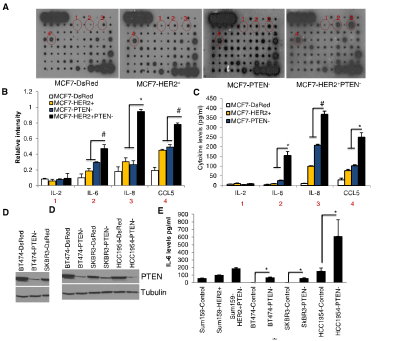
Figure 3. PTEN Downregulation in HER2-Overexpressing Cells Activates an IL6/NF-kB-Mediated Inflammatory Feedback Loop (A–E) MCF7-HER2+PTEN cells secreted 3- to 5-fold higher levels of IL6, IL8, and CCL5 compared to MCF7-HER2+ or MCF7-PTEN cells as determined by RayBio human cytokine antibody Array 5 (A). The intensity of each blot compared to control was determined by Kodak image analyzer (B) and confirmed by ELISA (C). Downregulation of PTEN in HER2-amplified breast cancer cells, BT474, SKBR3, HCC1954, and Sum159-HER2+ (D) results in increased levels of these cytokines in vitro (E). http://dx.doi.org/10.1016/j.molcel.2012.06.014

Array Capabilities:
  • Detects expression of 96 secreted proteins in a single sample, in a single day. 
  • If you can do a Western, you can use a C-Series Antibody Array. And get excellent results the first time!  
  • Sandwich ELISA pairs give C-Series arrays high sensitivity and specificity.  
  • C-Series arrays are compatible with practically ANY liquid sample. 
References have cited using membrane-based C-Series Cytokine Antibody Arrays with nearly every liquid sample type imaginable, including: cell-cultured and co cell cultured media, cell and tissue lysates, tissue/organ perfusates, serum, plasma, urine, and many other body fluids, including cyst, interstitial, synovial, blister, cerebrospinal, prostatic and amniotic fluids, as well as abscesses, broncho-alveolar lavage, sputum, saliva, tears, breath condensates, and even human milk and colostrum. Publications referencing use of our Antibody Arrays.

I will be posting more new developments here.

Tuesday, August 20, 2013

Proud Distributor of RayBiotech's Antibody Arrays and ELISAs

Pioneer and Market Leader

I am pleased to announce we are a distribution partner for RayBiotech. The solutions we offer include Antibody Arrays and ELISA Kits. With these additions, you now have many options for the either the qualitative, semi-quantitative or quantitative measurement of protein expression. From single antibody pair ELISAs to multiplexed arrays. The arrays enable you to simultaneously analyze up to 80 cytokine, inflammatory response, growth factor or angiogenesis related proteins.

In this posting, I focus on our new C-Series Antibody Array Kits. The kits utilize the sandwich immunoassay principle, wherein a panel of capture antibodies is printed on a nitrocellulose membrane solid support (usualy 2.5 cm x 3 cm). The array membranes are processed similarly to a Western blot (chemiluminescent readout). Signals are then visualized on x-ray film or a digital image, allowing densitometry data collection and calculation of fold-changes for each detected protein. The entire procedure can be completed in 1 day, and is simple enough to permit even the novice researcher to successfully collect data with very few pitfalls and little or no optimization.

They are proven and frequently published. You can now analyze the protein expression in you assays for a fraction of the cost of tradition methods. Here's an example: Simona Giorgini, Daniela Trisciuoglio, Chiara Gabellini, Marianna Desideri, Laura Castellini, Cristina Colarossi, Uwe Zangemeister-Wittke, Gabriella Zupi and Donatella Del Bufalo. Modulation of bcl-xL in Tumor Cells Regulates Angiogenesis through CXCL8 Expression. doi: 10.1158/1541-7786.MCR-07-0088. Mol Cancer Res August 2007 5; 7

Figure: bcl-xL overexpression in ADF glioblastoma and M14 melanoma cells increases CXCL8 expression. Image of membranes from a protein array. Membranes are probed with CM from the (A) glioblastoma control (AN8) and bcl-xL–overexpressing (AXL42 and AXL74) clones and (B) melanoma control (Mneo) and bcl-xL–overexpressing (MXL12 and MXL90) clones. In the negative control (NEG), the CM is replaced with an appropriate mock buffer according to array protocol. The intensity of protein signals for each membrane was compared with the relative positive signals by densitometric analysis. C. Schematic representation of proangiogenic factors that can be detected by the use of the membrane. The CXCL8, TGF-1β, TIMP1, TIMP2, and VEGF protein signals (two spots) are indicated by red, blue, yellow, violet, and green rectangles, respectively, in each image....The Human Angiogenesis Antibody Array I (RayBiotech. Inc.) was used according to the manufacturer's protocol to evaluate the secretion of 20 angiogenic factors into the CM of the different lines. A schematic representation of the proangiogenic factors that may be detected by the use of the array has been reported in the above figure. Membranes spotted in duplicate with antibodies against angiogenic factors were blocked with blocking buffer and then were incubated overnight with CM. Next, membranes were washed with wash buffer, incubated with biotin-conjugated antibodies against proangiogenic factors, washed with wash buffer, and incubated with horseradish peroxidase–conjugated streptavidin. The signals on the membranes were detected by chemiluminescence. Membranes, blocking and wash buffer, and antibodies against proangiogenic factors were all provided with the kit. The intensity of protein signal (two spots for each protein) was compared with the relative positive signals by densitometric analysis.

Here's an excellent video that provides more detail on Antibody Array Kit capabililities:
I will continue to post information on new addition with related pubs and data.