Showing posts with label NSE Antibody. Show all posts

Sunday, June 12, 2016

Neuroscience Cell Based Markers Applications

Progenitors, Neuronal, Astroglia and PNS Markers A to Z
Our Neuron/Synapse, Astrocytes, Glia, Microglia, Olidogodendrocytes, Progenitors and Schwann Cell Markers continue to widely used and frequently published.
Here're some recent examples: Cinzia Ambrosi , Cynthia Ren, Gaelle Spagnol, Gabriel Cavin, Angela Cone, Elena E. Grintsevich, Gina E. Sosinsky, Paul L. Sorgen. Connexin43 Forms Supramolecular Complexes through Non-Overlapping Binding Sites for Drebrin, Tubulin, and ZO-1. Published: June 9, 2016. http://dx.doi.org/10.1371/journal.pone.0157073...chicken anti-GFAP in blue (Neuromics, Catalog # CH22102)...
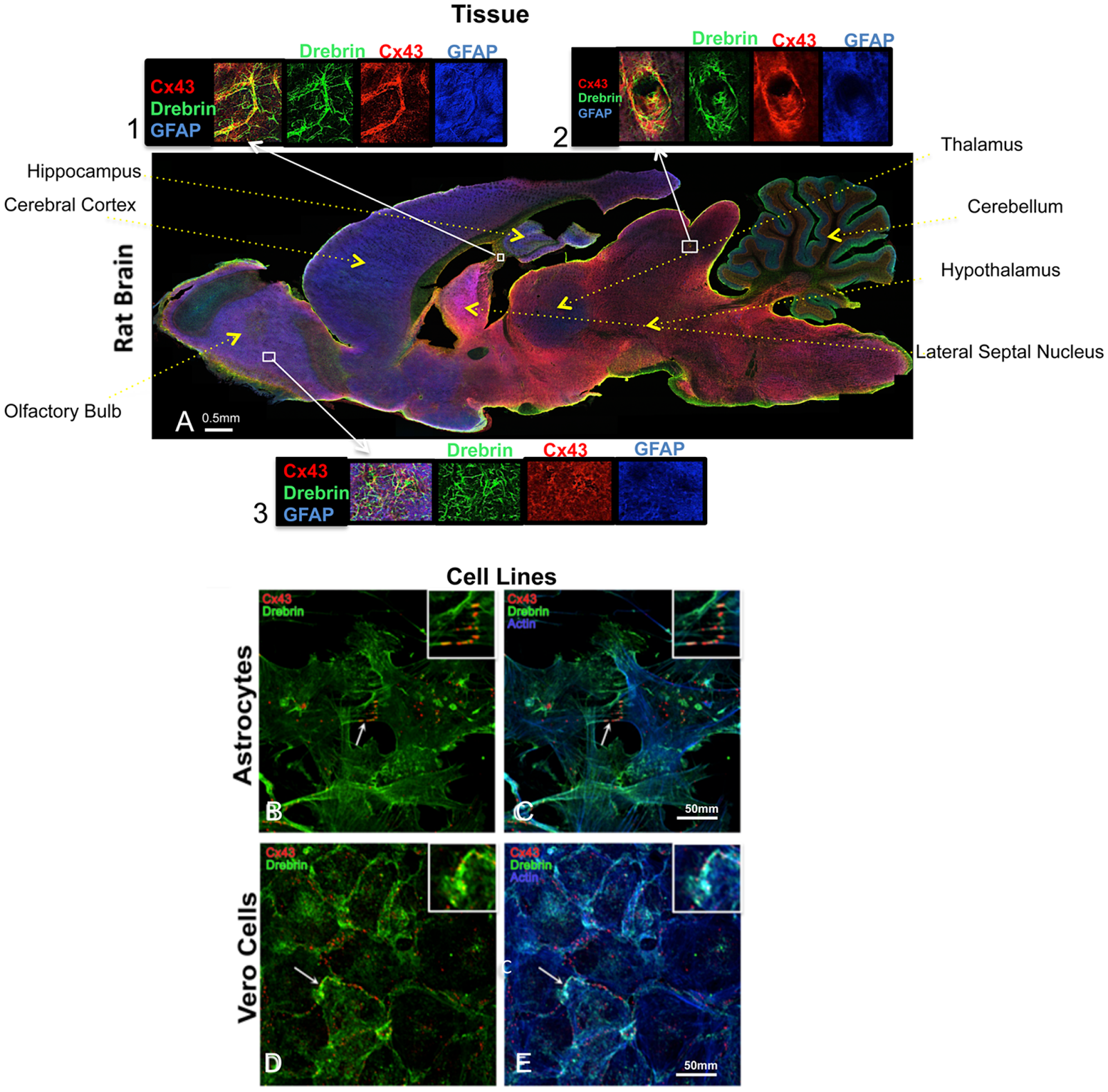
Figure 1. Cx43 and drebrin colocalization analysis in brain and cellular models. (A) Rat brain transversal slice mosaic shown after multiple immunolabeling with antibodies anti-Cx43 (red), anti-drebrin (green), and anti-GFAP (blue) as astrocytes marker. White boxes localize the area enlarged in insets 1, 2, and 3 (six fold enlargement). Colocalization of drebrin and Cx43 (yellow) is especially noticeable around the blood vessels (inset 2) and in regions rich of astrocytes (insets 1 and 3). The different regions of the brain were labeled. Cultured astrocytes (B and C) and Vero cells (D and E) were immunolabeled with anti-Cx43 (red), anti-drebrin (green), and anti-actin (blue). White arrows indicate zones of colocalization of Cx43, drebrin and actin that were enlarged in the insets (white boxes, three fold enlargement).

Xiangchen Li, Yu Guo, Yaxin Yao, Jinlian Hua, Yuehui Ma, Changqing Liu, Weijun Guan.Reversine Increases the Plasticity of Long-Term Cryopreserved Fibroblasts to Multipotent Progenitor Cells through Activation of Oct4. International Journal of Biological Sciences 2016; 12(1): 53-62. doi: 10.7150/ijbs.12199...anti-GFAP and NSE (1:200,Neuromics, MN, USA)...
Save 70 USD on High Titer Neuron/Synapse Markers-Only 225/100 ul (Through June 30, 2016).
We will continue to post news on our Neuroscience Research Solutions!

Thursday, December 17, 2015

Fibroblasts into Neurons

Via Activation of Oct4

We have a variety of stem cell, progenitor and neuron markers that are often referenced in publication. There represent a qualitative way to determine the state of stem cells as they differentiate.

Here're researchers take Fibroblasts and differentiate them into various progenitors. They were able to further differentiate these progenitors into astrocytes and neurons by: For neuronal differentiation, after 2 days of incubation with 20 nM reversine, cells were then treated with 0.5 μM all-trans-retinoic acid (RA) in serum-free DMEM/F-12 medium supplemented with the ITS for 2 days and switched into serum-free medium in the absence of RA with replacement of the medium every 2-3 days. 

Our NSE and GFAP were used to confirm this differentiation.
To learn more see: Xiangchen Li, Yu Guo, Yaxin Yao, Jinlian Hua, Yuehui Ma, Changqing Liu, Weijun Guan.Reversine Increases the Plasticity of Long-Term Cryopreserved Fibroblasts to Multipotent Progenitor Cells through Activation of Oct4. International Journal of Biological Sciences 2016; 12(1): 53-62. doi: 10.7150/ijbs.12199

Monday, October 19, 2009

NSE and TUJ-1 and Parkinson's Disease Research

I would like to thank Meghan Coakley from University College Cork for alerting me to a new publication referencing our Chicken NSE (Neuron-Specific Enolase) and Tuj 1 (Neuron-specific class III beta-tubulin) antibodies.

Here's her feedback: "Just letting you know we published our paper using the beta-III-Tubulin and NSE antibodies you supplied to us – Timmons et al., Neuroscience Letters, Oct 1, 2009 [Epub ahead of print]. The antibodies were excellent and I’m sure we’ll be using Neuromics again in the future."

Timmons S, Coakley MF, Moloney AM, O' Neill C. Akt signal transduction dysfunction in Parkinson's disease. Neurosci Lett. 2009 Oct 1. [Epub ahead of print].

Abstract: Significant attention has been drawn to the potential role of defective PI3-kinase-Akt (PKB) signalling in Parkinson's disease (PD) neurodegeneration and to the possibility that activation of Akt may provide neuroprotection in PD. However, little knowledge exists on the integrity of the Akt system in PD. Results of the present study show diminished levels of both total and active phospho(Ser473)-Akt in the brain in PD. This was evident by western blot analysis of midbrain fractions from PD compared to non-PD control brain, but more specifically by immunofluorescence microscopy of the substantia nigra pars compacta (SNpc). Here, double immunofluorescence microscopy found Akt and phospho(Ser473)-Akt to be expressed at high levels in tyrosine hydroxylase (TH) immunopositive dopaminergic neurons in control human brain. Selective loss of these neurons was accompanied by a marked decrease of Akt and phospho(Ser473)-Akt expression in the PD brain, however Akt and active phospho(Ser473)-Akt are still evident in degenerating dopaminergic neurons in the disease. This suggests that it may be possible to target neuronal Akt in advanced PD. Converse to the marked loss of neuronal Akt in PD, increased Akt and phospho(Ser473)-Akt levels were observed in small non-TH positive cells in PD SNpc, whose increased number and small nuclear size indicate they are glia. These findings implicate defective Akt as a putative signalling pathway linked to loss of dopaminergic neurons in PD.

Other Reagents to Consider:
Tuj 1 (Neuron-specific class III beta-tubulin)-Mouse Monoclonal
Neuron/Glial Marker Antibodies
Neurotrophins-Neuron/Glial Marker Proteins
Neurodegenerative Disease Research Antibodies
Neurodegenerative Disease Research Proteins
Stem Cell Research Reagents
