Our goal is to provide our customers and collaborators the tools they need to insure success. This is defined by having the specific Primary Neurons, Growth Factor plus the Markers to meet unique research needs.
The proof is in the results. Here are some highlights.
Images/Data: FIGURE 5. Microglial p38α MAPK-dependent TNFα is involved in LPS-induced neurite degeneration. (A) Photomicrographs of MAP-2 immunocytochemistry show the morphology of neurons after 72h of co-culture with microglia. The arrow points to the appearance of neurites that have been damaged by LPS-activated WT microglia. In contrast, the arrowhead points to the morphological appearance of healthy, undamaged neurites. (B) Diagram of the Sholl method for quantifying the total number of healthy neurites that intersect the concentric circles. (C) Quantification of healthy neurites by the Sholl analysis demonstrates that LPS stimulation of p38α WT microglia in co-culture causes neurite degeneration as seen by a significant reduction in the number of intersections by healthy neurites in the LPS-stimulated group compared to the unstimulated group (white bars). This degeneration can be attenuated by the addition of a blocking antibody to TNFα (5μg/ml), while the non-immune IgG control was not protective (gray bars). Microglia from p38α KO mice stimulated with LPS (black bar) also have significantly less neurite degeneration than the LPS-stimulated p38α WT microglia (white bar). However, by adding TNFα back to the p38α KO microglia co-culture, there is a significant decrease in the healthy neurite arborization compared to the p38α KO microglia stimulated with LPS alone (black bars). (***p<0.005; Bonferroni’s multiple comparison test). Data represents 2 independent experiments. Scale bar equals 25μm. Molecular Neurodegeneration 2011, 6:84 doi:10.1186/1750-1326-6-84
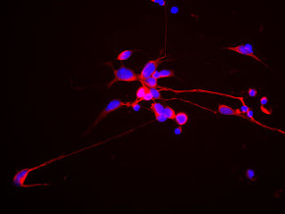
hN2 cells grown in culture for 4 days and stained with our chicken polyclonal to Neurofilament light or low molecular weight chain NF-L, a marker of neurons. Many of the differentiating cells show strong cytoplasmic and clearly fibrillar staining for NF-L. Blue stain is DAPI and reveals cell nuclei of some non neuronal cells in this culture.
We will continue to post relevant images and data that demonstrate our capabilities.
Scientists grow retina cells from skin-derived stem cells
-
WASHINGTON - University of Wisconsin-Madison researchers have successfully
grown multiple types of retina cells from two types of stem cells, giving
new ho...
16 years ago


No comments:
Post a Comment