Thursday, April 26, 2012

Primary Hippocampal Neurons Performing!

I advertise our Primary Neurons and Astrocytes as being easy to culture, grow and maintain. We confirm this via the data/images our customers generously share and the many publications referencing use of the cells.

I would like to thank George Kenneth Todd (Patterson Lab at UC Davis) for these wonderful images of our e18 Primary Rat Hippocampal Neurons. These were taken at day 67!


Prior to staining, the cells were treated with Wnt5 for 2 min, then Wnt5 + Wnt3 for an additional 2 min during Calcium Imaging experiments. The cells were then fixed and stained for IP3R (green), Frizzled2 (blue), and B-Catenin (red), and the confocal images were captured at 10x. Notice the parallel neurites formation in image 3.


For staining culture options, check out our neuron-glial-astrocyte markers.

GFAP Antibodies-Daily Double

I consider publications important for confirming our Neuronal-Glial Markers are working as advertised. These tools often used by our customers for lineages of differentiating Neural Progenitors. This publication references use of our Mouse Monoclonal GFAP and Rabbit Polyclonal GFAP Antibodies: Frances Y. Cheng, Xi Huang, Anuraag Sarangi, Tatiana Ketova, Michael K. Cooper, Ying Litingtung, Chin Chiang. Widespread Contribution of Gdf7 Lineage to Cerebellar Cell Types and Implications for Hedgehog-Driven Medulloblastoma Formation. PLoS ONE 7(4): e35541. doi:10.1371/journal.pone.0035541.
Abstract:The roof plate is a specialized embryonic midline tissue of the central nervous system that functions as a signaling center regulating dorsal neural patterning. In the developing hindbrain, roof plate cells express Gdf7 and previous genetic fate mapping studies showed that these cells contribute mostly to non-neural choroid plexus epithelium. We demonstrate here that constitutive activation of the Sonic hedgehog signaling pathway in the Gdf7 lineage invariably leads to medulloblastoma. Lineage tracing analysis reveals that Gdf7-lineage cells not only are a source of choroid plexus epithelial cells, but are also present in the cerebellar rhombic lip and contribute to a subset of cerebellar granule neuron precursors, the presumed cell-of-origin for Sonic hedgehog-driven medulloblastoma. We further show that Gdf7-lineage cells also contribute to multiple neuronal and glial cell types in the cerebellum, including glutamatergic granule neurons, unipolar brush cells, Purkinje neurons, GABAergic interneurons, Bergmann glial cells, and white matter astrocytes. These findings establish hindbrain roof plate as a novel source of diverse neural cell types in the cerebellum that is also susceptible to oncogenic transformation by deregulated Sonic hedgehog signaling.

Images: A subset of Gdf7-lineage cells express neural stem cell markers.(A–B″′) Many cells within the tumor tissue of Gdf7Cre/+;SmoM2 mice coexpress neural stem cell marker Nestin (red) and glial marker GFAP (green). Arrows indicate co-localization.


I will continue to post publication and customer data showing results with our Neuron-Glial Markers.

Wednesday, April 18, 2012

Slowing Hearing Loss in Diabetics and Hyperglycemics

Our Neuronal-Glial Markers are used in a variety of applications. Here our P0 or P-Zero antibody is used to show myelination levels in the cochlear ganglion. Demyelination is caused by hyperglycemia and type 2 diabetes and results in hearing loss and eventually deafness. Here's the related publication and highlights: Silvia Murillo-Cuesta, Guadalupe Camarero, Águeda González-Rodríguez, Lourdes Rodríguez-de la Rosa, Deborah J Burks, Carlos Avendaño,Ángela M Valverde and Isabel Varela-Nieto1. Insulin Receptor Substrate 2 (IRS2)-Deficient Mice Show Sensorineural Hearing Loss That Is Delayed by Concomitant Protein Tyrosine Phosphatase 1B (PTP1B) Loss of Function. Online address: http://www.molmed.org. doi: 10.2119/molmed.2011.00328
Highlights: The authors objective was to study the hearing function and cochlear morphology of Irs2-null mice and the impact of PTP1B deficiency. They have studied the auditory brainstem responses and the cochlear morphology of systemic Irs2–/–Ptpn1+/+, Irs2+/+Ptpn1–/– and Irs2–/–Ptpn1–/– mice at different postnatal ages. The results indicated that Irs2–/–Ptpn1+/+ mice present a profound congenital sensorineural deafness before the onset of diabetes and altered cochlear morphology with hypoinnervation of the cochlear ganglion and aberrant stria vascularis, compared with wild-type mice. Simultaneous PTP1B deficiency in Irs2–/–Ptpn1–/– mice delays the onset of deafness.


Images:  Cochlear ganglion and nerve fibers. (A–C) Cresyl violet staining of midmodiolar methacrylate sections of the cochlear ganglion at the cochlear basal turn in Irs2+/+Ptpn1+/+ (A), Irs2–/–Ptpn1+/+ (B) and Irs2–/–Ptpn1–/–mice (C) at postnatal wk 11. A slight reduction in the cellular density was more evident in both mutants (B, C) compared with wild-type (A). (D–F) Myelin P0 immunostaining of the cochlear ganglion shows less intense labeling in Irs2–/–Ptpn1+/+ (E) and Irs2–/–Ptpn1–/– (F) mice than in control mice (D). (G–I) Accordingly, myelin P0 immunostaining of nerve fibers projecting from the cochlear ganglion to the sensory cells is less intense in Irs2–/–Ptpn1+/+ and Irs2–/–Ptpn1–/– (arrows in H and I) than in wild-type mice (G). (J–L) Similarly, a weaker neurofilament 200-kDa immunostaining is observed in Irs2+/+Ptpn1–/– (K, white arrow) and Irs2–/–Ptpn1–/– (L, white arrow) mice compared with wild-type mice (J). Scale bars: A–I, 50 μm; J–L, 75 μm.

The results presented in this study demonstrate for the first time a unique tissue-specific role of IRS2 in cochlear development and hearing function; therefore, the Irs2–/– mouse could be a novel model for the in vivo study of hearing loss associated with altered glucose metabolism. The data also suggest that modulation of PTP1B activity could be a pharmacological target of interest for the sensory syndromes associated with diabetes.

Friday, April 13, 2012

Potent Mesenchymal Stem Cells and Media

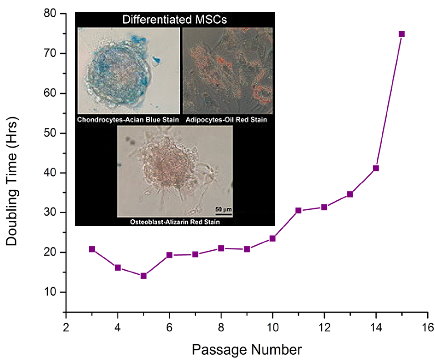
Researchers require a dependable and cost-effective source for Mesenchymal Stem Cells (hMSCs). These cells can be differentiated into cartilage, bone, fat, muscle and even neural cells using our MSCGro™ Media for growth and differentiation. Better cells and media means satisfaction with your Cell Based Assays
This makes them an ideal solution for research studies that include developmental biology, regenerative medicine, cell therapy, and tissue engineering.

These cells have the capabilities of passaging a minimum of 10-fold.

Cells and Media
Images: MSCs (Catalog #: SC00A1) Growth in MSCGroTM, Low Serum Medium (Catalog#: SC00B1). Inset: MSCs differentiatiated using MSCGro Differentiation Media.

I will continue to update you on customer data and pubs using these cells and media. I wish you exciting and rewarding discoveries.

Sunday, April 08, 2012

ApoTransferrin and the fate of Neural Stem Cell/Progenitors

Implications for De-Myelinating Diseases Like MS and ALS

Dr. Juana María Pasquini and her team at the University of Buenos Aires are ongoing users of our Neural Stem Cell-Progenitor (NSC-NP) Markers. In this study, they use these markers to determine the states and fates of NSCs and NPs as they proliferate and differentiate and the related role of ApoTransferrin (aTF). Here we learn aTf exposure during differentiating conditions favours OL maturation from OPCs by promoting OL morphological development. This evidence supports a key role of Tf on the generation of OL from NSC/NPCs and highlights its potential in demyelinating disorder treatment: Silvestroff L , Franco PG , Pasquini JM (2012) ApoTransferrin: Dual Role on Adult Subventricular Zone-Derived Neurospheres. PLoS ONE 7(3): e33937. doi:10.1371/journal.pone.0033937.

Proliferation rates under different conditions are shown in A–C. BrdU incorporation (red) during proliferation (CTLP, A) or differentiation (CTLPCTLD, B). BrdU+ cells are expressed as a percentage of total nuclei for either condition in C. Free floating NS during proliferation express Nestin (D, green) and GFAP (E, green). After dissociation, NS-derived cells continue to express Nestin (F, green). PDGFRα+ (G, green) and NG2+ cells (H, green). Few MBP+ (I, green) cells were found under proliferative conditions. A large proportion of BrdU incorporating cells (J, red) co-expressed with NG2 (J, green). Some BLBP+ cells (K, green) incorporated BrdU (K, red). After differentiation (L–O), MBP+ cells were found with a highly branched and complex morphology (L, green). Cells expressed GFAP (M, green), as well as the neuronal NF200 marker (N, green). BrdU incorporating (O, red) cells were mostly NG2+ (O, green) during differentiation conditions. BrdU+ cells co-expressing NG2, as a proportion of total BrdU+ cells, are shown in P for either culture condition. A representative Western Blot membrane in Q shows how MBP levels increase in whole cell protein extracts as cells differentiate. The densitometric analysis of the MBP isoforms/GAPDH ratio of 5 independent experiments was semi-quantitated in R. All 4 MBP isoforms were pooled and considered as a single value before normalizing to GAPDH values. Blue colour in images indicates Höechst nuclear dye. Scale bar in A represents 250 µm for A and B. Scale bar in D equals 100 µm in D–I and L–N, scale bar in J equals 250 µm in J and O, and scale bar in K represents 50 µm. Bars in P represent mean values of 2 independent experiments. Bars in C and R represent Mean + SD of 4 and 5 individual cultures, respectively. Student's t Test was used to analyze data in C, while a One Way ANOVA with an SNK Post-test was used to analyze data in R. * p<0.05, ** p<0.01, *** p<0.001

Note:  PDGFRα+ is a marker for oligodendrocytes (OLs).

Here's the pathway model that sumarizes authors' findings
I will keep you posted on research that could implications for the discovery of de-myelinating disorder therapies.

Wednesday, April 04, 2012

3-D Cell Based Assay Solutions

We want to help our customers, collaborators and friends bring their cell based assays to life! Neuromics began offering solutions in early 2011. We have extended our reach with our 3D Nanofibers. These 3-D environments are more biologically realistic leading to:
•more effective biomedical research.

•earlier breakthroughs.

•faster and cheaper time to market for drug development.

•improved stem cell expansion rates.

Here's a video showing green neurosphere (brain cancer cells)  migrating along our nanofibers aligned in the vertical direction. This enables cancer researchers to see cancer in ways never before possible!

Find more videos like this on TechLounge

I will update you as data and related publications become avaialable using these solutions.