Showing posts with label e-18 Primary Rat Cortical Neurons. Show all posts

Tuesday, January 31, 2017

Desperately Seeking Data

Answering the Bell
We continue to seek data using our cells. We offer a reward of 25 USD Starbucks' Gift Card.

We were pleased to receive a recently published study from Dr. Mahendran Subramanian of Keele University. In this study, researchers showed that oscillating nanomagnetic gene transfection could be used to successfully transfect SH‐SY5Y cells as well as our primary hippocampal and cortical neurons on different days in vitro. This novel technique was used to effectively deliver genetic material into various cell types, resulting in high transfection efficiency and viability. Mahendran Subramanian, Aimee‐Jayne Tyler, Eva Maria Luther, Elena Di Daniel, Jenson Lim and Jon Dobson. Oscillating Magnet Array−Based Nanomagnetic Gene Transfection: A Valuable Tool for Molecular Neurobiology Studies. Nanomaterials 2017, 7, 28; doi:10.3390/nano7020028...Primary rat hippocampal and cortical neurons were obtained from Neuromics (Edina, MN, USA) and disassociated using papain disassociation kit (Worthington, NJ, USA) according to the manufacturer’s instructions. Isolated neurons were maintained using neurobasal medium supplemented with 5% FBS, 0.5 mM Glutamax, 2% B27 supplement, 25 μM L‐glutamine and seeded onto poly‐D‐lysine–coated cells culture plates...
Figure 1. Oscillating magnet array−based nanomagnetic gene transfection experimental setup. (A) Representation of a 96‐well oscillating magnet array–based nanomagnetic transfection setup using NdFeB magnetic array (nanotherics); (B) Dimensions of the permanent magnets and magnetostatic (vectorpotential) algorithm based magnetic field density |B| distribution (T) contour plot for the NdFeB magnetic array.

Figure 2. Gene delivery by oscillating nanomagnetic gene transfection in primary cortical neurons. Images of pmaxGFP plasmid expressed in primary neurons using fluorescence microscopy and its corresponding Hoechst 33,342 stained counterpart of transfected DIV 1 (A,C) and DIV 5 (B,D) mature neurons were taken 48 h post transfection.

If you have data to share email it to me, pshuster@neuromics.com and we'll email you a 25 USD gift card. Thank you. Pete Shuster, CEO & Owner.

Wednesday, June 03, 2015

Neuromics' Neuronal Cultures in Action

Drug Discovery and Tox Assay Publications

Our Primary Human, Rat and Mouse Neurons are widely used and frequently cited in publications. Here's a pub hot of the presses referencing use of our e18 Rat Cortical Neurons to study the 
EPO-like cytoprotective effects cultures of these cells: James L. Miller, Timothy J. Church, Dmitri Leonoudakis, Karen Lariosa-Willingham, Normand L. Frigon, Connie S, Tettenborn, Jeffrey R. Spencer, and Juha Punnonen. Discovery and Characterization of Nonpeptidyl Agonists of The Tissue-Protective Erythropoietin Receptor. Molecular Pharmacology. May 27, 2015 mol.115.098400

...Cells were isolated from micro-surgically dissected embryonic day 18 rat cortices that were obtained from Neuromics (Edina, MN), and cultures were prepared according to the supplier's protocol.

Image: Neuromics' Cortical Neurons @ Day 6 in Culture.

I will new and unique applications for our neurons, astroglia and neural progenitors here. If you have questions, do not hesitate to contact me @ 612-801-1007 or pshuster@neuromics.com. Pete Shuster, CEO and Owner, Neuromics

Tuesday, August 06, 2013

Healthy and Happy Neuron/Astrocytes Cultures

A Track Record of Customer Success

Neuromics is  recognized for the quality of  hNP1™ Human Neural Progenitor,  hN2™ Neuron Discovery Kits, E18 and E20 Rat Primary Neurons and E18 Rat Primary Astroglia. As the company owner, it is important that I keep my finger on the pulse of how well they work for each and every unique application. I personally follow up with each user and if there are any issues, we replace the cells once free of charge. Your success is critical to our growth.

I wanted to share with you recent references. These give and excellent snapshot of the exciting ways our cells can used. Alexzander Asea, Punit Kaur, Alexander Panossian, Karl Georg Wikman, Evaluation of molecular chaperons Hsp72 and neuropeptide Y as characteristic markers of adaptogenic activity of plant extracts. Phytomedicine, Available online 6 August 2013, ISSN 0944-7113, http://dx.doi.org/10.1016/j.phymed.2013.07.001
...using trypan blue exclusion test and routinely found to contain less than ;5% dead cells. Primary human neurons were purchased from Neuromics (Edina, MN)...

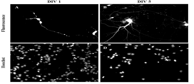
Images: Micropictograph of primary culture from micro-dissected hippocampus. (A) Neurons are round and healthy 1 h after plating on poly-d-lysine substrate. (B) Five days in culture, neurons remain healthy and have extended processes. Magnification 60×. http://dx.doi.org/10.1016/j.phymed.2013.07.001

Todd GK, Boosalis CA, Burzycki AA, Steinman MQ, Hester LD, et al. (2013) Towards Neuronal Organoids: A Method for Long-Term Culturing of High-Density Hippocampal Neurons. PLoS ONE 8(4): e58996. doi:10.1371/journal.pone.0058996
... a protocol that allows for culturing of E18 hippocampal neurons at high densities for more than 120 days. These cultured hippocampal neurons are (i) well differentiated with high numbers of synapses, (ii) anchored securely to their substrate, (iii) have high levels of functional connectivity, and (iv) form dense multi-layered cellular networks. We propose that our culture methodology is likely to be effective for multiple neuronal subtypes–particularly those that can be grown in Neurobasal/B27 media. This methodology presents new avenues for long-term functional studies in neurons...
Xiugong Gao, Hsiuling Lin, Radharaman Ray, Prabhati Ray. Toxicogenomic Studies of Human Neural Cells Following Exposure to Organophosphorus Chemical Warfare Nerve Agent VX. Neurochemical Research. February 2013.
...Human hN2 neurons were obtained from Neuromics...
Image: Staining of hN2 Human Neurons with Tuj 1 (Neuron-specific class III beta-tubulin) (red) and Nestin (green). Counter stained with DAPI (blue). hN2 Cells-Electro Phys Data. 

Xiufang Guo, Severo Spradling, Maria Stancescu, Stephen Lambert, James J. Hickman. Derivation of sensory neurons and neural crest stem cells from human neural progenitor hNP1. Biomaterials, In Press, Corrected Proof,Mar 2013.doi:10.1016/j.biomaterials.2013.02.061
...hNP1, were obtained from Neuromics (Edina, Minnesota)...
Wei Zhang , Radhia Benmohamed, Anthony C. Arvanites, Richard I. Morimoto, Robert J. Ferrante, Donald R. Kirsch, Richard B. Silverman. Cyclohexane 1,3-diones and their inhibition of mutant SOD1-dependent protein aggregation and toxicity in PC12 cells. Bioorganic & Medicinal Chemistry. Elsevier Ltd. All rights reserved.doi:10.1016/j.bmc.2011.11.039.
...Primary rat cortical tissue was purchased from Neuromics Inc., Edina, MN and used to initiate primary cortical neuron cultures. The tissue was isolated from micro-surgically dissected E18 embryonic Sprague/Dawley or Fischer 344 rat brain and shipped in a nutrient rich medium under refrigeration. To isolate neurons, the tissue was incubated with papain at a concentration of 2 mg/mL in Hibernate without calcium for 30 min at 37OC. The enzymatic solution was then removed, and 1 mL of culture media (Neurobasal, B27, 0.5 mM glutamine) was added. A sterile Pasteur pipette was used to gently disperse the cells, which were then washed, re-suspended and counted. The cells were plated on poly-D-lysine coated 96-well plates at a density of 20,000 cells/well and incubated at 37OC in a 5% CO2-humidified atmosphere for 5 days prior to use in compound testing. By microscopic inspection, the resulting cultures consisted of app. 90% neurons...
Majumder A, Dhara SK, Swetenburg R, Mithani M, Cao K, Medrzycki M, Fan Y, Stice SL. Inhibition of DNA methyltransferases and histone deacetylases induces astrocytic differentiation of neural progenitors. Stem Cell Res. 2013 Jul;11(1):574-86. doi: 10.1016/j.scr.2013.03.003. Epub 2013 Apr 2.
...Progenitor to Astrocytes Protocol: For astrocytic differentiation of hNP cells, neuronal differentiation media were supplemented with BMP2 (20 ng/mL) and combinations of Aza-C and TSA; Aza-C (500 nM), TSA (100 nM) and BMP2 (20 ng/mL) for 2 days, with one complete media change in between, followed by differentiation media supplemented with BMP2 but not with Aza-C or TSA. Cells were harvested prior to analysis at 5, 15 or 30 days of treatment or for cryopreservation at d6 or d10 of differentiation. For cryopreservation, cells were dissociated with Accutase™ and frozen in differentiation media containing10% DMSO. Viability was assessed at 30 days in Aza-C and TSA treated cultures by trypan blue exclusion, and datawas acquired using a Cellometer Auto T4® (Nexcelom Biosciences)...
Aparna Talekar, Antonello Pessi, and Matteo Porotto. Infection of primary neurons mediated by Nipah virus envelope proteins: Role of host target cells in antiviral action. J. Virol. doi:10.1128/JVI.00452-11.
...Hippocampus, Cortex and Ventricular Cells (Neuromics)...

I will continue posting results here.

Monday, September 17, 2012

Neuromics' Cortical Neurons & Kinetic NeuroTrack Assays

Providing tools that insure excellent Cell Based Assays is a cornerstone of our business strategy. Lauren McGillicuddy and her team at Essen Bioscience have been using our E18 Primary Rat Cortical Neurons to develop NeuroTrakTM assays enabling kinetic quantification of neurite dynamics (initiation, branching, extension, retraction). NeuroTrack is one of several CellPlayerTM assays that can be run in IncuCyte ZoomTM.

The proof is in the results and these show the both the potency of the cells and the powerful capablities of the IncuCyte ZOOM hardware and software:

Images:  Neurite outgrowth of rat E18 cortical neurons in a 96-well microplate at 24, 48 and 120 hours. top: 20x HD phase image of primary neurons  bottom: Image segmentation of neurites (light blue) and cell body cluster (raspberry).

Images: Neurite outgrowth of rat E18 cortical neurons in a 96-well microplate. Left: 20x HD phase image of primary neurons, 96 hours post plating ; Middle: Image segmentation of neurites (yellow) and cell body cluster (raspberry); Right: Concentration and time dependent inhibition of neurite outgrowth with the protein kinase C inhibitor, Ro-31-8220 (mean ± SD; n=6 per condition).

Check out this cool  assay animation!


I will continue to keep you posted on new assays and related methods using our primary neuron and astroglial cells.

Saturday, January 07, 2012

Primary Neurons vs PC12 cells for Compound Testing

This publication compares PC12 Cells vs E18 Primary Cortical Neurons. The cells showed permeability to some key compounds where the Neurons did not. This demonstrates the importance of including primary neurons in compound testing assays for Neuro-disease research: Wei Zhang , Radhia Benmohamed, Anthony C. Arvanites, Richard I. Morimoto, Robert J. Ferrante, Donald R. Kirsch, Richard B. Silverman. Cyclohexane 1,3-diones and their inhibition of mutant SOD1-dependent protein aggregation and toxicity in PC12 cells. Bioorganic & Medicinal Chemistry. Elsevier Ltd. All rights reserved.doi:10.1016/j.bmc.2011.11.039.
Abstract: Amyotrophic lateral sclerosis (ALS) is a fatal neurodegenerative disease characterized by the progressive loss of motor neurons. Currently, there is only one FDA-approved treatment for ALS (riluzole), and that drug only extends life, on average, by 2–3 months. Mutations in Cu/Zn superoxide dismutase (SOD1) are found in familial forms of the disease and have played an important role in the study of ALS pathophysiology. On the basis of their activity in a PC12-G93A-YFP high-throughput screening assay, several bioactive compounds have been identified and classified as cyclohexane-1,3-dione (CHD) derivatives. A concise and efficient synthetic route has been developed to provide diverse CHD analogs. The structural modification of the CHD scaffold led to the discovery of a more potent analog (26) with an EC50 of 700 nM having good pharmacokinetic properties, such as high solubility, low human and mouse metabolic potential, and relatively good plasma stability. It was also found to efficiently penetrate the blood–brain barrier. However, compound 26 did not exhibit any significant life span extension in the ALS mouse model. It was found that, although 26 was active in PC12 cells, it had poor activity in other cell types, including primary cortical neurons, indicating that it can penetrate into the brain, but is not active in neuronal cell potentially due to poor selective cell penetration. Further structural modification of the CHD scaffold was aimed at improving global cell activity as well as maintaining potency. Two new analogs (71 and 73)
were synthesized, which had significantly enhanced cortical neuronal cell permeability, as well as similar
potency to that of 26 in the PC12-G93A assay. These CHD analogs are being investigated further as novel
therapeutic candidates for ALS.
see: Bioorg. Med. Chem. 2011, 19, 613. and J. Med. Chem. 2012, in press

Related Links: Primary Neurons and Astrocytes-Primary human, rat and mouse neurons and astrocytes.

Image: E18 hippocampal neurons stained with Tau (red) and Doublecortin (green). The two proteins overlap in the proximal dendrites (yellow) Axons (low doublecortin content) are red. Blue staining is the nuclear DNA.


Thursday, September 15, 2011

Xona Microfluidics and Neurons

I am impressed with these Video from the Jeon Lab at UC Irvine. It represents a novel method for neuro-drug discovery:
Preparing E18 Cortical Rat Neurons for Compartmentalization in a Microfluidic Device.

This technology represents a way to separate axon from cell bodies.

Thursday, August 25, 2011

Primary Neurons and Cell Based Assays

The feedback I receive from Neuroscientists is consistent. To paraphrase, "gives us healthy, consistent and potent primary cells. I understand the hard work it takes to generate meaningful and publishable results from cell based assays. Our Primary Neurons and Astrocytes are merely inputs for these assays. The real cost is the time invested in culturing and time lost if they don't work.

I have numerous postings on success: Primary Neurons Postings. I wanted to share more data and feedback.

Primary DRGs-Culturing these can be tricky. I make it a point to work with labs to make sure the protocol options best match the desired outcome for assays. This includes replacing cells to make sure we can accurately troubleshoot. This approach insures I can pin point the issues and make sure they are all resolved in round two. Here's a representative testimonial: "Thanks for following up, the DRGs worked great and we were able to get excellent data from them. Thanks so much for working with us." Adam Ross, Dr. Chengji Zhou Lab, UC Davis

Image: DRGs cultured on Calf Skin Collagen.

Primary Hippocampal Neurons-I would like to thank Vimal Swarup, University of Utah for this excellent image.


The cells have been fixed after 48 hrs, they were grown over poly-lysine coated coverslips in the media supplied by Neuromics. Cells were imaged in phase contrast mode with 40x objective.

Put our primary cells to the test!


Monday, June 20, 2011

Transfection/Infection of Primary Neurons

Gene Expression Analysis of Neurons is an important tools in basic research and the study of neuropathologies. At the Neuromics' blog: "siRNA, DsiRNA and Plasmid Transfection Efficiency", I have posted many examples of successful tarnsfection of primary neurons and related cells using both our Transfection Kits/Reagents and others.

The other puzzle piece for these studies is having a fresh, pure and easy to use source of cells. Here, Neuromics has many options. These primary neurons and neural progenitors are widely referenced in key publications. Applications referenced include: transfection, pharmacology, electrophysiology, immunocytochemistry, and neuronal development studies.

This posting features infection of our e18 Primary Rat Combined Hippocampus, Cortex, and Ventricular Neurons using Nipah virus related components and HeV pseudotyped virions

Aparna Talekar, Antonello Pessi, and Matteo Porotto. Infection of primary neurons mediated by Nipah virus envelope proteins: Role of host target cells in antiviral action. J. Virol. doi:10.1128/JVI.00452-11.

Abstract: We have previously described heterotypic peptides from parainfluenza virus that potently inhibit Nipah virus in vitro, but are not efficacious in vivo. By contrast, our second-generation inhibitors, featuring a cholesterol moiety, are also efficacious in vivo. The difference between in vitro and in vivo results led us to investigate the basis for this discrepancy. Here we compare the activity of the compounds in standard laboratory cells and in cells relevant to the natural tropism of Nipah virus, i.e. primary neurons, and show that while our first generation inhibitors are poorly active in primary neurons, the cholesterol-conjugated compounds are highly potent. These results highlight the advantage of evaluating antiviral potency in cells relevant to natural host target tissue.

Customer Data: Transfection of functional HeV glycoproteins and infection with HeV pseudotyped virions.In order to establish the feasibility of carrying out the proposed experiments in primary neurons, we show (figure ) that our assays are amenable to use in primary neurons. In the experiment, Combined Hippocampus, Cortex, and Ventricular -E18 (Neuromics) were plated, and at 3 days were transfected with plasmids encoding HeV G/F as well as YFP. On the following day, these cells were infected with HeV or VSV pseudotyped viruses bearing RFP. In the figure, (A) the phase contrast photos show the differentiated neurons; (B) upon excitation for RFP, the red fluorescence indicates neurons infected by HeV pseudotyped virions; (C) upon excitation for YFP, and the green fluorescence shows the efficiency of transfection in neurons. This experiment indicates that the proposed experiments can be carried out in primary neurons, which are transfectable and infectable in our systems, and thus supports all the proposed aims. Data Courtesy of Dr. Matteo Porotto, Weill Cornell Medical College. Larger Image

We will continue to keep you updated.

Sunday, August 16, 2009

Link Between Inflammation and Tau Pathology in Alzheimer's Disease

Providing reagents for Alzheimer's Disease Researchers is a strategic focus area for us. This includes making a variety of Primary Neuronal Cultures available to these researchers.

We evolve the strategy based on Researcher input and how the cultures are being referenced in publications. This reference is especially satisfying because of the vangaurd findings:

Lisette T. Arnaud, Natura Myeku and Maria E. Figueiredo-Pereira. Proteasome–caspase–cathepsin sequence leading to tau pathology induced by prostaglandin J2 in neuronal cells. Journal of Neurochemistry. Volume 110 Issue 1, Pages 328 - 342.

"Our data suggest a potential sequence of events triggered by the neurotoxic product of inflammation PGJ2 leading to tau pathology. The accumulation of Ub proteins is an early response. If cells fail to overcome the toxic effects induced by PGJ2, including accumulation of Ub proteins, apoptosis kicks in triggering caspase activation and tau cleavage, the clearance of which by cathepsins could be compromised culminating in tau pathology. Our studies are the first to provide a mechanistic link between inflammation and tau pathology."

Featured Reagent:
E18 Primary Rat Cortical Neurons

Related Reagents:

Images: Neural phenotypes derived from hN2 cell lines. (A) Phase contrast image of differentiated culture. (B) Network including post-mitotic motoneurons (HB9). (C) Cholinergic neuron. (D) Tuj-1 positive cells that are DAT-positive (dopamine transporter; closed arrow) and DAT-negative (open arrow). (E) Gabaergic neurons, inset illustrates GABA in axon, but not the dendrites (arrow).
We will continue to post "whats new" in AD research and potential uses of our reagents.