Monday, March 26, 2012

Pain Research-Pubs Update

I am pleased to update you in recent publications and videos referencing use of our reagents for pain research. Check them out.
Andrew C. Eschenroeder, Allison A. Vestal-Laborde, Emilse S. Sanchez, Susan E. Robinson, Carmen Sato-Bigbee. Oligodendrocyte responses to buprenorphine uncover novel and opposing roles of μ-opioid- and nociceptin/orphanin FQ receptors in cell development: Implications for drug addiction treatment during pregnancy. Glia Volume 60, Issue 1, pages 125–136, January 2012....The MOR and NOP receptor antibodies were from Neuromics (Edina, MN) and used for immunocytochemistry (1:100)and western blotting (1:500)...
TRPV1s

FiFigure. Sensory axons, but not soma, from Type III Nrg1+/− mice show reduced capsaicin responsiveness compared to axons from WT mice. (A) Representative traces of intracellular calcium along sensory axons in response to 1 µM capsaicin or 56 mM KCl. The change in intracellular calcium from baseline over time ([(F−F0)/F0]*100) is shown for WT (left) and Type III Nrg1+/− (right) axons. Hatched diagonal lines indicate where the time course was non-continuous. (B) Quantification of the maximum change in intracellular calcium in response to application of 1 µM capsaicin or 56 mM KCl by genotype. Averages of 5 animals per genotype were compared using a Student's t-test. Type III Nrg1+/− axons showed a significantly decreased response to capsaicin (p<0.05), but not to KCl, relative to WTs. Graph shows mean±SEM. (C) Type III Nrg1+/− sensory soma show normal response to capsaicin. Quantification of maximal change in fluorescence from baseline ([(F−F0)/F0]*100) in WT or Type III Nrg1+/− sensory neuron soma in response to 1 µM capsaicin or 56 mM KCl. Average responses from 4 WT and 4 Type III Nrg1+/− animals to application of capsaicin or KCl were compared by genotype using a Student's t-test. There was no statistically significant difference between genotypes. Graphs show mean±SEM. (D) Type III Nrg1 (green) and TRPV1 (red) are co-expressed along P21 WT cultured sensory neuron axons identified with a pan-axonal (PA) marker (blue). White arrows indicate examples where Type III Nrg1 and TRPV1 are in close proximity. Scale bar equals 10 µm. (E) P21 WT and Type III Nrg1+/− sensory neuron cultures have equivalent levels of total TRPV1 protein. Total TRPV1 protein measurement by immunoblot. The 95 kD TRPV1 band and the 35 kD GAPDH band are shown from a representative experiment comparing protein from P21 WT and Type III Nrg1+/− cultures. Quantification of fold change in intensity of TRPV1:GAPDH normalized to WT average. There was no statistically significant change in the ratio of TRPV1 to GAPDH between genotypes (WT, Type III Nrg1+/−, n = 3 animals). Genotype comparisons were made using a Student's t-test. Graph shows mean±SEM. doi:10.1371/journal.pone.0025108.g004
In this study, researchers successfully transfect DRG cultures with IKAP-shRNA using our pn-Fect kit. The own regulation of IKAP in these cultures support findings that helped explain the potential pathology of Familial Dysautonomia (FD; Hereditary Sensory Autonomic Neuropathy; HSAN III): Hunnicutt BJ , Chaverra M , George L , Lefcort F , 2012 IKAP/Elp1 Is Required In Vivo for Neurogenesis and Neuronal Survival, but Not for Neural Crest Migration. PLoS ONE 7(2): e32050. doi:10.1371/journal.pone.0032050.

Related Links:
I will continue to post updates related to this important research area.

Sunday, March 18, 2012

Leptins and Obesity

Our Leptin and Leptin Receptor Markers have proven effective for researchers studying root causes of obesity. In this reference the authors show an interesting twist regarding the impact of endothial vs neuronal leptin signaling: Weihong Pan, Hung Hsuchou, Germaine G. Cornelissen-Guillaume, Bhavvani Jayaram, Yuping Wang, Hong Tu, Franz Halberg, Xiaojun Wu, Streamson C. Chua Jr., and Abba J. Kastin. Endothelial leptin receptor mutation provides partial resistance to diet-induced obesity. Published online before print February 2012, doi: 10.​1152/​japplphysiol.​00590.​2011.
...chicken anti-ObRb (1:100, Neuromics)...

Highlights: Leptin, a polypeptide hormone produced mainly by adipocytes, has diverse effects in both the brain and peripheral organs, including suppression of feeding. Other than mediating leptin transport across the blood-brain barrier, the role of the endothelial leptin receptor remains unclear. We recently generated a mutant mouse strain lacking endothelial leptin receptor signaling, and showed that there is an increased uptake of leptin by brain parenchyma after its delivery by in-situ brain perfusion. Here, we tested the hypothesis that endothelial leptin receptor mutation confers partial resistance to diet-induced obesity. These ELKO mice had similar body weight and percent fat as their wildtype littermates when fed with rodent chow, but blood concentrations of leptin were significantly elevated. In response to a high fat diet, wildtype mice had a greater gain of body weight and fat than ELKO mice . As shown by metabolic chamber measurement, the ELKO mice had higher oxygen consumption, carbon dioxide production, and heat dissipation, although food intake was similar to that of the wildtype mice and locomotor activity was even reduced. This indicates that the partial resistance to diet-induced obesity was mediated by higher metabolic activity in the ELKO mice. Since neuronal leptin receptor knockout mice show obesity and diabetes, the results suggest that endothelial leptin signaling shows opposite effects from that of neuronal leptin signaling, with a facilitatory role in diet-induced obesity.


Obesity is a growing health problem and will continue to drive up costs. Research like this could help find effective and log term solutions for helping with weight loss.

Thursday, March 08, 2012

P2X3 Receptors and Cool Science

Our P2X3 Receptor Antibodies are widely used and frequently published. This publication references use of our P2X3 Guinea Pig Antibody.

I like the "cool factor" in this study: Ji Z-G , Ito S , Honjoh T , Ohta H , Ishizuka T , et al. 2012 Light-evoked Somatosensory Perception of Transgenic Rats That Express Channelrhodopsin-2 in Dorsal Root Ganglion Cells. PLoS ONE 7(3): e32699. doi:10.1371/journal.pone.0032699.-"We have recently generated several transgenic lines of rat in which channelrhodopsin-2 (ChR2) transgene is driven by the Thy-1.2 promoter. In one of them, W-TChR2V4, some neurons were endowed with photosensitivity by the introduction of the ChR2 gene, coding an algal photoreceptor molecule. The DRG neurons expressing ChR2 were immunohistochemically identified using specific antibodies to the markers of mechanoreceptive or nociceptive neurons. Their peripheral nerve endings in the plantar skin as well as the central endings in the spinal cord were also examined. We identified that ChR2 is expressed in a certain population of large neurons in the DRG of W-TChR2V4. On the basis of their morphology and molecular markers, these neurons were classified as mechanoreceptive but not nociceptive. ChR2 was also distributed in their peripheral sensory nerve endings, some of which were closely associated with CK20-positive cells to form Merkel cell-neurite complexes or with S-100-positive cells to form structures like Meissner's corpuscles. These nerve endings are thus suggested to be involved in the sensing of touch. Each W-TChR2V4 rat showed a sensory-evoked behavior in response to blue LED flashes on the plantar skin. It is thus suggested that each rat acquired an unusual sensory modality of sensing blue light through the skin as touch-pressure. This light-evoked somatosensory perception should facilitate study of how the complex tactile sense emerges in the brain."

The researchers used Blue LED light to fire neurons involved somatosensory or tactile response!


Images. Distribution of ChR2V in the dorsal part of the spinal cord. A–C. Immunohistochemical localizationof ChR2V with the cell-type specific markers, NF200 (A), CGRP (B) or P2X3 (C). Scale bars indicate 40 µm.

Note that the receptors involved in Nociceptive Pain Sensing do not overlap with ChR2V. From this the authors conclude that ChR2V is involved in mechanoreception. This rat model should facilitate future study of how complex tactile perception, such as for texture, size and shape, is generated. We will keep you posted.

In the meantime check out our markers and antibodies for studying Neurotransmission and Synaptic Mechanisms.

Friday, February 24, 2012

TRPV1, Gut Inflammation and Pain

"Where there's smoke there's fire" or in this study, where there's inflammation there is pain: Robert P. Watson, Elliot Lilley, Moh Panesar, Gurdip Bhalay, Steven Langridge, Shin-Shay Tian, Conor McClenaghan, Anna Ropenga, Fanning Zeng, Mark S. Nash. Increased prokineticin 2 expression in gut inflammation: role in visceral pain and intestinal ion transport. Neurogastroenterology & Motility. Volume 24, Issue 1, pages 65–e12, January 2012....Formalin-fixed, wax-embedded tissues with no overt signs of pathology were purchased from Asterand (Detroit, MI, USA); informed consent had been sought and received from all donors. All tissues were used in accordance with the Human Tissue Act 2004 (UK)...guinea pig anti-TRPV1 antibody (GP14100 – Neuromics, Edina, MN, USA)...


Images: expression of prokineticin receptors in human and rat tissues. (A) Immunohistochemical analysis of PKR1 expression in the myenteric and submucosal ganglia of human stomach, ileum, and colon. Pkr1 (B) and Pkr2(C) expression in a range of human tissues determined using qRT-PCR. (D, E) Immunohistochemical of PKR1 distribution in rat DRG showing an absence of expression in large diameter, NF200 positive neurons (D), but co-expression with TRPV1 in presumptive nociceptive sensory neurons (E). (F) Distribution of Pkr2 mRNA by in situ hybridization in rat DRG. The IHC/ISH images shown are representative of the data obtained from n = 3 donors/animals.


Key Results Prok2 gene expression was up-regulated in biopsy samples from ulcerative colitis patients, and similar elevations were observed in rodent models of inflammatory colitis. Prokineticin receptor 1 (PKR1) was localized to the enteric neurons and extrinsic sensory neurons, whereas Pkr2 expression was restricted to sensory ganglia. In rats, PROK2-increased intracellular calcium levels in cultured enteric and dorsal root ganglia neurons, which was blocked by Compound 3. Moreover, PROK2 acting at prokineticin receptors stimulated intrinsic neuronally mediated ion transport in rat ileal mucosa. In vivo, Compound 3 reversed intracolonic mustard oil-induced referred allodynia and TNBS-induced visceral hypersensitivity, but not non-inflammatory, stress-induced visceral pain.

Check out our Pain and Inflammation Research Antibodies.

Wednesday, February 15, 2012

Guinea Pig P2X3 Antibody-It's Back!

Our Guinea Pig P2X3 Antibody has historically been one of our most popular Purinergic Receptor Antibodies. It has been frequently cited in publications.

We exhausted our supply of the antibody in the spring of 2011 and it took us many rounds with multiple partners and a degree of disappointing results. Well our efforts have finally yielded the positive testing results required to again provide users this important antibody. Here're related images from our internal testing:
P2X3 staining in rat Dorsal Root Ganglia.
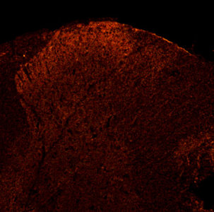
P2X3 in rat Dorsal Horn.
Check out our Purinergic Receptor Antibodies today.


Sunday, February 12, 2012

Building Muscle-TRIM32

What causes Muscular Dystrophies like Limb girdle muscular dystrophy type 2H (LGMD2H)? Here researchers demonstrate for the first time that TRIM32 is expressed in the skeletal muscle stem cell lineage of adult mice, and that in the absence of TRIM32, myogenic differentiation is disrupted. Moreover, they show that the ubiquitin ligase TRIM32 controls this process through the regulation of c-Myc, a similar mechanism to that previously observed in neural progenitors. Importantly, they show that loss of TRIM32 function induces a LGMD2H-like phenotype and strongly affects muscle regeneration in vivo. These studies implicate that the loss of TRIM32 results in dysfunctional muscle stem cells which could contribute to the development of LGMD2H: Nicklas S , Otto A , Wu X , Miller P , Stelzer S , et al. 2012 TRIM32 Regulates Skeletal Muscle Stem Cell Differentiation and Is Necessary for Normal Adult Muscle Regeneration. PLoS ONE 7(1): e30445. doi:10.1371/journal.pone.0030445.

Stem Cell Markers  like PAX7 are important for types of studies. PAX7 is a marker for muscle progenitors or stem cells. If these cells fail to differentiate the level of PAX7 expression will remain high. MyoD is an indicator for successfully differentiating cells. As a consequence, MyoD expression levels increase within the cell and the ratio of Pax7:MyoD is reduced, thus inhibiting proliferation and inducing differentiation through Myogenin expression. In the absence of TRIM32, this balance of Pax7:MyoD is dysregulated, because degradation of c-Myc is not initiated, MyoD levels do not increase and Myogenin expression is not activated.


Figure . Differentiation is strongly reduced in satellite cell progeny on TRIM32−/− myofibers. (a), (c) and (e) Immunostainings of satellite cells on freshly isolated myofibers from wild type (+/+) and TRIM32−/− (−/−) mice cultured for 24 h (a), 48 h (c) and 72 h (e) and labelled with the indicated markers (upper grey boxes). (b), (d) and (f) Diagrams showing the relative frequency of Pax7+/MyoD− cells on fibers cultured for 24 h (b), Pax7+/Myogenin− cells on fibers cultured for 48 h (d) and Pax7−/Myogenin+ cells on fibers cultured for 72 h (f). In all cases cells on myofibers from wild type mice and TRIM32−/− mice are compared. (mean ± std; *P<0.001 compared to wild type).


Check out our more publications referencing use of our PAX7.

Saturday, February 04, 2012

Neurotrophins-Growth Factors Update

Our Neurotrophins and Growth Factor Antibodies and Recombinant Proteins help support a wide span of research areas. These areas include: neuroscience, immunology, cardiac disease research and cancer.

I would like to update you on recent publications highlighting use of some of these reagents: Aiko Sada, Kazuteru Hasegawa, Pui Han Pin, Yumiko Saga. NANOS2 Acts Downstream of Glial Cell Line-Derived Neurotrophic Factor Signaling to Suppress Differentiation of Spermatogonial Stem Cells. DOI: 10.1002/stem.790. Copyright © 2011 AlphaMed Press...anti-GFRA1 (1:200, Neuromics, Edina, MN)...


Images: Ngn3-Cre targets GFRA1-negative cells. (A-F): At P7, Rosa-YFP; Ngn3-Cre double transgenic testes were immunostained with the indicated markers. Most of YFP-positive spermatogonia (Ngn3-lineage cells) were not stained with GFRA1. Scale bar, 100 μm. (G): Quantification of YFP-positive cells per GFRA1-positive cells at P7 and 6 weeks (N=3).

Karine Bédard, Stéphanie Segura, Stéphanie Mahaut, Catherine Tardivel, Guylaine Ferland, Bruno Lebrun, Pierrette Gaudreau. Effects of aging and caloric restriction on brainstem satiety center signals in rats. Mechanisms of Ageing and Development. dx.doi.org/10.1016/j.mad.2012.01.004...goat serum and 0.3% Triton-X-100) (Sigma–Aldrich), overnight at 4 °C with chicken anti-rat BDNF antibody (Neuromics, Edina, MN; diluted 1/200 in blocking buffer), rinsed 3× 10 min in fresh PBS and incubated for 2 h, in the dark...

Tali Kobilo, Qing-Rong Liu, Kriti Gandhi, Mohammed Mughal, Yavin Shaham and Henriette van Praag. Running is the neurogenic and neurotrophic stimulus in environmental enrichment. doi: 10.1101/lm.2283011. Learn. Mem. 2011. 18: 605-609... human recombinant BDNF (0.1 µg) monomer (Neuromics)...

Masamichi Shinoda, Masatake Asano, Daisuke Omagari, Kuniya Honda, Suzuro Hitomi, Ayano Katagiri, and Koichi Iwata. Nerve Growth Factor Contribution via Transient Receptor Potential Vanilloid 1 to Ectopic Orofacial Pain. The Journal of Neuroscience, 11 May 2011, 31(19):7145-7155; doi:10.1523/JNEUROSCI.0481-11.2011....expression immunohistochemically in TG- anti-p75 goat antibody (1:200; Neuromics)...


Images: Mandibular nerve fibers were labeled by β-NGF which was administrated into the lower lip with CFA (A) or saline (F). On day 1 after β-NGF administration into the lower lip with CFA or saline, β-NGF-positive whisker pad or lower lip TG neurons defined by FG or DiI, respectively. B, G, β-NGF-positive TG neurons. C, H, DiI-labeled TG neurons. D, I, FG-labeled TG neurons. E, J, DiI- and FG-labeled β-NGF-positive TG neurons. Mandibular nerve fibers (K) and TG neurons (L) on day 1 after labeled BSA administration into the lower lip with CFA. Open arrow, β-NGF-positive nerve fibers. Arrow, DiI-labeled β-NGF-positive TG neurons. Arrowhead, FG-labeled β-NGF-positive TG neurons. Scale bar, 50 μm. M, Frequency of β-NGF-positive neurons in FG- or DiI-labeled TG neurons after CFA or saline injection into the lower lip (n = 5 in CFA-injected group; n = 4 in saline-injected group; Student's t test).

F. R. Carreño, J. D. Walch, M. Dutta, T. P. Nedungadi, J. T. Cunningham. Brain-Derived Neurotrophic Factor-Tyrosine Kinase B Pathway Mediates NMDA Receptor NR2B Subunit Phosphorylation in the Supraoptic Nuclei Following Progressive Dehydration. Journal of Neuroendocrinology. Volume 23, Issue 10, pages 894–905, October 2011. DOI: 10.1111/j.1365-2826.2011.02209.x...TrkB receptor (Neuromics, Edina, MN, USA)...

Jie Shen, Yoko Ishii, Guihua Xu, Thanh Chung Dang, Takeru Hamashima, Takako Matsushima, Seiji Yamamoto, Yuichi Hattori, Yusuke Takatsuru, Junichi Nabekura and Masakiyo Sasahara. PDGFR-β as a positive regulator of tissue repair in a mouse model of focal cerebral ischemia. Journal of Cerebral Blood Flow & Metabolism , (28 September 2011). doi:10.1038/jcbfm.2011.136...goat polyclonal anti-PDGFR-β antibody (1:100; Neuromics, Edina)...

Thomas Karsten Kilvaer, Andrej Valkov, Sveinung W. Sorbye, Tom Donnem, Eivind Smeland, Roy Martin Bremnes,and Lill-Tove Busund. Platelet-Derived Growth Factors in Non-GIST Soft-Tissue Sarcomas Identify a Subgroup of Patients with Wide Resection Margins and Poor Disease-Specific Survival. Volume 2010 (2010), Article ID 751304, 10 pages doi:10.1155/2010/751304...PDGF-CC (goat polyclonal; GT15151; Neuromics; 1 : 80)...

I will keep you posted on new developments.

Friday, February 03, 2012

Aging-Caloric Restriction and BDNF-Leptin

As we age, selective BDNF receptors increase in the the dorsal vagal complex (DVC), the brainstem center mediating the satiety reflex. Aging also affects the suppressor of cytokine signaling-3. This results in abnormal responses to BDNF and Leptin Signaling.

In this study, the investigators found that by restricting calories in rats, DVC BDNF immunoreactive concentrations were elevated and resulting in satiety threshold stability.  This indicates functional desensitization of the DVC to these signals: Karine Bédard, Stéphanie Segura, Stéphanie Mahaut, Catherine Tardivel, Guylaine Ferland, Bruno Lebrun, Pierrette Gaudreau. Effects of aging and caloric restriction on brainstem satiety center signals in rats. Mechanisms of Ageing and Development. dx.doi.org/10.1016/j.mad.2012.01.004.

The authors used our BDNF Antibody to determine expression in the DVC.....goat serum and 0.3% Triton-X-100) (Sigma–Aldrich), overnight at 4 °C with chicken anti-rat BDNF antibody (Neuromics, Edina, MN; diluted 1/200 in blocking buffer), rinsed 3× 10 min in fresh PBS and incubated for 2 h, in the dark...

These findings could lead to potential therapies based on modulating BDNF expression in the DVC.

Thursday, January 26, 2012

Lineage Selection-Neural Stem Cells for SC Grafts

Our Neural Progenitor Markers keep moving up in the "hit parade". These markers are important for lineage selection. This selection is essential to circumvent the possibility of tumor formation and facilitate the safe translation of ES-based therapies to humans.

Here's a recent pub referencing use of several of our markers for selecting or confirming lineage: J. Simon Lunn, Crystal Pacut, Emily Stern, Stacey A. Sakowski, J. Matthew Velkey, Sue O'Shea, Eva Feldman. Intraspinal transplantation of neurogenin-expressing stem cells generates spinal cord neural progenitors. dx.doi.org/10.1016/j.nbd.2011.12.044...Tuj1 (Neuromics, 1:1000), Nestin (Neuromics, 1:500)...
Highlights: Expression of appropriate transcription factors is one approach to direct the differentiation of ES cells towards a specific lineage and stop proliferation. Neural differentiation can be initiated in ES cells by expression of Neurogenin1 (Ngn1). In this study we investigate the effects of controlled Ngn1 expression on mouse ES (mES) cell differentiation in vitro and following grafting into the rat spinal cord. In vitro, Ngn1 expression in mES cells leads to rapid and specific neural differentiation, and a concurrent decrease in proliferation. Similarly transplantation of Ngn1-expressing mES cells into the spinal cord lead to in situ differentiation and spinal precursor formation. These data demonstrate that Ngn1 expression in mES cells is sufficient promote neural differentiation and inhibit proliferation, thus establishing an approach to safely graft ES cells into the spinal cord.

Image: Neural progenitors were labeled with anti-rat Nestin polyclonal antibody (Cat#:GT15114) and stained with conjugated donkey anti-goat secondary anti-body (green). Differentiated neurons were labeled with neuron-specific mouse anti-β-III tubulin/ Tuj1-(Cat#MO15013 monoclonal antibody (red). Nuclei were stained with DAPI (blue).

I will continue to post updates on the application of Neuromics' Stem Cell Markers

Sunday, January 22, 2012

Early Diagnosis of Diabetic Retinopathy

The earlier the diagnosis the better the outcome. This is especially true with autoimmune diseases like Diabetic Retinopathy (DR). DR is the leading cause of blindness among persons of working age in the industrialized world. Here I feature a publication that shows axoglial alterations at the distal portion of the optic nerve could be the first structural change in the diabetic visual pathway. This could prove good news for discovering better therapies thus preventing blindness: Diego C. Fernandez, Laura A. Pasquini, Damián Dorfman, Hernán J. Aldana Marcos, Ruth E. Rosenstein. Early Distal Axonopathy of the Visual Pathway in Experimental Diabetes. doi:10.1016/j.ajpath.2011.09.018

Oligodendrocytes are responsible for insulating axons. Disruptions in the formation of oligodendrocytes could initiate the domino effect that leads to decreasing and eventual total loss of vision. The authors, for example, discovered that in diabetic rats, oligodendrocyte lineage (OL) cells showed hypertrophic somas and a high number of processes.


Images/Data: OL linage evaluation. Immature OL (O1+ cells) and OL precursor (PDGFR-α+ cells) were evaluated by immunostaining and measured as optical density (OD) per section. In the distal ON from animals that were diabetic for 6 weeks, significantly increased O1 and PDGFR-α immunostaining was observed, with the presence of disorganized and hypertrophic cells. Data are mean ± SEM (n = 5 animals per group); *P < 0.01 versus age-matched controls, by Student′s t-test. Scale bar = 50 μm.

At the ultrastructural level, alterations and loss of larger axons were observed in the distal ON from animals that were diabetic for 6 weeks. In these fibers, myelin was highly disorganized, and frequent lamellar membranous bodies were observed.

I will track new develops in this research and post relevant results here.

Monday, January 16, 2012

Primary Neuron Assays for Studying Neurodegeneration

Our goal is to provide our customers and collaborators the tools they need to insure success. This is defined by having the specific Primary Neurons, Growth Factor plus the Markers to meet unique research needs.

The proof is in the results. Here are some highlights.
Images/Data: FIGURE 5. Microglial p38α MAPK-dependent TNFα is involved in LPS-induced neurite degeneration. (A) Photomicrographs of MAP-2 immunocytochemistry show the morphology of neurons after 72h of co-culture with microglia. The arrow points to the appearance of neurites that have been damaged by LPS-activated WT microglia. In contrast, the arrowhead points to the morphological appearance of healthy, undamaged neurites. (B) Diagram of the Sholl method for quantifying the total number of healthy neurites that intersect the concentric circles. (C) Quantification of healthy neurites by the Sholl analysis demonstrates that LPS stimulation of p38α WT microglia in co-culture causes neurite degeneration as seen by a significant reduction in the number of intersections by healthy neurites in the LPS-stimulated group compared to the unstimulated group (white bars). This degeneration can be attenuated by the addition of a blocking antibody to TNFα (5μg/ml), while the non-immune IgG control was not protective (gray bars). Microglia from p38α KO mice stimulated with LPS (black bar) also have significantly less neurite degeneration than the LPS-stimulated p38α WT microglia (white bar). However, by adding TNFα back to the p38α KO microglia co-culture, there is a significant decrease in the healthy neurite arborization compared to the p38α KO microglia stimulated with LPS alone (black bars). (***p<0.005; Bonferroni’s multiple comparison test). Data represents 2 independent experiments. Scale bar equals 25μm. Molecular Neurodegeneration 2011, 6:84 doi:10.1186/1750-1326-6-84
hN2 cells grown in culture for 4 days and stained with our chicken polyclonal to Neurofilament light or low molecular weight chain NF-L, a marker of neurons. Many of the differentiating cells show strong cytoplasmic and clearly fibrillar staining for NF-L. Blue stain is DAPI and reveals cell nuclei of some non neuronal cells in this culture.

We will continue to post relevant images and data that demonstrate our capabilities.

Friday, January 13, 2012

TRPV1s in Action

Our TRPV1s continue to be widely used and published. This recent publication features use out TRPV1-C guinea pig polyclonal for immunohistochemistry and TRPV1-mouse specific for Western Blotting: Sarah E. Canetta, Edlira Luca, Elyse Pertot, Lorna W. Role, David A. Talmage. Type III Nrg1 Back Signaling Enhances Functional TRPV1 along Sensory Axons Contributing to Basal and Inflammatory Thermal Pain Sensation. PLoS ONE 6(9): e25108. doi:10.1371/journal.pone.0025108...IHC: TRPV1 (guinea pig, 1:1000, GP14100 Neuromics); WB: TRPV1 (rabbit, 1:1000, RA14113 Neuromics).


Figure. Sensory axons, but not soma, from Type III Nrg1+/− mice show reduced capsaicin responsiveness compared to axons from WT mice. (A) Representative traces of intracellular calcium along sensory axons in response to 1 µM capsaicin or 56 mM KCl. The change in intracellular calcium from baseline over time ([(F−F0)/F0]*100) is shown for WT (left) and Type III Nrg1+/− (right) axons. Hatched diagonal lines indicate where the time course was non-continuous. (B) Quantification of the maximum change in intracellular calcium in response to application of 1 µM capsaicin or 56 mM KCl by genotype. Averages of 5 animals per genotype were compared using a Student's t-test. Type III Nrg1+/− axons showed a significantly decreased response to capsaicin (p<0.05), but not to KCl, relative to WTs. Graph shows mean±SEM. (C) Type III Nrg1+/− sensory soma show normal response to capsaicin. Quantification of maximal change in fluorescence from baseline ([(F−F0)/F0]*100) in WT or Type III Nrg1+/− sensory neuron soma in response to 1 µM capsaicin or 56 mM KCl. Average responses from 4 WT and 4 Type III Nrg1+/− animals to application of capsaicin or KCl were compared by genotype using a Student's t-test. There was no statistically significant difference between genotypes. Graphs show mean±SEM. (D) Type III Nrg1 (green) and TRPV1 (red) are co-expressed along P21 WT cultured sensory neuron axons identified with a pan-axonal (PA) marker (blue). White arrows indicate examples where Type III Nrg1 and TRPV1 are in close proximity. Scale bar equals 10 µm. (E) P21 WT and Type III Nrg1+/− sensory neuron cultures have equivalent levels of total TRPV1 protein. Total TRPV1 protein measurement by immunoblot. The 95 kD TRPV1 band and the 35 kD GAPDH band are shown from a representative experiment comparing protein from P21 WT and Type III Nrg1+/− cultures. Quantification of fold change in intensity of TRPV1:GAPDH normalized to WT average. There was no statistically significant change in the ratio of TRPV1 to GAPDH between genotypes (WT, Type III Nrg1+/−, n = 3 animals). Genotype comparisons were made using a Student's t-test. Graph shows mean±SEM. doi:10.1371/journal.pone.0025108.g004
All TRPV1 Publications.

Saturday, January 07, 2012

Primary Neurons vs PC12 cells for Compound Testing

This publication compares PC12 Cells vs E18 Primary Cortical Neurons. The cells showed permeability to some key compounds where the Neurons did not. This demonstrates the importance of including primary neurons in compound testing assays for Neuro-disease research: Wei Zhang , Radhia Benmohamed, Anthony C. Arvanites, Richard I. Morimoto, Robert J. Ferrante, Donald R. Kirsch, Richard B. Silverman. Cyclohexane 1,3-diones and their inhibition of mutant SOD1-dependent protein aggregation and toxicity in PC12 cells. Bioorganic & Medicinal Chemistry. Elsevier Ltd. All rights reserved.doi:10.1016/j.bmc.2011.11.039.
Abstract: Amyotrophic lateral sclerosis (ALS) is a fatal neurodegenerative disease characterized by the progressive loss of motor neurons. Currently, there is only one FDA-approved treatment for ALS (riluzole), and that drug only extends life, on average, by 2–3 months. Mutations in Cu/Zn superoxide dismutase (SOD1) are found in familial forms of the disease and have played an important role in the study of ALS pathophysiology. On the basis of their activity in a PC12-G93A-YFP high-throughput screening assay, several bioactive compounds have been identified and classified as cyclohexane-1,3-dione (CHD) derivatives. A concise and efficient synthetic route has been developed to provide diverse CHD analogs. The structural modification of the CHD scaffold led to the discovery of a more potent analog (26) with an EC50 of 700 nM having good pharmacokinetic properties, such as high solubility, low human and mouse metabolic potential, and relatively good plasma stability. It was also found to efficiently penetrate the blood–brain barrier. However, compound 26 did not exhibit any significant life span extension in the ALS mouse model. It was found that, although 26 was active in PC12 cells, it had poor activity in other cell types, including primary cortical neurons, indicating that it can penetrate into the brain, but is not active in neuronal cell potentially due to poor selective cell penetration. Further structural modification of the CHD scaffold was aimed at improving global cell activity as well as maintaining potency. Two new analogs (71 and 73)
were synthesized, which had significantly enhanced cortical neuronal cell permeability, as well as similar
potency to that of 26 in the PC12-G93A assay. These CHD analogs are being investigated further as novel
therapeutic candidates for ALS.
see: Bioorg. Med. Chem. 2011, 19, 613. and J. Med. Chem. 2012, in press

Related Links: Primary Neurons and Astrocytes-Primary human, rat and mouse neurons and astrocytes.

Image: E18 hippocampal neurons stained with Tau (red) and Doublecortin (green). The two proteins overlap in the proximal dendrites (yellow) Axons (low doublecortin content) are red. Blue staining is the nuclear DNA.


Friday, December 23, 2011

Salivary Neuropeptides Y2s and Satiety

A possible new slant for combating obesity

The researchers in this study acheived a sustained increased PYY3-36 (a neuropeptide) expression via viral vector-mediated gene delivery targeting salivary glands and the good news: this increase resulted in a significant long-term reduction in food intake (FI) and body weight (BW). This is evidence for new functions of the previously characterized gut peptide PYY3-36 suggesting a potential simple and efficient alternative therapeutic approach for the treatment of obesity: Andres Acosta1, Maria D. Hurtado1, Oleg Gorbatyuk, Michael La Sala, David Duncan, George Aslanidi, Martha Campbell-Thompson, Lei Zhang, Herbert Herzog, Antonis Voutetakis, Bruce J. Baum, Sergei Zolotukhin. Salivary PYY: A Putative Bypass to Satiety. PLoS ONE 6(10): e26137. doi:10.1371/journal.pone.0026137. Received: July 22, 2011; Accepted: September 20, 2011; Published: October 10, 2011...Rabbit anti-Y2R (Dilution: 1:3000) using TSA...

Images: A) Immunolocalization of Y2R-positive cells in the hippocampus of C57Bl/6J mouse (WT), a (+) control. (B) Immunolocalization of Y2R in the tongue epithelia of Y2R KO mouse, a (-) control. VEG – von Ebner's gland. (C) Immunolocalization of Y2R-positive cells in the CV area of the tongue of a C57Bl/6J mouse. (D) close-up of (C). (E), and (F) close ups of (D), top and bottom rectangles, respectively. doi:10.1371/journal.pone.0026137.g003

These findings could prove a first step in identifying new targets for obesity fighting therapies. Increasing PYY salivary output would provide satiety with less food intake. This would give a whole new perspective on dieting. If we are satsified with less food intake is this really dieting? Stay tuned.

Saturday, December 03, 2011

Opioid Addiction During Pregnancy-Implications for Neuro-development

I would like to thank Dr. Carmen Sato-Bigbee, Virginia Commonwealth University School of Medicine, for kindly sharing this important study. The publication also references use of our Mu Opioid and Nociceptin/Orphanin FQ Receptor Antibodies: Andrew C. Eschenroeder, Allison A. Vestal-Laborde, Emilse S. Sanchez, Susan E. Robinson, Carmen Sato-Bigbee. Oligodendrocyte responses to buprenorphine uncover novel and opposing roles of μ-opioid- and nociceptin/orphanin FQ receptors in cell development: Implications for drug addiction treatment during pregnancy. Glia Volume 60, Issue 1, pages 125–136, January 2012.

Highlights: Oligodendrocytes are responsible for making myelin in the CNS. The authors have shown We have shown previously that rat brain myelination is significantly altered by buprenorphine, an opioid analogue currently used in clinical trials for managing pregnant opioid addicts. In this study, perinatal exposure to low levels of this drug induced accelerated and increased expression of myelin basic proteins (MBPs), cellular and myelin components that are markers of mature oligodendrocytes. In contrast, supra-therapeutic drug doses delayed MBP brain expression and resulted in a decreased number of myelinated axons. We have now found that this biphasic-dose response to buprenorphine can be attributed to the participation of both the l-opioid receptor (MOR) and the nociceptin/orphanin FQ receptor (NOP receptor) in the oligodendrocytes. This is the first study showing the potential role of the NOP receptor in myelination.
High levels of opiate exposure could negatively disrupt the normal interplay between these two systems altering the developmental pattern of brain myelination. Understanding this pathway, could help researchers find ways to favorably modulate myelination and protect neuro-development of fetuses exposed to high levels of opiates during pregnancy.

Related Data:
Direct treatment of immature oligodendrocytes with buprenorphine alters MBP expression in a dose-specific manner. Cells isolated from 9-day-old rat brains were incubated for 4 days in CDM with or without 0.25, 0.5, 1.0, and 3.0 lM buprenorphine. MBP levels were determined by western blotting using b-actin levels as loading controls. Figures correspond to representative experiments. Results in the bar graph are expressed as percentage of controls (0 lM buprenorphine) 6 SEM from five experiments and correspond to the combined scanning of the four major MBP isoforms. **P <0.005 and ***P<0.0001.
Pre-oligodendrocytes express both MOR and the NOP receptor. Cells isolated from 9-day-old rat brain were allowed to fully attachon the culture plates by overnight incubation and stained by double
immunocytochemistry with O4 (green) together with anti-MOR or anti-NOP receptor antibodies (red). Scale bar: 20 lm. The western blot shows MOR and NOP receptor expression in two different samples of developing oligodendrocytes directly isolated from 9-day-old rat brains.

I will be posting future studies investigating the molecular mechanisms by which buprenorphine and methadone affect myelination and neuron-glial interactions. These should provide deeper understanding into these developmental processes and new and better strategies for the managing of both pregnant addicts and drug addiction in adolescence.

Friday, November 25, 2011

IF Staining of Human Primary Neurons

Primary Neurons are inputs or raw materials for cell based assays. When cells do not work as promised, there are multiple costs including lost time and potentially flawed data. Neuromics strives to provide easy to culture, potent and cost effective cells. Proving these capabilities is an ongoing activity for us. This includes testing these cells using our markers.

I wanted to share new immunofluorescence images. Here is a link to the protocol: staining primary neurons.

hN2 cells stained with our chicken polyclonal antibody to Vimentin, in red. Islands of Hn2 cells form after 4 days in culture forming beautiful flower like structures. Vimentin is a well established marker of early differentiating neuronal lineage cells. Taken with a 10X objective lens. Blue staining is the nuclear DNA.
hN2 cells grown in culture for 4 days and stained with our chicken polyclonal to MAP2, a marker of neurons. Differentiating cells show strong cytoplasmic staining for MAP2 . Blue stain is DAPI and reveals cell nuclei of some non neuronal cells in this culture.
hN2 cells grown in culture for 4 days and stained with our chicken polyclonal to Neurofilament light or low molecular weight chain NF-L, a marker of neurons. Many of the differentiating cells show strong cytoplasmic and clearly fibrillar staining for NF-L. Blue stain is DAPI and reveals cell nuclei of some non neuronal cells in this culture.

Sunday, November 20, 2011

Fear Changes Hippocampus Neuropns

Fear in mice catalyzes rapid accumalation of EphrinB2 pyramidal neurons of the CA1 area.

This study suggests that rapid accumulation of EphrinB2 in hippocampal CA1 neurons is involved in the behavioural and cellular modifications induced by contextual fear conditioning. A similar mechanism does not appear to occur in lateral amygdala neurons, in spite of the robust behavioural and cellular modifications induced in such structure by cued fear conditioning: Antonio Trabalzaa, Sandra Colazingaria, Carmelo Sgobiob, Arturo Bevilacqua. Contextual learning increases dendrite complexity and EphrinB2 levels in hippocampal mouse neurons. Behavioural Brain Research. doi:10.1016/j.bbr.2011.11.008.

Friday, November 11, 2011

Diabetic retinopathy blindness-root causes

Diabetic retinopathy is a leading cause of acquired blindness. This publication from our friends at University of Buenos Aires touches on potential root causes: Diego C. Fernandez, Laura A. Pasquini, Damián Dorfman, Hernán J. Aldana Marcos, Ruth E. Rosenstein. Early Distal Axonopathy of the Visual Pathway in Experimental Diabetes. doi:10.1016/j.ajpath.2011.09.018
" In animals that had been diabetic for 6 weeks, a large increase in astrocyte reactivity occurred in the distal (but not the intraorbital) portion, which coincided with significant axon loss. Moreover, profound myelin alterations and altered morphologic features of oligodendrocyte lineage were observed at the distal (but not the proximal) optic nerve portion. The present results suggest that axoglial alterations at the distal portion of the optic nerve could be the first structural change in the diabetic visual pathway."
The authors used our PDGFR Alpha/CD140A Marker to Study the change in Oligodendrocyte Lineage precursor cells. Expression of the protein was increased in these cells with the presence of disorganized and hypertrophic cells. This could disrupt formation of myelin resulting the pathological alteration at the distal portion.

Tuesday, November 08, 2011

Opioid Induced Itch

Our widely used and frequently published Opioid Receptors Antibodies are used for a spectrum of pain research. Here is an interesting study on the root causes of opioid induced itch.

Xian-Yu Liu, Zhong-Chun Liu, Yan-Gang Sun, Michael Ross1, Seungil Kim, Feng-Fang Tsai, Qi-Fang Li, Joseph Jeffry, Ji-Young Kim, Horace H. Loh, Zhou-Feng Chen. Unidirectional Cross-Activation of GRPR by MOR1D Uncouples Itch and Analgesia Induced by Opioids. Cell, Volume 147, Issue 2, 14 October 2011, Pages 261-262.


Root Causes: Spinal opioid-induced itch, a prevalent side effect of pain management, has been proposed to result from pain inhibition. We now report that the μ-opioid receptor (MOR) isoform MOR1D is essential for morphine-induced scratching (MIS), whereas the isoform MOR1 is required only for morphine-induced analgesia (MIA). MOR1D heterodimerizes with gastrin-releasing peptide receptor (GRPR) in the spinal cord, relaying itch information. We show that morphine triggers internalization of both GRPR and MOR1D, whereas GRP specifically triggers GRPR internalization and morphine-independent scratching. Providing potential insight into opioid-induced itch prevention, we demonstrate that molecular and pharmacologic inhibition of PLCβ3 and IP3R3, downstream effectors of GRPR, specifically block MIS but not MIA. In addition, blocking MOR1D-GRPR association attenuates MIS but not MIA. Together, these data suggest that opioid-induced itch is an active process concomitant with but independent of opioid analgesia, occurring via the unidirectional cross-activation of GRPR signaling by MOR1D heterodimerization.

Wednesday, October 26, 2011

Mouse epiSCs Into Myelinating Cells

This study published recently in Nature Methods hit my radar scope becaused it referenced use of our widely used and frequently published stem cell marker Tuj 1 (Neuron-specific class III beta-tubulin): Fadi J Najm, Anita Zaremba, Andrew V Caprariello, Shreya Nayak, Eric C Freundt, Peter C Scacheri, Robert H Miller & Paul J Tesar. Rapid and robust generation of functional oligodendrocyte progenitor cells from epiblast stem cells. Nature Methods (2011) doi:10.1038/nmeth.1712.

Dr. Paul Tesar and his team at Case Western University demonstrated the ability to convert pluripotent epiblast stem cells into pure populations of myelinating cells, called oligodendrocyte progenitor cells (OPCs). First, stem cells in a petri dish are treated with molecules to direct them to become the most primitive cells in the nervous system. To produce OPCs, these primitive cells are treated with a defined set of proteins. The cells were cultured on laminin and treated withh apporopriate growth factors. The OPCs were nearly homogenous and could be multiplied to obtain more than a trillion cells.

The OPCs were treated with thyroid hormone, which is key to regulating the transition of the OPCs to oligodendrocytes. The result was the OPCs stopped proliferating and turned into oligodendrocytes within four days.

These methods could used to potentially produce stable and pure populations of human OPCs in a significant enough number to treat patients with demyelinating diseases such as multiple sclerosis and cerebral palsy.

Thursday, October 20, 2011

Immunostaining Neurons and Glia

I would like to thank Dr. Gerry Shaw, University of Florida for his excellent work with our Primary Neurons and Astrocytes and Neuronal-Glial Markers. Here's an example image with many more to follow:

Image: E18 hippocampal neurons stained with MAPT (red) and Doublecortin (green). The two proteins overlap in the proximal dendrites, but doublecortin is more abundant in the growth cones and periphery. As a result, the periphery appears green while the more proximal regions of the cells are yellow. The single longer process of this cell, presumably an axon, has a low doublecortin content and so appears red. Blue staining is the nuclear DNA. Protocol on datasheet.

Sunday, October 09, 2011

BDNF and Exercise Study-Running Mice

Get fit and get smart. There is increasing evidence that vigorous exercise increases secretion of Brain-Derived Neurotrophic Factor (BDNF) Protein.  BDNF is a catalyst of processes that increase growth of neurons especially in the hippocampus.

In this study researchers show new cell proliferation, survival, neuron number, and neurotrophin levels were enhanced only when running was accessible to mice. They conclude that exercise is the critical factor mediating increased BDNF levels and adult hippocampal neurogenesis: Tali Kobilo, Qing-Rong Liu, Kriti Gandhi, Mohammed Mughal, Yavin Shaham and Henriette van Praag. Running is the neurogenic and neurotrophic stimulus in environmental enrichment. doi: 10.1101/lm.2283011. Learn. Mem. 2011. 18: 605-609... human recombinant BDNF (0.1 µg) monomer (Neuromics)...
BDNF, CF Recombinant Protein
Related Reagents:
Neuron-Glial Expressed-Includes Neurotrophin Proteins
Neurotrophins and Growth Factor Antibodies
Primary Neurons and Astrocytes-Primary human, rat and mouse neurons and astrocytes.

Keep your brain healthy.

Monday, September 19, 2011

Immune-Inflammatory Response and Pain Research

Our Pain and Inflammation Related Research Antibodies are increasingly being used to study the root causes of immune/inflammatory related pain induction. Here're related publications: Lintao Qu, Pu Zhang, Robert H. LaMotte, Chao Ma. Neuronal Fc-gamma receptor I mediated excitatory effects of IgG immune complex on rat dorsal root ganglion neurons. Brain, Behavior, and Immunity. Volume 25, Issue 7, October 2011, Pages 1399-1407......rabbit-anti-TRPV1, 1:1000, Neuromics...

Highlights: Pain often accompanies antigen-specific immune-related disorders though little is known of the underlying neural mechanisms. A common feature among these disorders is the elevated level of antigen-specific immunoglobulin (Ig) G in the serum and the presence of IgG immune complex (IC) in the affected tissue. We hypothesize that IC may directly activate the Fc-gamma receptor type I (FcγRI) expressed in nociceptive dorsal root ganglion (DRG) neurons and increase neuronal excitability thus potentially contributing to pain. Immunofluorescent labeling indicated that FcγRI, but not FcγRIIB or FcγRIII, was expressed in a subpopulation of rat DRG neurons including those expressing nociceptive markers. Calcium imaging revealed that the IC, but neither of the antibody (IgG) or antigen alone, produced an increase in intracellular calcium. This effect was abolished by the removal of the IgG Fc portion in the IC or the application of an anti-FcγRI antibody, suggesting a key role of the FcγRI receptor. Removal of extracellular calcium or depletion of intracellular calcium stores prevented the IC-induced calcium response. In whole-cell current-clamp recordings, IC depolarized the resting membrane potential, decreased the rheobase, and increased the number of action potentials evoked by a depolarizing current at 2× rheobase. In about half of the responsive neurons, IC evoked action potential discharges. These results suggest that a subpopulation of nociceptive neurons expresses functional FcγRI and that the activation of this receptor by IC increases neuronal excitability.

B. Huanga, X. Zhaoc, L.-B. Zhengb, L. Zhanga, B. Nia. Different expression of tissue inhibitor of metalloproteinase family members in rat dorsal root ganglia and their changes after peripheral nerve injury. Neuroscience, Volume 193, 13 October 2011, Pages 421-428....anti-P2X3 (rabbit, Neuromics, MN, USA)...

Thursday, September 15, 2011

Xona Microfluidics and Neurons

I am impressed with these Video from the Jeon Lab at UC Irvine. It represents a novel method for neuro-drug discovery:
Preparing E18 Cortical Rat Neurons for Compartmentalization in a Microfluidic Device.

This technology represents a way to separate axon from cell bodies.

Tuesday, August 30, 2011

Opioid Receptor Antibodies Trifecta

This publication proposes a role for opioid receptors in treating cancers. It also references use of our μ, δ, and κ opioid receptor antibodies.

Kohei Yamamizu1, Sadayoshi Furuta, Shiori Katayama, Michiko Narita, Naoko Kuzumaki, Satoshi Imai, Hiroshi Nagase, Tsutomu Suzuki, Minoru Narita, and Jun K. Yamashita. The κ opioid system regulates endothelial cell differentiation and pathfinding in vascular development. Blood July 21, 2011 vol. 118 no. 3 775-785.

Highlights: The opioid system is, thus, a new regulator of vascular development that simultaneously modifies 2 distinct vascular properties, EC differentiation and vascular pathfinding. We confirmed that KOR, but not MOR, was highly expressed in various ECs such as HUVECs (data not shown), suggesting that KOR agonists could directly act on tumor ECs to suppress VEGF receptor expression, similar to the effects observed in embryonic ECs. If so, a combination therapy including an MOR agonist, morphine, and a KOR agonist (such as TRK820, a clinically approved drug in Japan for uremic pruritus) may prove useful for cancer therapy through the suppression of tumor angiogenesis by dual inhibition of VEGF ligands and receptors, extending the therapeutic benefits beyond pain relief.

Images: KOR was highly expressed in Flk1+ vascular progenitors. (A) RT-PCR showing mRNA expression of MOR, DOR, and KOR in ES cells, Flk1+ cells, cells after 1 or 3 days of Flk1+ cell culture (Flk-d1 or Flk-d3), CD31-positive cells (ECs) and CD31-negative cells (MCs) at Flk-d3. (B) Fluorescent staining for MOR, DOR, and KOR at Flk-d1. Nuclei are stained with DAPI (blue). Left, MOR; middle, DOR; right, KOR. Scale bars, 100 μm. (C) Double fluorescent staining for MOR, DOR, and KOR with CD31 (red) at Flk-d3. Nuclei are stained with DAPI (blue). Top, opioid (green) receptors (green); middle, CD31 (red); bottom, merged. Scale bars, 100 μm.

Saturday, August 27, 2011

Is Neuropathy Really Gliopathy?

I found this excellent website from posting by Dr. Jan M. Keppel Hesselink, Professor molecular pharmacology, director Institute neuropathic pain: http://www.neuropathie.nu/. It represents a new way of understanding root causes and potential therapies for Neuropathic Pain. Here're highlights:

Gliopathic pain: is a brand new term for what we always thought to be neuropathic pain. It refers to pain related to neuropathic pain, however, the primary driver of this pain is most probably more linked to glia and asterocytes. The mechanism of gliopathic pain is the hyperactivation of glia cells, which results in neuropathic pain.

The role of Glia and Astrocytes:Glia and astrocytes play a central role in neuropathic pain, and gliopathic pain, or asteropathic pain will become new synonyms for neuropathic pain. In a recent hallmark paper the term 'Gliopathic pain' was coined.

This is a reason to put our magnifying glass on glia. Gliamodulating drugs will become a new class of neuropathic drugs, the so called gliopathic modulating drugs, and the first prototype, the endogenous fatty acid palmitoylethanolamide, has already been explored in positive proof of principle studies.

For more than a century doctors are aware of the special properties of glia in response to injury. In Germany in 1894 professor Franz Nissl decribed the reaction of glial cells in relation to the nerve fibers in the spinal cord and highlighted their morphological changes after injury. Microglia becomes mores bigger and more abundant after injury and these glial responses can be seen as a biological reponse to promote nerve repair after injury. However, this response can go berzerk and might be one of the most important mechanisms leading to neuropathic pain.

This is a short synopsis. There is a wealth of more information on the website. That said, I will be posting more on Gliopathic Pain.

Thursday, August 25, 2011

Primary Neurons and Cell Based Assays

The feedback I receive from Neuroscientists is consistent. To paraphrase, "gives us healthy, consistent and potent primary cells. I understand the hard work it takes to generate meaningful and publishable results from cell based assays. Our Primary Neurons and Astrocytes are merely inputs for these assays. The real cost is the time invested in culturing and time lost if they don't work.

I have numerous postings on success: Primary Neurons Postings. I wanted to share more data and feedback.

Primary DRGs-Culturing these can be tricky. I make it a point to work with labs to make sure the protocol options best match the desired outcome for assays. This includes replacing cells to make sure we can accurately troubleshoot. This approach insures I can pin point the issues and make sure they are all resolved in round two. Here's a representative testimonial: "Thanks for following up, the DRGs worked great and we were able to get excellent data from them. Thanks so much for working with us." Adam Ross, Dr. Chengji Zhou Lab, UC Davis

Image: DRGs cultured on Calf Skin Collagen.

Primary Hippocampal Neurons-I would like to thank Vimal Swarup, University of Utah for this excellent image.


The cells have been fixed after 48 hrs, they were grown over poly-lysine coated coverslips in the media supplied by Neuromics. Cells were imaged in phase contrast mode with 40x objective.

Put our primary cells to the test!


Monday, August 22, 2011

MOR and NMDAR Interplay-Implications in Pain Control

Our Opioid Receptor Antibodies continue to be referenced in publications by Pain Researchers. Many of these studies provide a greater understanding of how opioids alleviate pain and what modulates this ability.

For example, the capacity of opioids to alleviate inflammatory pain is negatively regulated by the glutamate-binding N-methyl-D-aspartate receptor (NMDAR). This study drills down into the specifics of this regulation and references use of Neuromics' MOR1C Antibody: María Rodríguez-Muñoz, Pilar Sánchez-Blázquez, Ana Vicente-Sánchez, Esther Berrocoso and Javier Garzón. María Rodríguez-Muñoz, Pilar Sánchez-Blázquez, Ana Vicente-Sánchez, Esther Berrocoso and Javier Garzón. The Mu-Opioid Receptor and the NMDA Receptor Associate in PAG Neurons: Implications in Pain Control. Neuropsychopharmacology , (3 August 2011) | doi:10.1038/npp.2011.155.

Abstract: The capacity of opioids to alleviate inflammatory pain is negatively regulated by the glutamate-binding N-methyl-D-aspartate receptor (NMDAR). Increased activity of this receptor complicates the clinical use of opioids to treat persistent neuropathic pain. Immunohistochemical and ultrastructural studies have demonstrated the coexistence of both receptors within single neurons of the CNS, including those in the mesencephalic periaqueductal gray (PAG), a region that is implicated in the opioid control of nociception. We now report that mu-opioid receptors (MOR) and NMDAR NR1 subunits associate in the postsynaptic structures of PAG neurons. Morphine disrupts this complex by protein kinase-C (PKC)-mediated phosphorylation of the NR1 C1 segment and potentiates the NMDAR–CaMKII, pathway that is implicated in morphine tolerance. Inhibition of PKC, but not PKA or GRK2, restored the MOR–NR1 association and rescued the analgesic effect of morphine as well. The administration of N-methyl-D-aspartic acid separated the MOR–NR1 complex, increased MOR Ser phosphorylation, reduced the association of the MOR with G-proteins, and diminished the antinociceptive capacity of morphine. Inhibition of PKA, but not PKC, CaMKII, or GRK2, blocked these effects and preserved morphine antinociception. Thus, the opposing activities of the MOR and NMDAR in pain control affect their relation within neurons of structures such as the PAG. This finding could be exploited in developing bifunctional drugs that would act exclusively on those NMDARs associated with MORs.

I will continue to post these studies. They give hope for pain sufferers as many propose potential new druggable targets.

Thursday, August 18, 2011

Plasma netrin-1 is a diagnostic biomarker of human cancers

I am pleased to report broadening application for our Stem Cell Markers as a diagnostic for cancers. This publication references use of our Netrin-1 antibody.

Ganesan Ramesh, Arthur Berg, and Calpurnia Jayakumar. Plasma netrin-1 is a diagnostic biomarker of human cancers. Biomarkers. Author manuscript; available in PMC 2011 July 26. Published in final edited form as: Biomarkers. 2011 March; 16(2): 172–180. Published online 2011 February 8. doi: 10.3109/1354750X.2010.541564.
Objectives: To determine whether plasma netrin-1 can be used as a diagnostic biomarker of human cancer.

Materials and Methods: A total of 300 cancer plasma samples from breast, renal, prostate, liver, meningioma, pituitary adenoma, glioblastoma, lung, pancreatic and colon cancer patients were compared against 138 control plasma samples. Netrin-1 levels were quantified by ELISA and immunohistochemistry.

Results: Plasma netrin-1 levels were significantly increased in breast, renal, prostate, liver, meningioma, pituitary adenoma, and glioblastoma cancers as compared to control samples.

Discussion and Conclusion: Our results suggest that plasma netrin-1 can be used as a diagnostic biomarker for many human cancers.

Image: Immunohistochemical localization of netrin-1 in renal cell carcinoma (RCC) tissues. A. Secondary antibody control showing no staining. B. Normal adjacent tissues do not show any staining for netrin-1. C. Stage I RCC shows staining for netrin-1. D. Stage II RCC shows staining for netrin-1. E-F.


Netrin-1 Immunohistochemistry: Stage I–III renal cell carcinoma and normal tissue section (Tissue Array) was obtained from Biomax to immunolocalize netrin-1, as described previously (29). Briefly, tissue sections were dewaxed and rehydrated with graded ethanol (100%, 90%, 70% and 30%) and then washed with PBS. Antigen retrieval was carried out using citrate buffer and steamer. The tissue section was permeabilized with 0.2% Triton X-100 in PBS, and washed and blocked with PBS containing 5% donkey serum and 1% BSA. Primary antibodies included a chicken anti-netrin-1 polyclonal antibody (Neuromics cat # CH23002). Primary antibodies were detected using secondary antibodies conjugated with biotin, which was followed by incubation with streptavidin-horseradish peroxidase (Pierce). Slides were mounted in permount and photographed using an Olympus microscope attached to a CCD camera.
Potent diagnostic tools for cancers save lives. This is especially true of markers that can diagnose them in early stages. I will post these important studies as the cross my radar scope.

Friday, August 12, 2011

SOX2 and Initiation of Breast Tumors

I consider it a feather in Neuromics' cap when our reagent(s) are referenced in a Nature Journal. More importantly it enables me to keep the pulse on novel and important discovery.

This pub references use of one of our SOX2 Antibody and comes from Dr. Angel García Martín and his Team at INBIOMED: O Leis, A Eguiara, E Lopez-Arribillaga, M J Alberdi, S Hernandez-Garcia, K Elorriaga, A Pandiella, R Rezola and A G Martin. Sox2 expression in breast tumours and activation in breast cancer stem cells. Oncogene , (8 August 2011) | doi:10.1038/onc.2011.338.

The important insight from this study is: "Over-expression of Sox2 increased mammosphere formation, effect dependent on continuous Sox2 expression; furthermore, Sox2 knockdown prevented mammosphere formation and delayed tumour formation in xenograft tumour initiation models. Induction of Sox2 expression was achieved through activation of the distal enhancer of Sox2 promoter upon sphere formation, the same element that controls Sox2 transcription in pluripotent stem cells. These findings suggest that reactivation of Sox2 represents an early step in breast tumour initiation, explaining tumour heterogeneity by placing the tumour-initiating event in any cell along the axis of mammary differentiation."

Could these findings ultimately lead to a better diagnostic for Breast Cancer? I'll keep you posted.

Lab Highlights: Breast cancer is the most frequent cancer in women making up to 20% of all tumours diagnosed in women, with 1 million new cases diagnosed every year worldwide (16,000 new cases only in Spain). Worlwide it causes over 350,000 deaths with an increasing tendency. Breast cancer stem cells show the phenotype CD44+/CD24low/-Lin- though only a fraction of this population has the capacity to initiate tumours. Therefore a complete and precise description of the breast cancer stem cells is lacking.

The focus of this laboratory is to identify, isolate and culture breast cancer stem cells from natural breast tumours and compare at the molecular level with normal mammary stem cells. This research involves the stablishment of both in vitro and in vivo functional assays and the molecular characterization (both genomic and proteomic) of breast cancer stem cells to define the mechanisms responsible for its transformed phenotype.

Sunday, August 07, 2011

High Content and High Throughput Toxicity Screening

Kits designed for Drug Discovery and Development

Our customers have been impressed with the capablities of our in vivo and in vitro apoptosis and toxicity kits. Here's a recent pub referencing use of one of our FLICA™ in vitro Caspase Kits: Giovanna Grandinetti, Nilesh P. Ingle, and Theresa M. Reineke. Interaction of Poly(ethylenimine)–DNA Polyplexes with Mitochondria: Implications for a Mechanism of Cytotoxicity. Mol. Pharmaceutics, Article ASAP Publication Date (Web): June 23, 2011 Copyright © 2011 American Chemical Society...inhibitor of caspases (FLICA) specific for caspase-9 (Neuromics, Inc)...
Images: Jurkat cells were treated with 1 µM staurosporine for 3 hours to induce caspase 9 activity (top), or were treated with a control (bottom). Both populations were incubated with ICT’s green FAM-LEHD-FMK FLICA™ caspase 9 reagent. DIC images were taken of both samples. Almost all cells in the induced sample (top) fluoresce green therefore they have activated caspase 9. None of the control cells (bottom) fluoresce green, therefore they do not have activated caspase 9 Courtesy of Dr. Brian Lee, ICT

Neuromics is pleased to announce the addition of HemoGenix® Predictive in vitro Toxicity and Apoptosis Kits:

Solutions for predictive in vitro toxicity and apoptosis are now important than ever. These kits enable you to do high throughput and high content screening of stem cells, progenitors and primary cells.
Kit options include:

•LumiSTEM™-96 iPS and LumiSTEM™-iPS HT Assays to Study Induced Pluripotent Stem Cells (iPS) and Toxicity to iPS Cells and Cells Derived from iPS Cells.

•LUMENESC™-Tox HT (LUMENESC™-96 Tox and LUMENESC™-384 HT). A Toxicity Screening and Testing Platform for Cells of the Mesenchymal Stem/Stromal Cell System.

•HALO®-Tox HT Predictive Hemotoxicity Platform using HALO®-96 Tox and HALO®-384 HT. A Highly Predictive, In Vitro Stem and Progenitor Cell Hemotoxicity Screening and Testing Platform for all Stages of Drug Development and Xenobiotic.

I will continue to post regarding progress.