Showing posts with label Dorsal Root Ganglia. Show all posts

Saturday, August 17, 2019

FBS-Data/Pubs/Reviews

Only $339 USD/500 ml.
We are pleased with the increasing amount of Data, Pubs and Reviews. Here's some samples:
DRGs cultured in media supplemented with Neuromics' FBS

Gabriela Fernandes, Stephen T Vanyo, Shahad Bakheet Alsharif, Sebastiano Andreana, Michelle B Visser, Rosemary Dziak, Ph.D. Strontium Effects on Human Gingival Fibroblasts. https://doi.org/10.1563/aaid-joi-D-18-00253.

"I looked around for a good price on FBS and Neuromics has the best price and great selection." Tracy Doebler - Aug 08, 2019 - Rating: 5.0

You can request a sample by emailing Rose Ludescher, Manager, Customer Satisfaction rose@neuromics.com.

Thursday, March 24, 2016

Epigenetics and Pain Research

i-Fect Used to Study Impacts

Our i-Fect siRNA, miRNA and shRNA Trasfection Kit was recently used to study the impact of G9a-specific siRNA (AGUAACGGGCAUCAAUGC) on Mu Opioid Receptors: Yuhao Zhang, Shao-Rui Chen, Geoffroy Laumet, Hong Chen and Hui-Lin Pan. Nerve Injury Diminishes Opioid Analgesia through Lysine Methyltransferase-Mediated Transcriptional Repression of µ-Opioid Receptors in Primary Sensory Neurons. First Published on February 25, 2016, doi: 10.1074/jbc.M115.711812... In some SNL rats, G9a-specific siRNA (4 µg) or the negative control siRNA was administered intrathecally. G9a-specific siRNA(AGUAACGGGCAUCAAUGC) or universal negative control siRNA (#SIC001, Sigma-Aldrich) was mixed with i-Fect (Neuromics, Edina, MN) to a final concentration of 400 mg/L for the intrathecal injections...

Figures: G9a knockdown with siRNA reverses the MOR expression in the DRG and the morphine analgesic effect diminished by nerve injury. (A,B) Quantitative PCR (A) and Western blotting (B) analyses show the mRNA and protein levels of MORs in the DRGs of sham and SNL rats treated with control or G9a-specific siRNA (n = 10 rats in each group). The ipsilateral L5 and L6 DRG tissues were removed 24 h after the last siRNA injection. The amount of MOR mRNA and protein was normalized to GAPDH in the same samples, and the mean value of MOR levels in sham control rats was considered to be 1. (C) Time course of the intrathecal morphine effects on the tactile and pressure withdrawal thresholds in sham and SNL rats treated with G9a-specific siRNA or negative control siRNA (n = 9 rats in each group). The withdrawal thresholds after the last siRNA injection were plotted as the baseline control (BL).

Summary: The findings provide new insight into the epigenetic mechanism regulating MOR expression in primary sensory neurons in neuropathic pain. This multidisciplinary approach provides conclusive evidence for G9a as a key chromatin regulator responsible for MOR downregulation in the DRG and the analgesic efficacy of opioids reduced by nerve injury. A better understanding of the epigenetic mechanisms underlying nerve injury-induced downregulation of MORs in primary sensory neurons could help improve the analgesic efficacy of opioids for treating chronic neuropathic pain. G9a inhibitors could be used to enhance the opioid analgesic effect and reduce opioid consumption in patients with chronic neuropathic pain.

Sunday, April 14, 2013

P2X3 Receptors and Migraine

P2X3 Receptors of Trigeminal Sensory Neurons and Familial Hemiplegic Migraine Type 1 (FHM-1).

Our P2X Receptor Markers continue to be used in interesting and novel ways. Here researchers use our P2X3 Receptor Antibody to study expression using primary rat ganlia cultures: Swathi K. Hullugundi,Michel D. Ferrari, Arn M. J. M. van den Maagdenberg, Andrea Nistri. Andrea Nistri. The Mechanism of Functional Up-Regulation of P2X3 Receptors of Trigeminal Sensory Neurons in a Genetic Mouse Model of Familial Hemiplegic Migraine Type 1 (FHM-1). PLoS ONE 8(4): e60677. doi:10.1371/journal.pone.0060677

Abstract: A knock-in (KI) mouse model of FHM-1 expressing the R192Q missense mutation of the Cacna1a gene coding for the α1 subunit of CaV2.1 channels shows, at the level of the trigeminal ganglion, selective functional up-regulation of ATP -gated P2X3 receptors of sensory neurons that convey nociceptive signals to the brainstem. Why P2X3 receptors are constitutively more responsive, however, remains unclear as their membrane expression and TRPV1 nociceptor activity are the same as in wildtype (WT) neurons. Using primary cultures of WT or KI trigeminal ganglia, we investigated whether soluble compounds that may contribute to initiating (or maintaining) migraine attacks, such as TNFα, CGRP, and BDNF, might be responsible for increasing P2X3 receptor responses. Exogenous application of TNFα potentiated P2X3 receptor-mediated currents of WT but not of KI neurons, most of which expressed both the P2X3 receptor and the TNFα receptor TNFR2. However, sustained TNFα neutralization failed to change WT or KI P2X3 receptor currents. This suggests that endogenous TNFα does not regulate P2X3 receptor responses. Nonetheless, on cultures made from both genotypes, exogenous TNFα enhanced TRPV1 receptor-mediated currents expressed by a few neurons, suggesting transient amplification of TRPV1 nociceptor responses. CGRP increased P2X3 receptor currents only in WT cultures, although prolonged CGRP receptor antagonism or BDNF neutralization reduced KI currents to WT levels. Our data suggest that, in KI trigeminal ganglion cultures, constitutive up-regulation of P2X3 receptors probably is already maximal and is apparently contributed by basal CGRP and BDNF levels, thereby rendering these neurons more responsive to extracellular ATP.

Images: Examples of TNFR2 and P2X3 co-exexpression in (wildtype) WT and R192Q (knockin) KI neurons. Left panel shows P2X3 expression (green), and right panel shows TNFR2 staining (red). B, Histograms quantifying % of cells co-expressing TNFR2 and P2X3: both WT and KI cultures show similar TNFR2 and P2X3 co-expression. N = 3 independent experiments (6 mice). C, Representative traces of currents induced by application of α,β-meATP (10 µM, 2 s) to WT or R192Q KI neurons in control conditions or after 4 h TNFα application. D, Histograms show average peak amplitudes of P2X3 receptor-mediated currents: WT control (open bar), n = 30; WT TNFα (stippled bar), n = 38; KI control (grey bar), n = 34; KI TNFα (stippled gray bar), n = 34; ** = p<0 .006="" i="" nbsp="" p="">doi:10.1371/journal.pone.0060677.g001.

Understanding the interplay between TNFR2 and P2X3 could lead to a better understanding of the root causes of migraines. This could open up yet more potential drug targets for this insidious condition.

Check out these related reagent categories:
All Purinergic Receptor Antibodies
Pain and Inflammation Research Antibodies 
Neurotransmission Research Antibodies
Primary Neurons and Astrocytes-Primary human, rat and mouse neurons and astrocytes

Thursday, April 04, 2013

Science Behind Elecroacupuncture for Treating Shingles

Postherpetic neuralgia (PHN) or shingles, caused by caused by herpes zoster, causes nerve damage in the skin and results in abnormal electrical signals to the brain,  and may persist or recur for months, years or for life.

Electroacupuncture (EA) is effective in relieving pain in patients with PHN. Researchers in this study determined the beneficial effect of EA and the potential mechanisms in a rat model of PHN. They use our TRPV1 antibody to track expression in the Dorsal Root Ganglia (DRG) and Dorsal Horn (DH): Cai-hua Wu, Zheng-tao Lv, Yin Zhao, Yan Gao, Jia-qing Li, Fang Gao, Xian-fang Meng, Bo Tian, Jing Shi, Hui-lin Pan and Man Li. Electroacupuncture improves thermal and mechanical sensitivities in a rat model of postherpetic neuralgia. Molecular Pain 2013, 9:18 doi:10.1186/1744-8069-9-18


Conclusions: EA treatment improves thermal perception by recovering TRPV1-positive sensory neurons
and nerve terminals damaged by RTX. EA Also reduces RTX-induced tac tile allodynia by attenuating the damage of myelinated afferent nerves and their abnormal sprouting into the spinal lamina II. Our study provides new information about the mechanisms of the therapeutic actions of EA in the treatment of PHN.
Figure 2:  Effect of EA on RTX-induced deletion of TRPV1-immunoreactive neuron s in the DRG. A, Representative images showing TRPV1-immunoreactive neurons in the lumbar DRG of vehicle ( a ) , RTX ( b ), RTX plus 2 Hz EA ( c ), RTX plus 15 Hz EA ( d ), RTX plus 100 Hz EA ( e ), and RTX plus sham EA ( f ) groups. Scale bar, 50 μ m. B, Summary data show the number of TRPV1 immunoreactive neurons in different groups. Data are expresse d as means ± SEM (n = 6 rats in each group). *P < 0.05, compared with the vehicle group; # P< 0.05, compared with the sham EA group.
Figure 3: Effect of EA on RTX-induced deletion of TRPV1 immunoreactive central terminals in the spinal dorsal horn. A, Representative images showing TRPV1 immunoreactive central terminals of afferent fibers in the spinal dorsal horn of vehicle (a) ,RTX (b), RTX plus 2 Hz EA (c), RTX plus 15 Hz EA (d), RTX plus 100 Hz EA (e), and RTX plus sham EA (f) groups. Scale bar, 50 μm. B, Summary data show the area of TRPV1 immunoreactive central terminals in different groups. Data are expressed as means ± SEM (n= 6 rats in each group). *P < 0.05, compared with the vehicle group; # P < 0.05, compared with the sham EA group. 
I am always interested in how our Pain and Inflammation Research Markers are used to help understand the science behind pain therapies and also discovery of new therapies. There are multiple postings on these subjects with many more to come.

Thursday, January 10, 2013

Pain and the Interplay Between P2Y Receptors with P2X3

P2Y2-P2X3 crosstalk in DRG neurons

Alfredo Ribeiro-da-Silva and his team use our Purinergic Receptor Antibodies to the study role of their expression in Nociceptive and Neuropathic Pain. They referenced their use in 8 publications.

Based on their research, they suspect changes in P2X3 function under pathological conditions are more complex than simple up- or down-regulation of expression at the protein level. This would have profound implications for the dicovery of drugs that target P2X3 expression levels: Gary Mo, Jennifer C. Peleshok, Chang-Qing Cao, Alfredo Ribeiro-da-Silva and Philippe Séguéla. Control of P2X3 channel function by metabotropic P2Y2 UTP receptors in primary sensory neurons. Molecular Pharmacology Fast Forward. Published on December 18, 2012 as doi:10.1124/mol.112.082099.
Images: P2X3 and P2Y2 receptors are co-expressed in rat DRG sensory neurons.(A) DRG sections labeled for P2Y2 (green) and P2X3 (red) and merged (yellow). P2Y2 immunoreactivity is broadly distributed in small-diameter neuronal somata and co-localized with P2X3 (arrows). P2Y2 immunoreactivity can be detected in cells negative for P2X3 (arrow heads). (B) P2Y2 (green) and P2X3 (red) are also colocalized (yellow) in peripheral nerve fibers.
The two receptors were found to be colocalized both in cell bodies and nerve fibers, indicating co-trafficking to peripheral and central terminals. The immunolocalization evidence presented here provides valuable information on the physiological relevance of crosstalks between ionotropic and metabotropic ATP receptors in sensory pathways, yet much work is still needed to fully comprehend the role of nucleotide signaling in pain, especially in pathological conditions. Changes in P2Y2 receptor expression in response to inflammation have been documented (Malin et al., 2008), therefore it will be worth investigating the functional impact of P2Y2-P2X3 interactions on ATP signaling under chronic pain conditions.

I will keep you posted on any new developments.

Wednesday, September 12, 2012

ASIC3 and Osteoarthritis

ASIC3 modulates pain and disease progression

Neuromics' foundation is built on providing reagents for pain researchers. I have posted the twists and turns via key publications and related data. Here's yet another success story with one of our Pain and Inflammation Research Antibodies.

Acid sensing ion channels (ASICs) are sodium-selective ion channels activated by low extracellular pH, and belong to the degenerin/epithelial Na+ channel superfamily. ASIC3  is the most sensitive to such a pH change [2,3], abundantly expressed in dorsal root ganglia (DRG) [4], and strongly correlated with pain. Here researchers show the role of ASIC3 in osteoarthritis: Masashi Izumi, Masahiko Ikeuchi, Qinghui Ji, Toshikazu Tani. Local ASIC3 modulates pain and disease progression in a rat model of osteoarthritis. Journal of Biomedical Science 2012, 19:77 doi:10.1186/1423-0127-19-77.
Highlights: OA was induced via intra-articular mono-iodoacetate (MIA) injection, and pain related behaviors were evaluated including weight bearing measured with an incapacitance tester and paw withdrawal threshold in a von Frey hair test. OA rats showed not only weight-bearing pain but also mechanical hyperalgesia outside the knee joint (secondary hyperalgesia). ASIC3 expression in knee joint afferents was significantly upregulated approximately twofold at Day 14. Continuous intra-articular injections of APETx2 inhibited weight distribution asymmetry and secondary hyperalgesia by attenuating ASIC3 upregulation in knee joint afferents. Histology of ipsilateral knee joint showed APETx2 worked chondroprotectively if administered in the early, but not late phase.

Images: Fast Blue labeling and immunohistochemistry staining for ASIC3 : (a-b) Naïve- model, (c-d) OA-model, (e-f) APETx2 administration to OA-model in early phase. Photos in each row are the same DRG. In (b),(d),(f), large arrows indicate Fast Blue labeled, ASIC3 immunoreactive (ASIC3-ir) DRG cells, while ASIC3-ir cells that were not labeled by Fast Blue are indicated by small arrowheads. More than 100 FB-labeled neurons were analyzed from 4 rats in each group. The percentage of ASIC3-ir knee joint afferents was 18 ± 3% (mean ± SD) in naïve models, 46 ± 4% in OA-models (p = 0.003), and 20 ± 5% in the early-phase APETx2 group (p = 0.006), respectively. Scale bar: 50 μm

Protocol: The [DRG] sections were blocked in 3% normal goat serum for 1 h, then incubated in primary antibody of ASIC3 (Neuromics; Edina, MN, GP 14015, 1:500) overnight in a humid chamber. The next day, the sections were incubated in the secondary antibody (Vector; Burlingame, CA, FI-7000, 1:500, FITC tagged) for 2 h. All antisera used were diluted in PBS containing 1% normal goat serum and 0.05% Triton X-100. Before, between, and after each incubation step, the sections were washed 3 times for 5 min in PBS. Finally, all sections were mounted with Vectashield (Vector, Burlingame, CA).
1. Waldmann R, Champigny G, Bassilana F, Heurteaux C, Lazdunski M: A proton-gated cation channel involved in acid-sensing. Nature 1997, 386:173–177. 2. Lingueglia E: Acid-sensing ion channels in sensory perception. J Biol Chem 2007, 282:17325–17329. note: see http://neuromics.net/weblog/post/tag/dr-eric-lingueglia/ for research using our siRNA transfectio reagent for ASIC3 gene expression analysis. 
3. Wemmie JA, Price MP, Welsh MJ: Acid-sensing ion channels: advances, questions and therapeutic opportunities. Trends Neurosci 2006, 29:578–586.
4. Voilley N, de Weille J, Mamet J, Lazdunski M: Nonsteroid anti-inflammatory drugs inhibit both the activity and the inflammation-induced expression of acid-sensing ion channels in nociceptors. J Neurosci 2001, 21:8026–8033.

Sunday, May 20, 2012

Cannabinoid Type-1 Receptors and Chemotherapy Related Pain

Chemotherapy can induce painful peripheral neuropathy and also have a toxic effect on peripheral nerves. The authors here show that that cisplatin produces hyperalgesia and toxicity to sensory neurons as indicated by neurochemical, morphological, and functional measures. Increasing AEA signaling at CB1 receptors not only reduced the hyperalgesia but reduced the neurotoxicity of cisplatin as well:  Iryna A. Khasabova,Sergey Khasabov, Justin Paz, Catherine Harding-Rose, Donald A. Simone, and Virginia S. Seybold. Cannabinoid Type-1 Receptor Reduces Pain and Neurotoxicity Produced by Chemotherapy. The Journal of Neuroscience, 16 May 2012, 32(20): 7091-7101; doi: 10.1523/​JNEUROSCI.0403-12.2012.

The authors use our guinea pig TRPV1 antibody to measure cisplatin hyperalgesia vs treated and control mice.

Images: URB597 attenuated effects of cisplatin on protein expression in DRGs. A, TRPV1- and ATF-3-ir were detected by immunofluorescence in L3–L5 DRGs from mice treated with vehicle, cisplatin, or cisplatin plus URB597. Cisplatin (1 mg/kg of body weight, daily for 7 d, i.p.) increased the occurrence of TRPV1- and ATF3-ir in neurons. Co-injection of URB597 (0.3 mg/kg daily, i.p.) with cisplatin attenuated the effect cisplatin on protein-ir. Scale bars: 10 μm (for images within each antigen). B, Quantitative summary of the effect of treatments on TRPV1-ir in neurons. Data are expressed as the mean ± SEM. aSignificantly different from each other group (p < 0.05, one-way ANOVA with Student–Newman–Keuls test; n = 4 mice/treatment). C, Quantitative summary of the effect of treatments on ATF-ir in neurons. Data are expressed as the median and 25th and 75th percentile range. *Significantly different from vehicle control and cisplatin plus URB597 groups (n = 6 mice/treatment; p < 0.001, Kruskal–Wallis ANOVA on ranks test).

Related Reagents:

VR1 N-Terminus (TRPV1)
VR1 (TRPV1)-Goat
VR1 C-Terminus (TRPV1) - mouse specific
All TRP Antibodies
Pain and Inflammation Research Antibodies
Neurotransmission -Neurotransmission Research Antibody Categories

Thursday, March 08, 2012

P2X3 Receptors and Cool Science

Our P2X3 Receptor Antibodies are widely used and frequently published. This publication references use of our P2X3 Guinea Pig Antibody.

I like the "cool factor" in this study: Ji Z-G , Ito S , Honjoh T , Ohta H , Ishizuka T , et al. 2012 Light-evoked Somatosensory Perception of Transgenic Rats That Express Channelrhodopsin-2 in Dorsal Root Ganglion Cells. PLoS ONE 7(3): e32699. doi:10.1371/journal.pone.0032699.-"We have recently generated several transgenic lines of rat in which channelrhodopsin-2 (ChR2) transgene is driven by the Thy-1.2 promoter. In one of them, W-TChR2V4, some neurons were endowed with photosensitivity by the introduction of the ChR2 gene, coding an algal photoreceptor molecule. The DRG neurons expressing ChR2 were immunohistochemically identified using specific antibodies to the markers of mechanoreceptive or nociceptive neurons. Their peripheral nerve endings in the plantar skin as well as the central endings in the spinal cord were also examined. We identified that ChR2 is expressed in a certain population of large neurons in the DRG of W-TChR2V4. On the basis of their morphology and molecular markers, these neurons were classified as mechanoreceptive but not nociceptive. ChR2 was also distributed in their peripheral sensory nerve endings, some of which were closely associated with CK20-positive cells to form Merkel cell-neurite complexes or with S-100-positive cells to form structures like Meissner's corpuscles. These nerve endings are thus suggested to be involved in the sensing of touch. Each W-TChR2V4 rat showed a sensory-evoked behavior in response to blue LED flashes on the plantar skin. It is thus suggested that each rat acquired an unusual sensory modality of sensing blue light through the skin as touch-pressure. This light-evoked somatosensory perception should facilitate study of how the complex tactile sense emerges in the brain."

The researchers used Blue LED light to fire neurons involved somatosensory or tactile response!


Images. Distribution of ChR2V in the dorsal part of the spinal cord. A–C. Immunohistochemical localizationof ChR2V with the cell-type specific markers, NF200 (A), CGRP (B) or P2X3 (C). Scale bars indicate 40 µm.

Note that the receptors involved in Nociceptive Pain Sensing do not overlap with ChR2V. From this the authors conclude that ChR2V is involved in mechanoreception. This rat model should facilitate future study of how complex tactile perception, such as for texture, size and shape, is generated. We will keep you posted.

In the meantime check out our markers and antibodies for studying Neurotransmission and Synaptic Mechanisms.

Wednesday, February 15, 2012

Guinea Pig P2X3 Antibody-It's Back!

Our Guinea Pig P2X3 Antibody has historically been one of our most popular Purinergic Receptor Antibodies. It has been frequently cited in publications.

We exhausted our supply of the antibody in the spring of 2011 and it took us many rounds with multiple partners and a degree of disappointing results. Well our efforts have finally yielded the positive testing results required to again provide users this important antibody. Here're related images from our internal testing:
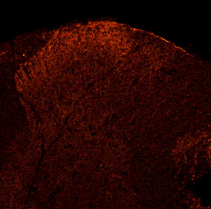
P2X3 staining in rat Dorsal Root Ganglia.
P2X3 in rat Dorsal Horn.
Check out our Purinergic Receptor Antibodies today.


Monday, September 19, 2011

Immune-Inflammatory Response and Pain Research

Our Pain and Inflammation Related Research Antibodies are increasingly being used to study the root causes of immune/inflammatory related pain induction. Here're related publications: Lintao Qu, Pu Zhang, Robert H. LaMotte, Chao Ma. Neuronal Fc-gamma receptor I mediated excitatory effects of IgG immune complex on rat dorsal root ganglion neurons. Brain, Behavior, and Immunity. Volume 25, Issue 7, October 2011, Pages 1399-1407......rabbit-anti-TRPV1, 1:1000, Neuromics...

Highlights: Pain often accompanies antigen-specific immune-related disorders though little is known of the underlying neural mechanisms. A common feature among these disorders is the elevated level of antigen-specific immunoglobulin (Ig) G in the serum and the presence of IgG immune complex (IC) in the affected tissue. We hypothesize that IC may directly activate the Fc-gamma receptor type I (FcγRI) expressed in nociceptive dorsal root ganglion (DRG) neurons and increase neuronal excitability thus potentially contributing to pain. Immunofluorescent labeling indicated that FcγRI, but not FcγRIIB or FcγRIII, was expressed in a subpopulation of rat DRG neurons including those expressing nociceptive markers. Calcium imaging revealed that the IC, but neither of the antibody (IgG) or antigen alone, produced an increase in intracellular calcium. This effect was abolished by the removal of the IgG Fc portion in the IC or the application of an anti-FcγRI antibody, suggesting a key role of the FcγRI receptor. Removal of extracellular calcium or depletion of intracellular calcium stores prevented the IC-induced calcium response. In whole-cell current-clamp recordings, IC depolarized the resting membrane potential, decreased the rheobase, and increased the number of action potentials evoked by a depolarizing current at 2× rheobase. In about half of the responsive neurons, IC evoked action potential discharges. These results suggest that a subpopulation of nociceptive neurons expresses functional FcγRI and that the activation of this receptor by IC increases neuronal excitability.

B. Huanga, X. Zhaoc, L.-B. Zhengb, L. Zhanga, B. Nia. Different expression of tissue inhibitor of metalloproteinase family members in rat dorsal root ganglia and their changes after peripheral nerve injury. Neuroscience, Volume 193, 13 October 2011, Pages 421-428....anti-P2X3 (rabbit, Neuromics, MN, USA)...

Thursday, August 25, 2011

Primary Neurons and Cell Based Assays

The feedback I receive from Neuroscientists is consistent. To paraphrase, "gives us healthy, consistent and potent primary cells. I understand the hard work it takes to generate meaningful and publishable results from cell based assays. Our Primary Neurons and Astrocytes are merely inputs for these assays. The real cost is the time invested in culturing and time lost if they don't work.

I have numerous postings on success: Primary Neurons Postings. I wanted to share more data and feedback.

Primary DRGs-Culturing these can be tricky. I make it a point to work with labs to make sure the protocol options best match the desired outcome for assays. This includes replacing cells to make sure we can accurately troubleshoot. This approach insures I can pin point the issues and make sure they are all resolved in round two. Here's a representative testimonial: "Thanks for following up, the DRGs worked great and we were able to get excellent data from them. Thanks so much for working with us." Adam Ross, Dr. Chengji Zhou Lab, UC Davis

Image: DRGs cultured on Calf Skin Collagen.

Primary Hippocampal Neurons-I would like to thank Vimal Swarup, University of Utah for this excellent image.


The cells have been fixed after 48 hrs, they were grown over poly-lysine coated coverslips in the media supplied by Neuromics. Cells were imaged in phase contrast mode with 40x objective.

Put our primary cells to the test!


Saturday, January 15, 2011

Upregulations of P2X3 and ASIC3 involve in hyperalgesia

It gets my attention when several of our ion channel markers are referenced in the title of a publication.

Kiyomi Hori, Noriyuki Ozaki, Shigeyuki Suzuki, Yasuo Sugiura. Upregulations of P2X3 and ASIC3 involve in hyperalgesia induced by cisplatin administration in rats. PAIN 149 (2010) 393–405

Findings: "We explored the role of ion channels expressed in DRG neurons in the painful neuropathy associated with cisplatin administration. Upregulations of TRPV2, P2X3 and ASIC3 may play important roles in the mechanical hyperalgesia induced by cisplatin. In addition to cutaneous hyperalgesia, cisplatin treatment might also induce muscle hyperalgesia associated with upregulations of P2X3 and
ASIC3. Interfering with these channels may prove to be a promising therapeutic target for treating painful symptoms of cisplatin neuropathy, and may further be able to ensure the continuation of anticancer therapy."

Images: ASIC3 (Dilution 1:10) amd P2X3 (Dilution 1:500) satining of rat Dorsal Root Ganglia (DRGs) of cisplatin-treated animals. After dilution in 0.1 M phosphate-buffered saline (PBS) containing 1.5% normal goat serum and 0.3% Triton X-100 (Sigma), DRG sections were incubated with either guinea pig polyclonal antiserum against synthetic rat ASIC3 and rabbit polyclonal antiserum against synthetic rat P2X3 (1:500; Neuromics). The sections for ASIC3 were reacted with reagents for 2 days at room temperature and others at 4OC. After being rinsed with 0.1 M PBS, the sections were reacted in PBS with fluorescein-isothiocyanate (FITC)-conjugated goat anti-guinea pig or - rabbit IgG antibody (Vector Laboratories, Burlingame, CA, USA) at a concentration of 1:100. After being rinsed with 0.1 M PBS, the sections were cover-slipped in mounting medium (Immunon, Pittsburgh, PA, USA) and examined under a fluorescence microscope equipped with a digital camera

Wednesday, November 12, 2008

DRG Neurons Now Available.

Primary Rat DRGs are live neurons isolated from micro-surgically dissected regions of day 18 embryonic Sprague/Dawley rat brain. These cells are prepared fresh each week and shipped in a nutrient rich medium that keeps the cells alive for up to 14 days under refrigeration.
Please note: It is important to review Protocol/Datasheet prior to ordering. There is a unique step for making the dissociation enzyme solution. Do not hesitate to call or e-mail me (612-801-1007 or pshuster@neuromics.com) should you have questions.

Image: DRGs cultured on Calf Skin Collagen.

References:
-A dissection and Tissue Culture Manual of the Nervous System (1989). A. Shahar, J.D. Vellis, A. Vernadakis, B. Haber (Eds.), Dissociated Spinal Cord - Dorsal Root Ganglion Cultures on Plastic Tissue Culture Dishes and Glass Coverslips and Wells (pp.219-222). Wiley-Liss, Inc. J.L. Werth, -S.A. Thayer (1994) Mitrochondria Buffer Physiological Calcium Loads in Cultured Rat Dorsal Root Ganglion Neurons, The Journal of Neuroscience, 14(1), 348-356