Showing posts with label Mu Opioid Receptor antibody. Show all posts

Wednesday, December 28, 2016

Dopamine and Morphine Tolerance

Dopamine Identified as Key Player

Our Mu Opioid Receptor antibody is used to show that blocking dopamine decreases morphine tolerance: Wen-Ling Dai, Feng Xiong, Bing Yan, Zheng-Yu Cao, Wen-Tao Liu, Ji-Hua Liu1, Bo-Yang Yu. Blockade of neuronal dopamine D2 receptor attenuates morphine tolerance in mice spinal cord. Scientific Reports 6, Article number: 38746 (2016). doi:10.1038/srep38746.

Images: (A) Double immunofluorescence staining showed that MOR (green) and D2DR (red) were co-localized in the mice spinal cord (20X magnification). Chronic morphine treatment increased the co-localization of MOR and D2DR in the spinal cord, and D2DR antagonist sulpiride (8 μg/10 μl, i.t.) reduced the increased co-expression of D2DR with MOR (n = 4). (B) Co-IP experiments showed that D2DR could interact with MOR, and the MOR/D2DR interactions were increased in the spinal dorsal horn after chronic morphine treatment for 7 days while D2DR antagonist sulpiride (8 μg/10 μl, i.t.) disrupted the interactions of the MOR/D2DR (n = 3).

Blockade of D2DR in spinal cord can disrupt the interactions between MOR and D2DR to attenuate morphine tolerance. These findings highlight the possibility of a new clinical strategy to prevent morphine antinociceptive tolerance.

Sunday, January 24, 2016

More and More OR Pubs

Potent, Proven and Frequently Published

Many labs have used our Opioid Receptor antibodies over the past 10+ years. We are grateful for the many publications and testimonals referencing successful use of these antibodies.

Here's a sampling of more recent publications: Wendy M. Walwyn, Wenling Chen, Hyeyoung Kim, Ani Minasyan, Helena S. Ennes, James A. McRoberts, and Juan Carlos G. Marvizón. Sustained Suppression of Hyperalgesia during Latent Sensitization by μ-, δ-, and κ-opioid receptors and α2A Adrenergic Receptors: Role of Constitutive Activity. The Journal of Neuroscience, 6 January 2016, 36(1): 204-221; doi: 10.1523/JNEUROSCI.1751-15.2016
...probed with antibodies to MOR (RA10104; Neuromics), p-Ser375-MOR (pMOR, RA18001; Neuromics) and p-Tyr416-Src family kinase (pSFK...serine-phosphorylated MOR (p-Ser375-MOR, RA18001; Neuromics) and a different MOR antibody (RA10104...
Jan Fraessdorf, Markus W. Hollmann , Iris Hanschmann, André Heinen, Nina C. Weber, Benedikt Preckel, Ragnar Huhn. Role of Endogenous Opioid System in Ischemic-Induced Late Preconditioning. Published: July 30, 2015DOI: 10.1371/journal.pone.0134283.
...Antibodies for μ-OR and δ-OR were purchased from Neuromics (Minneapolis, USA)...
Ying-Biao Chen, Fen-Sheng Huang, Ban Fen, Jun-Bin Yin, Wei Wang and Yun-Qing L nhibitory effects of endomorphin-2 on excitatory synaptic transmission and the neuronal excitability of sacral parasympathetic preganglionic neurons in young rat. Journal Name: Frontiers in Cellular Neuroscience, ISSN: 1662-5102, Article type: Original Research Article,Received on: 02 Apr 2015, Accepted on: 12 May 2015, Provisional PDF published on: 12 May 2015.
... guinea pig antiserum against MOR (1:1000, GP10106, Neuromics, Edina, Minnesota, USA)...
Sherika N. Smith, Candler Paige, Kandy T. Velazquez, Terika P. Smith, Srinivasa N. Raja, Steven P. Wilson, Sarah M. Sweitzer. Injury-specific promoters enhance herpes simplex virus mediated gene therapy for treating neuropathic pain in rodents. doi:10.1016/j.jpain.2014.12.007
...: MOR (Neuromics, RA10104, 1:1000)...


Figure: MOR in primary sensory neuron and its terminals were decreased in neuropathic mice. (A, B) Western blot shows that MOR protein were markedly decreased in sciatic nerve (A) and hindpaw skin (B) on day 7 after nerve injury when compared with sham-operated mice. The ratio of MOR/GAPDH in sham mice was set at 1 for quantifications. Zhou et al. Molecular Pain 2014, 10:51 http://www.molecularpain.com/content/10/1/51

We will continue to post information on our ORs.


Thursday, February 20, 2014

More MOR Publications

Latest Mu Opioid Publications

Our Mu Opioid Receptor Antibodies have set the standard for the study of Pain Mechanisms. We have posted >40 publications referencing use of these antibodies.

Here's several published in 2014:

Charlie H.T. Kwok, Ian M. Devonshire, Andrew J. Bennett, Gareth J. Hathway. Postnatal maturation of endogenous opioid systems within the periaqueductal grey and spinal dorsal horn of the rat. PAIN - January 2014 (Vol. 155, Issue 1, Pages 168-178, DOI: 10.1016/j.pain.2013.09.0220. ...rabbit anti-MOR (Neuromics, Edina, MN, USA; 1:1000 with tyramide signal amplification protocol)...


Images: Immunohistochemical expression of opioid peptides and receptors in the DH (spinal cord dorsal horn) during postnatal development. (A) POMC (pro-opiomelanocortin) immunoreactivity in the dorsal horn in postnatal day (P)10, P21, and adult rats. White arrows depict where cell staining was found. Interestingly, fibre staining was also observed in the superficial dorsal horn (lamina I) of adult rats, but not in the younger ages. (B) Since both cell and fibre staining were observed, staining intensity was used to quantify the immunoreactivity of POMC in the DH. Quantified staining intensity for POMC in the DH significantly decreased as the animals aged, with highest immunoreactivity found at P10. (C) Enkephalin immunoreactivity in the DH was restricted to the superficial laminae and only fibre staining was observed. (D) Quantified staining intensity for enkephalin illustrate an age-dependent increase in immunoreactivity, which was highest at adult P10.(C) Enkephalin immunoreactivity in the DH was restricted to the superficial laminae and only fibre staining was observed. (D) Quantified staining intensity for enkephalin illustrate an age-dependent increase in immunoreactivity, which was highest at adult. (E) MOR (μ-opioid receptor) immunoreactivity in the DH, cell staining was found throughout the superficial and deeper laminae in all ages. (F) Cell count of MOR staining in the DH, which showed a significant increase as the animals aged (∗∗P<0 .01="" adult="" i="" p21="" vs="">


J. Desroches, J.-F. Bouchard, L. Gendron, P. Beaulieu. Involvement of cannabinoid receptors in peripheral and spinal morphine analgesia ☆ Neuroscience, Volume 261, 7 March 2014, Pages 23–42. http://dx.doi.org/10.1016/j.neuroscience.2013.12.030. ...The floating sections were then incubated in 1% sodium borohydride in PBS for 30 min, rinsed twice with PBS, and incubated for 30 min at room temperature in a blocking solution containing 3% normal goat serum (NGS) and 0.3% Triton X-100 in PBS. The sections were then incubated overnight at 4 °C with the guinea pig anti-MOP primary antibody (cat# GP10106; Neuromics, Minneapolis, MN, USA) diluted 1:1000 in the blocking solution. The floating sections were then washed in PBS and incubated with a goat anti-guinea pig secondary antibody conjugated with Alexa Fluor 488 (Molecular Probes, Invitrogen, Carlsbad, CA, USA) at a concentration of 1:1000 in PBS for 2 h at room temperature...

We will continue posting customer data and publications that give new insights into the mechanisms of pain.

Tuesday, January 28, 2014

Pot and Pain

The pressure for states to legalize Marijuana for medical and recreational use is building. The tax benefits are self evident.

The debate for many centers of "true medical benefits". That's why research on understanding analgesic pathways is so important. Ironically, this study was conducted by my friends at Université de Montréal and Université de Sherbrooke in Quebec Canada: J. Desroches, J.-F. Bouchard, L. Gendron, P. Beaulieu. Involvement of cannabinoid receptors in peripheral and spinal morphine analgesia ☆ Neuroscience, Volume 261, 7 March 2014, Pages 23–42. http://dx.doi.org/10.1016/j.neuroscience.2013.12.030.

These teams have proven expert is using our Opioid Receptor Antibodies in their pain research. Here's a synopsis:
•Analgesia is the most common feature shared by the cannabinoid and opioid systems.
•The role of the cannabinoid system in the morphine-induced analgesia is uncertain.
•Peripheral and intrathecal morphine analgesia is altered in cnr1KO and cnr2KO mice.
•This attenuation is neither caused by a MOP malfunction nor by its downregulation.


Images: Deletion of the CB1 or CB2 receptors has no effect on the expression of MOP in the spinal cord. Immunofluorescence of spinal MOP revealed that the expression of MOP in laminae I and II of the dorsal horn of the spinal cord did not differ between cnr1WT (A) and cnr1KO (B) mice or between cnr2WT (C) and cnr2KO mice (D).


Observations here further support the existence of interactions between the cannabinoid and opioid systems. The loss of peripheral and spinal morphine analgesia is apparently caused neither by a decrease in MOP spinal expression nor by altered binding properties or G protein coupling of this receptor in cnr1KO and cnr2KO mice. The mechanisms underlying the loss of morphine analgesia are not clear but could include the release of endogenous cannabinoids in structures along the pain pathway or a disrupted endocannabinoid tone.

It is important funding that enables researchers to understand the analgesic pathways of marijuana continues to grow. This research could yield better control of pain with reduced side effects.

Tuesday, November 08, 2011

Opioid Induced Itch

Our widely used and frequently published Opioid Receptors Antibodies are used for a spectrum of pain research. Here is an interesting study on the root causes of opioid induced itch.

Xian-Yu Liu, Zhong-Chun Liu, Yan-Gang Sun, Michael Ross1, Seungil Kim, Feng-Fang Tsai, Qi-Fang Li, Joseph Jeffry, Ji-Young Kim, Horace H. Loh, Zhou-Feng Chen. Unidirectional Cross-Activation of GRPR by MOR1D Uncouples Itch and Analgesia Induced by Opioids. Cell, Volume 147, Issue 2, 14 October 2011, Pages 261-262.


Root Causes: Spinal opioid-induced itch, a prevalent side effect of pain management, has been proposed to result from pain inhibition. We now report that the μ-opioid receptor (MOR) isoform MOR1D is essential for morphine-induced scratching (MIS), whereas the isoform MOR1 is required only for morphine-induced analgesia (MIA). MOR1D heterodimerizes with gastrin-releasing peptide receptor (GRPR) in the spinal cord, relaying itch information. We show that morphine triggers internalization of both GRPR and MOR1D, whereas GRP specifically triggers GRPR internalization and morphine-independent scratching. Providing potential insight into opioid-induced itch prevention, we demonstrate that molecular and pharmacologic inhibition of PLCβ3 and IP3R3, downstream effectors of GRPR, specifically block MIS but not MIA. In addition, blocking MOR1D-GRPR association attenuates MIS but not MIA. Together, these data suggest that opioid-induced itch is an active process concomitant with but independent of opioid analgesia, occurring via the unidirectional cross-activation of GRPR signaling by MOR1D heterodimerization.

Tuesday, August 30, 2011

Opioid Receptor Antibodies Trifecta

This publication proposes a role for opioid receptors in treating cancers. It also references use of our μ, δ, and κ opioid receptor antibodies.

Kohei Yamamizu1, Sadayoshi Furuta, Shiori Katayama, Michiko Narita, Naoko Kuzumaki, Satoshi Imai, Hiroshi Nagase, Tsutomu Suzuki, Minoru Narita, and Jun K. Yamashita. The κ opioid system regulates endothelial cell differentiation and pathfinding in vascular development. Blood July 21, 2011 vol. 118 no. 3 775-785.

Highlights: The opioid system is, thus, a new regulator of vascular development that simultaneously modifies 2 distinct vascular properties, EC differentiation and vascular pathfinding. We confirmed that KOR, but not MOR, was highly expressed in various ECs such as HUVECs (data not shown), suggesting that KOR agonists could directly act on tumor ECs to suppress VEGF receptor expression, similar to the effects observed in embryonic ECs. If so, a combination therapy including an MOR agonist, morphine, and a KOR agonist (such as TRK820, a clinically approved drug in Japan for uremic pruritus) may prove useful for cancer therapy through the suppression of tumor angiogenesis by dual inhibition of VEGF ligands and receptors, extending the therapeutic benefits beyond pain relief.

Images: KOR was highly expressed in Flk1+ vascular progenitors. (A) RT-PCR showing mRNA expression of MOR, DOR, and KOR in ES cells, Flk1+ cells, cells after 1 or 3 days of Flk1+ cell culture (Flk-d1 or Flk-d3), CD31-positive cells (ECs) and CD31-negative cells (MCs) at Flk-d3. (B) Fluorescent staining for MOR, DOR, and KOR at Flk-d1. Nuclei are stained with DAPI (blue). Left, MOR; middle, DOR; right, KOR. Scale bars, 100 μm. (C) Double fluorescent staining for MOR, DOR, and KOR with CD31 (red) at Flk-d3. Nuclei are stained with DAPI (blue). Top, opioid (green) receptors (green); middle, CD31 (red); bottom, merged. Scale bars, 100 μm.

Thursday, July 29, 2010

Let-7 microRNAs and Nociceptive Pain

Our Opioid Receptor Antibodies have set a potent standard for studying Nociceptive and Neuropathic Pain. Related Publications.

We want to recognize Dr. Zaijie Jim Wang and his team for being the first to use our Mu Opioid Receptor for studying the potential role of microRNAs in Nociception.

Ying He, Cheng Yang, Chelsea M. Kirkmire, and Zaijie Jim Wang. Regulation of Opioid Tolerance by let-7 Family MicroRNA Targeting the µ Opioid Receptor. The Journal of Neuroscience, July 28, 2010, 30(30):10251-10258; doi:10.1523/JNEUROSCI.2419-10.2010
Abstract: MicroRNA has emerged as a critical regulator of neuronal functions. This study aimed to test whether let-7 microRNAs can regulate the µ opioid receptor (MOR) and opioid tolerance. Employing bioinformatics, we identified a let-7 binding site in the 3'-untranslated region (UTR) of MOR mRNA, which was experimentally confirmed as a direct target of let-7. The repressive regulation of MOR by let-7 was revealed using a LNA-let-7 inhibitor to knockdown let-7 in SH-SY5Y cells. Conversely, morphine significantly upregulated let-7 expression in SH-SY5Y cells and in a mouse model of opioid tolerance. The LNA-let-7 inhibitor decreased brain let-7 levels and partially attenuated opioid antinociceptive tolerance in mice. Although chronic morphine treatment did not change overall MOR transcript, polysome-associated mRNA declined in a let-7-dependent manner. let-7 was identified as a mediator translocating and sequestering MOR mRNA to P-bodies, leading to translation repression. These results suggest that let-7 plays an integral role in opioid tolerance.

  • Western blot analysis. Western blot analysis was performed as previously described (Tang et al., 2006) using the anti-µ opioid receptor antibody (1:1000; Neuromics). The expression of β-actin was similarly determined from the same blots using a monoclonal antibody (1:10,000; Sigma).
  • For immunofluorescence analysis, the antibody for hDcp1a (Santa Cruz Biotechnology) and MOR were used at 1:500 and 1:5000 dilutions, respectively. Secondary anti-goat and anti-mouse antibodies labeled with Alexa 488 and Alexa 594 fluorochromes (Invitrogen), respectively, were used at 1:500 dilutions.

Related Reagent Links:
All Opioid Receptor Antibodies

Pain and Inflammation
       
Neurotransmission Research Antibodies
-GPCRs, Ligand Gated Ion Channels,
Biogenic Amines and more
i-Fect Transfection Kit
-gene silencing of DOR,
NaV1.8 tetrodotoxin-resistant sodium channel, NTS2 and more in-vitro and in vivo
Primary Neurons and Astrocytes
-Primary human,
rat and mouse neurons and astrocytes 


Monday, July 26, 2010

δ- and μ-opioid receptors co-expression and Nociceptive Pain

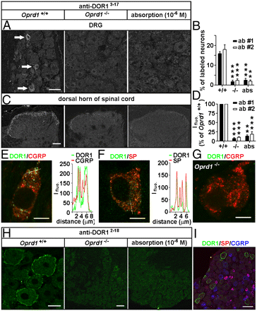
Dr. Tomas Hokfelt and his team at Karolinska Institute recently published use of our Opioid Receptor Antibodies and Substance P Antibody.They show the interplay of DOR and MOR in modulation of nociceptive afferent transmission and opioid analgesia.

Hai-Bo Wanga, Bo Zhaoa, Yan-Qing Zhonga, Kai-Cheng Li, Zi-Yan Li, Qiong Wang, Yin-Jing Lua, Zhen-Ning Zhang, Shao-Qiu He, Han-Cheng Zheng, Sheng-Xi Wu, Tomas G. M. Hökfelt, Lan Baob, and Xu Zhanga. Coexpression of δ- and μ-opioid receptors in nociceptive sensory neurons. PNAS July 20, 2010 vol. 107 no. 29 13117-13122.

Immunostaining. Adult rats, mice, and Oprd1 exon 1-deleted mice were fixed. Cryostat sections of L4 and L5 DRGs and spinal cord segments were processed for immunofluorescence staining (13) with Rb anti-DOR13–17 (1:2,000–1:60,000; DiaSorin and 1:4,000–1:60,000; Neuromics), Rb anti-DOR12–18 (1:30,000–1:120,000; Alomone), Rb anti-DOR1358–372 (1:1,000–1:2,000; Lifespan Biosciences), Rb anti-MOR (1:1,000; Neuromics); guinea pig anti-SP (1:500; Neuromics), and mouse anti-CGRP (1:1,000; Biogenesis) antibodies. IB4-labeling was carried out with fluorescein-labeled GSL I-IB4 (1:200). The Myc-DOR1–transfected HEK293 cells and neurons were fixed and processed with mouse anti-Myc antibodies (1:500; DSHB). Nuclear DAPI staining was used to indicate HEK293 cells in control experiments.

Images: Distinct distribution patterns of DORs in subsets of DRG neurons of mice. Immunostaining with antibodies against DOR13–17 [A: 1:30,000, antibody 1 (ab #1); DiaSorin and C: antibody 2 (ab #2); Neuromics] shows DORs in small DRG neurons and afferent fibers in spinal laminae I–II. This immunostaining pattern is abolished by the antiserum preabsorption or the deletion of Oprd1 exon 1. Reduction in immunostaining is quantitatively assayed by determining the percentage of positive DRG neurons (B; n = 6) and fluorescence intensity (Ifluo.) in the laminae I–II (D; n = 5). **P < 0.01; ***P < 0.001. (Scale bars: A and C, 40 μm.). DOR labeling (anti-DOR13–17, 1:30,000; DiaSorin) associated with vesicles in peptidergic small DRG neurons (E and F) is absent in Oprd1 exon 1-deleted mice (G). Colocalization of DORs and neuropeptides is shown by correlated peaks of Ifluo. measured along lines. (Scale bar: 8 μm.) (H) Immunostaining with antibodies against DOR12–18 (1:60,000; Alomone) shows the presence of DORs on the cell surface of large DRG neurons of mice. (Scale bar: 25 μm.) This staining pattern is abolished by preabsorption and is absent in Oprd1 exon 1-deleted mice. (Scale bar: 80 μm.) (I) Triple-immunostaining shows that DOR+ large DRG neurons contain neither SP nor CGRP. (Scale bar: 80 μm.)

Immunoblotting.The samples were processed for SDS/PAGE, transferred, probed with Rb antibodies against MOR (1:500; Neuromics), phospho-DOR1 (1:1,000; Neuromics), phospho-MOR (1:1,000; Neuromics), Myc (1:500; DSHB), Flag (1:1,000; Sigma), or actin (1:50,000; Chemicon) and visualized with enhanced chemiluminescence (19).

Featured and Related Reagents:

Mu Opioid Receptor-Rabbit


Mu Opioid Receptor-Guinea Pig


Delta Opioid Receptor 3-17


Delta Opioid Receptor 358-372


Delta Opioid Receptor 358-372


Kappa Opioid Receptor


phospho-Mu Opioid Receptor (Ser375)


MOR-1C
 

ORL 1-Pure


ORL1-Whole Serum


All Opioid Receptor Antibodies


Pain and Inflammation


Neurotransmission Research Antibodies
-GPCRs, Ligand Gated Ion
Channels, Biogenic Amines and more

i-Fect Transfection Kit
-gene silencing
of DOR, NaV1.8 tetrodotoxin-resistant sodium channel, NTS2 and more
in-vitro and in vivo

Primary Neurons and Astrocytes
-Primary
human, rat and mouse neurons and astrocytes