Showing posts with label Purinergic Receptor Antibodies. Show all posts

Saturday, October 12, 2013

P2X3 Receptor and Inflammatory Nociception

P2X3 Activates TRPA1, 5-HT3 and 5-HT1A Receptors

Endogenous ATP via activation of P2X3 Receptors contributes to inflammatory nociception in different models, including the formalin injected in subcutaneous tissue of the rat's hind paw. In this study, researchers evaluated whether TRPA1, 5-HT3 and 5-HT1A receptors, whose activation is essential to formalin-induced inflammatory nociception, are involved in the nociception induced by activation of P2X3 receptors on subcutaneous tissue of the rat's hind paw: Suzy Krimon, Dionéia Araldi, Filipe César do Prado, Cláudia Herrera Tambeli, Maria Cláudia G. Oliveira-Fusaro, Carlos Amílcar Parada. P2X3 receptors induced inflammatory nociception modulated by TRPA1, 5-HT3 and 5-HT1A receptors. Pharmacology Biochemistry and Behavior, Available online 8 October 2013. http://dx.doi.org/10.1016/j.pbb.2013.09.017. Our widely used and frequently published P2X3 R Antibody places a central role in measuring the expression of the protein...containing 5% non-fat dry milk at room temperature, followed by incubation with P2X3 rabbit polyclonal antibody (1:500; Neuromics) overnight at 4 °C, rinsed six times with TBST, and then incubated for 40 minutes in goat anti-rabbit IgG peroxidase...
Image: Neuromics' P2X3 R WB Example: Sequence‐specific siRNA‐mediated repression of P2X3. (A) P2X3 mRNA inhibition by 200 nM siRNA duplexes. Twenty‐four hours after transfection of CHO‐rP2X3 cells, P2X3‐specific mRNA was measured with Q‐PCR and plotted as percentage of mRNA detected in the control treated with Oligofectamine alone. Sequences and modifications are shown in Figure 1B and Table 1. (B) P2X3 protein reduction by 200 nM siRNA‐8646/8647, but not by its mismatch analogue siRNA‐MM‐7558/7559 or the unrelated siRNA‐7126/7127. Twenty‐four hours after transfection, protein was extracted and analysed by western blotting. P2X3‐specific immunodetection reveals expression levels as shown below (an average value from two experiments). Time points as indicated at the top. Molecular weights of two glycosylated forms of P2X3 are shown on the left. 

Conclusions: Nociceptive response intensity was measured by observing the rat's behavior and considering the number of times the animal reflexively raised its hind paw (flinches) in 60 min. Local subcutaneous administration of the selective TRPA1, 5-HT3 or 5-HT1A receptor antagonists HC 030031, tropisetron and WAY 100,135, respectively, prevented the nociceptive responses induced by the administration in the same site of the non-selective P2X3 receptor agonist αβmeATP. Administration of the selective P2X3 and P2X2/3 receptor antagonist A-317491 or pretreatment with oligonucleotides antisense against P2X3 receptor prevented the formalin-induced behavioral nociceptive responses during the first and second phases. Also, the co-administration of a subthreshold dose of αβmeATP with a subthreshold dose of formalin induced nociceptive behavior, which was prevented by local administration of tropisetron, HC 030031 or WAY 100, 135. These findings have demonstrated that the activation of P2X3 receptors induces inflammatory nociception modulated by TRPA1, 5-HT3 and 5-HT1A receptors. Also, they suggest that inflammatory nociception is modulated by the release of endogenous ATP and P2X3 receptor activation, which in turn, increases primary afferent nociceptor susceptibility to the action of inflammatory mediators via interaction with TRPA1, 5-HT3 and 5-HT1A receptors in the peripheral tissue.

I will continue to post pain and inflammation related studies that reference the use of our antibodies.

Sunday, April 14, 2013

P2X3 Receptors and Migraine

P2X3 Receptors of Trigeminal Sensory Neurons and Familial Hemiplegic Migraine Type 1 (FHM-1).

Our P2X Receptor Markers continue to be used in interesting and novel ways. Here researchers use our P2X3 Receptor Antibody to study expression using primary rat ganlia cultures: Swathi K. Hullugundi,Michel D. Ferrari, Arn M. J. M. van den Maagdenberg, Andrea Nistri. Andrea Nistri. The Mechanism of Functional Up-Regulation of P2X3 Receptors of Trigeminal Sensory Neurons in a Genetic Mouse Model of Familial Hemiplegic Migraine Type 1 (FHM-1). PLoS ONE 8(4): e60677. doi:10.1371/journal.pone.0060677

Abstract: A knock-in (KI) mouse model of FHM-1 expressing the R192Q missense mutation of the Cacna1a gene coding for the α1 subunit of CaV2.1 channels shows, at the level of the trigeminal ganglion, selective functional up-regulation of ATP -gated P2X3 receptors of sensory neurons that convey nociceptive signals to the brainstem. Why P2X3 receptors are constitutively more responsive, however, remains unclear as their membrane expression and TRPV1 nociceptor activity are the same as in wildtype (WT) neurons. Using primary cultures of WT or KI trigeminal ganglia, we investigated whether soluble compounds that may contribute to initiating (or maintaining) migraine attacks, such as TNFα, CGRP, and BDNF, might be responsible for increasing P2X3 receptor responses. Exogenous application of TNFα potentiated P2X3 receptor-mediated currents of WT but not of KI neurons, most of which expressed both the P2X3 receptor and the TNFα receptor TNFR2. However, sustained TNFα neutralization failed to change WT or KI P2X3 receptor currents. This suggests that endogenous TNFα does not regulate P2X3 receptor responses. Nonetheless, on cultures made from both genotypes, exogenous TNFα enhanced TRPV1 receptor-mediated currents expressed by a few neurons, suggesting transient amplification of TRPV1 nociceptor responses. CGRP increased P2X3 receptor currents only in WT cultures, although prolonged CGRP receptor antagonism or BDNF neutralization reduced KI currents to WT levels. Our data suggest that, in KI trigeminal ganglion cultures, constitutive up-regulation of P2X3 receptors probably is already maximal and is apparently contributed by basal CGRP and BDNF levels, thereby rendering these neurons more responsive to extracellular ATP.

Images: Examples of TNFR2 and P2X3 co-exexpression in (wildtype) WT and R192Q (knockin) KI neurons. Left panel shows P2X3 expression (green), and right panel shows TNFR2 staining (red). B, Histograms quantifying % of cells co-expressing TNFR2 and P2X3: both WT and KI cultures show similar TNFR2 and P2X3 co-expression. N = 3 independent experiments (6 mice). C, Representative traces of currents induced by application of α,β-meATP (10 µM, 2 s) to WT or R192Q KI neurons in control conditions or after 4 h TNFα application. D, Histograms show average peak amplitudes of P2X3 receptor-mediated currents: WT control (open bar), n = 30; WT TNFα (stippled bar), n = 38; KI control (grey bar), n = 34; KI TNFα (stippled gray bar), n = 34; ** = p<0 .006="" i="" nbsp="" p="">doi:10.1371/journal.pone.0060677.g001.

Understanding the interplay between TNFR2 and P2X3 could lead to a better understanding of the root causes of migraines. This could open up yet more potential drug targets for this insidious condition.

Check out these related reagent categories:
All Purinergic Receptor Antibodies
Pain and Inflammation Research Antibodies 
Neurotransmission Research Antibodies
Primary Neurons and Astrocytes-Primary human, rat and mouse neurons and astrocytes

Sunday, January 27, 2013

Epidermal Nerve Fibers in Neuropathic Pain Model

Re-innervation in spared nerve injury (SNI) vs normal rats
Neuromics' Pain and Inflammation Research Antibodies are frequently used to help researchers find root causes of neuropathic pain. This is a fascinating study of re-innervation patterns of epidermal and dermal nerve fibers in a rat neuropathic pain model (Note: our Guinea Pig Polyclonal P2X3 Antibody was used in this study): Liron S. Durakua, Mehdi Hossaini, Barthold N. Schüttenhelm, b, Joan C. Holstege, Martijn Baas, Tom J.H. Ruigrok, Erik T. Walbeehm. Re-innervation patterns by peptidergic Substance-P, non-peptidergic P2X3, and myelinated NF-200 nerve fibers in epidermis and dermis of rats with neuropathic pain. Experimental Neurology Volume 241, March 2013, Pages 13–24. doi.org/10.1016/j.expneurol.2012.11.029.
Non-footpad area vs. footpad.The distribution pattern between the center non-footpad area and footpad is compared for the different subgroups of sensory skin fibers. For epidermal Sub P-IR and upper dermal NF-200-IR fibers there is an equal density of fibers in the center and footpad area suggesting a homogenous distribution over the foot sole of a rat. Whereas for epidermal P2X3-IR fibers there is a significant lower number of P2X3-IR fibers in the footpads as compared to the center non-footpad area, suggesting a heterogeneous distribution in the foot sole for this type of sensory fibers. N = 5 per group. Unpaired t-test. ***: p < 0,001. Scale bar: 250 μm. doi.org/10.1016/j.expneurol.2012.11.029

Illustration of the skin innervation in normal and SNI situation.Illustration showing the innervation pattern of subgroups of sensory skin fibers in the normal situation and in the SNI model 10 weeks PO. In the naïve animals the peptidergic CGRP and non-peptidergic P2X3 fibers have a lower density in the footpads as compared to the non-footpad areas, while the Substance P and NF-200 fibers have an equal distribution over the complete foot sole. In the SNI model there is a significant increase of CGRP epidermal fibers in the medial and lateral area of the foot sole and there is a complete re-innervation of the center area. In addition the footpads in the SNI model are hyper-innervated with CGRP fibers. Substance P and P2X3 and NF-200 fibers show no increased density in the uninjured medial and lateral area after 10 weeks PO. In addition, Substance P and P2X3 fibers hardly re-innervate the center area; however, the NF-200 fibers have the same density of fibers in the denervated area as in the normal situation. In the SNI model the medial, center and lateral area have all an increase in LC's, with the center area being the most prominent one. In addition the epidermis thickness of the SNI model has decreased significantly in all the three areas after 10 weeks PO.
Related Postings: http://neuromics.blogspot.com/search/label/Neuropathic%20Pain.

Thursday, January 10, 2013

Pain and the Interplay Between P2Y Receptors with P2X3

P2Y2-P2X3 crosstalk in DRG neurons

Alfredo Ribeiro-da-Silva and his team use our Purinergic Receptor Antibodies to the study role of their expression in Nociceptive and Neuropathic Pain. They referenced their use in 8 publications.

Based on their research, they suspect changes in P2X3 function under pathological conditions are more complex than simple up- or down-regulation of expression at the protein level. This would have profound implications for the dicovery of drugs that target P2X3 expression levels: Gary Mo, Jennifer C. Peleshok, Chang-Qing Cao, Alfredo Ribeiro-da-Silva and Philippe Séguéla. Control of P2X3 channel function by metabotropic P2Y2 UTP receptors in primary sensory neurons. Molecular Pharmacology Fast Forward. Published on December 18, 2012 as doi:10.1124/mol.112.082099.
Images: P2X3 and P2Y2 receptors are co-expressed in rat DRG sensory neurons.(A) DRG sections labeled for P2Y2 (green) and P2X3 (red) and merged (yellow). P2Y2 immunoreactivity is broadly distributed in small-diameter neuronal somata and co-localized with P2X3 (arrows). P2Y2 immunoreactivity can be detected in cells negative for P2X3 (arrow heads). (B) P2Y2 (green) and P2X3 (red) are also colocalized (yellow) in peripheral nerve fibers.
The two receptors were found to be colocalized both in cell bodies and nerve fibers, indicating co-trafficking to peripheral and central terminals. The immunolocalization evidence presented here provides valuable information on the physiological relevance of crosstalks between ionotropic and metabotropic ATP receptors in sensory pathways, yet much work is still needed to fully comprehend the role of nucleotide signaling in pain, especially in pathological conditions. Changes in P2Y2 receptor expression in response to inflammation have been documented (Malin et al., 2008), therefore it will be worth investigating the functional impact of P2Y2-P2X3 interactions on ATP signaling under chronic pain conditions.

I will keep you posted on any new developments.

Sunday, December 16, 2012

P2X Receptor Markers-Pubs Update

2012 has been a record year for publications referencing use of our Purinergic Receptor Antibodies. These publications demonstrate use of these markers in a variety of assays and applications. Examples range from analysis of P2X2 expression in human bladder epithelial cells to showing P2X3 expressing nerve fiber boutons in rat horizontal spinal cord sections by immunofluorescence and much more.

Here's a summary of publications: F.C. Pradoa, D. Araldia, A.S. Vieiraa, M.C.G. Oliveira-Fusarob, C.H. Tambelia, C.A. Parada. Neuronal P2X3 receptor activation is essential to the hyperalgesia induced by prostaglandins and sympathomimetic amines released during inflammation. http://dx.doi.org/10.1016/j.neuropharm.2012.11.011. non-fat dry milk at room temperature, followed by incubation with P2X3 or PKCɛ rabbit polyclonal antibody (1:500; Neuromics) overnight at 4 °C, rinsed six times with TBST, and then incubated for 40 min in goat anti-rabbit IgG peroxidase conjugate...

Min Liu, PhD, MD, Yun-fei Xu, PhD, MD, Yuan Feng, PhD, MD, Fengqiang Yang, MD, Jun Luo, MD, Wei Zhai, PhD, MD, Jian-ping Che, MD, Guang-chun Wang, MD, Jun-hua Zheng, PhD. Epigallocatechin gallate attenuates interstitial cystitis in human bladder urothelium cells by modulating purinergic receptors. Journal of Surgical Research. http://dx.doi.org/10.1016/j.jss.2012.11.041
...P2X2 (Neuromics, Northfield, MN)...

Abeer W Saeed, Alfredo Ribeiro-da-Silva. Non-peptidergic primary afferents are presynaptic to neurokinin-1 receptor immunoreactive lamina I projection neurons in rat spinal cord. Molecular Pain 2012, 8:64 doi:10.1186/1744-8069-8-64Anna M.W. Taylora, Maria Osikowicza, Alfredo Ribeiro-da-Silva. Consequences of the ablation of nonpeptidergic afferents in an animal model of trigeminal neuropathic pain. PAIN. Volume 153, Issue 6, June 2012, Pages 1311–1319. http://dx.doi.org/10.1016/j.pain.2012.03.023.
Confocal images at high power obtained from horizontal spinal cord sections. In a confocal optical section from lamina I adjacent to the white matter (A), note the relatively abundant P2X3-IR fibers with varicosities (boutons). CGRP-IR fibers and boutons were considerably more abundant in this lamina. In a confocal optical section from inner lamina II (B), note the very high density of P2X3-IR fibers and varicosities, higher than that of CGRP-IR fibers in lamina I. Note that most varicosities display either P2X3 or CGRP immunoreactivity, although some co-localization is observed (yellow). Scale bar (A, B) = 20 μm.
T. Cho, V. V. Chaban. Interaction Between P2X3 and Oestrogen Receptor (ER)α/ERβ in ATP-Mediated Calcium Signalling In Mice Sensory Neurones. Journal of Neuroendocrinology Volume 24, Issue 5, pages 789–797, May 2012...with polyclonal rabbit antibody against P2X3 receptor (dilution 1 : 1000; Neuromics)...

Anna M.W. Taylora, Maria Osikowicza, Alfredo Ribeiro-da-Silva. Consequences of the ablation of nonpeptidergic afferents in an animal model of trigeminal neuropathic pain. PAIN. Volume 153, Issue 6, June 2012, Pages 1311–1319. doi.org/10.1016/j.pain.2012.03.023...Sections were then incubated for 48 hours at 4°C with a guinea pig polyclonal anti-P2X3 antibody (1:25,000; Neuromics, Edina, MN, USA), diluted in PBS-T. Following primary antibody incubation, sections were treated with a biotin-conjugated...

Ji Z-G , Ito S , Honjoh T , Ohta H , Ishizuka T , et al. 2012 Light-evoked Somatosensory Perception of Transgenic Rats That Express Channelrhodopsin-2 in Dorsal Root Ganglion Cells. PLoS ONE 7(3): e32699...guinea-pig anti-P2X3 (1:1,000, GP10108, Neuromics, Edina, MN, USA)...
Distribution of ChR2V in the dorsal part of the spinal cord. A–C. Immunohistochemical localization of ChR2V with the cell-type specific markers, NF200 (A), CGRP (B) or P2X3 (C). Scale bars indicate 40 µm. doi:10.1371/journal.pone.0032699.g003.

I will continue to post new publications and data in the coming year. Stay tuned. 

Sunday, October 21, 2012

P2X3 Receptor and CGRP Antibodies Immunostaining

Dr. Alfredo Ribeiro-da-Silva, McGill University, is a serial publisher of studies using our pain and inflammation research antibodies.

Here, use of our rabbit anti-CGRP and guinea pig anti-P2X3 is referenced. Please note the high titer of these antibodies (dilution is 1:25,000): Abeer W Saeed, Alfredo Ribeiro-da-Silva. Non-peptidergic primary afferents are presynaptic to neurokinin-1 receptor immunoreactive lamina I projection neurons in rat spinal cord. Molecular Pain 2012, 8:64 doi:10.1186/1744-8069-8-64.


Images: CGRP, IB4 and P2X3 staining in transverse spinal cord sections. A and B show low magnification confocal images of CGRP-IR and IB4 positive (A) or P2X3-IR (B) fibers. C and D represent high magnification confocal images from the middle third of the lateromedial extent of the superficial dorsal horn. In C, note that there is limited co-localization of IB4 and CGRP (in yellow). Arrowheads show axonal varicosities (boutons) from nonpeptidergic fibers in lamina I, which do not co-localize CGRP immunoreactivity. The framed regions in A and B indicate the approximate regions from where C and D, respectively, were obtained (the latter originate from other sections). CGRP (in green); IB4 (in red); P2X3 (in red). Scale bar (A, B) = 200 μm; scale bar (C, D) = 20 μm

Tissue processing: The injection site at the level of the parabrachial nucleus was examined by cutting serial, 100 μm-thick coronal sections of the relevant brain region. The dorsal aspect of the L4-L5 spinal cord segment was cut into serial, 50 μm-thick horizontal sections (n = 10), 50 μm-thick parasagittal sections (n = 4) or 50 μm-thick transverse sections (n = 4). All sections were cut using a freezing sledge microtome (Leica, Richmond Hill, Ontario) and collected as freefloating in phosphate-buffered saline (PBS) with 0.2% Triton-X 100 (PBS + T). To block unspecific staining, all spinal cord sections were incubated, for one hour, in 10% normal donkey serum (NDS) (Jackson, West Grove, PA) in PBS + T at room temperature. Subsequently, the sections were placed in primary antibodies (or conjugated lectin IB4 - see below) for 48 hours at 4 °C. We used a mixture of 2 or 4 primary antibodies (each raised in a different species), or IB4, in PBS + T containing 5% NDS. Next, the sections were washed in PBS + T and then incubated in species-specific secondary antibodies that were raised in donkey and conjugated to either AlexaFluor 488, AlexaFluor 405, Rhodamine RedX or biotin. The sections were incubated in 3 different cocktails: #1) rabbit anti-CGRP at a 1:200 dilution (Sigma, St Louis, MO) and lectin IB4 conjugated to AlexaFluor 568 at a 1:200 dilution (Molecular Probes); #2) rabbit anti-CGRP and guinea pig anti-P2X3 at a 1:25,000 dilution (Neuromics, Edina, MN); #3) goat anti-CTb at a 1:5000 dilution (List Biological), rabbit anti-NK-1r at a 1:10000 dilution (Sigma, St Louis, MO), guinea pig anti-CGRP at a 1:8000 dilution (Peninsula, San Carlos, CA) and lectin IB4 conjugated to AlexaFluor 647 at a 1:200 dilution (Molecular Probes). All the sections were washed with PBS + T and then (for #1) incubated for 2 hours at room temperature with donkey anti-rabbit AlexaFluor 488; (for #2) incubated for 90 minutes in a biotin conjugated donkey anti-guinea pig IgG (Jackson Immunoresearch, West Grove, PA, 1:200). Further signal amplification was achieved by treating the sections with 1 hour incubation in an avidin-biotin (A + B) complex (Vectastain Elite ABC kit, Vector Laboratories) followed by tyramide (Perkin-Elmer, Norwalk, CT, 1:75) for 7 minutes. Sections were then incubated in streptavidin conjugated to AlexaFluor 568 (Molecular Probes, Eugene, OR, 1:200) and donkey anti-rabbit AlexaFluor 488; or (for #3) incubated for 2 hours at room temperature with secondary antibodies: donkey anti-goat Rhodamine Red X, donkey anti-rabbit AlexaFluor 488, and donkey anti-guinea pig AlexaFluor 405. Finally, sections were washed with PBS, mounted on gelatin-subbed slides and coverslipped with an anti-fading mounting medium (Aqua Polymount; Polysciences, Warrington, PA). Slides were stored at −4 °C pending further processing.

I will continue to publish outstanding customer data/images using our natibodies/markers.

Thursday, May 17, 2012

Pinpointing Neuropathic Pain

I am pleased to feature our Guinea Pig P2X3 Antibody as a tool for studying the root causes of Neuropathic Pain.

In this study, the authors showed ablation of nonpeptidergic fibers in a chronic constriction injury model caused significant sympathetic and parasympathetic P2X3 fiber sprouting, and led to an exacerbated pain response. This was an unexpected finding, as it has been suggested that nonpeptidergic fibers play a major role in mechanical pain, and suggests that these fibers play a complex role in the development of neuropathic pain: Anna M.W. Taylora, Maria Osikowicza, Alfredo Ribeiro-da-Silva. Consequences of the ablation of nonpeptidergic afferents in an animal model of trigeminal neuropathic pain. PAIN. Volume 153, Issue 6, June 2012, Pages 1311–1319. doi.org/10.1016/j.pain.2012.03.023....Sections were then incubated for 48 hours at 4°C with a guinea pig polyclonal anti-P2X 3 antibody (1:25,000; Neuromics, Edina, MN, USA), diluted in PBS-T. Following primary antibody incubation, sections were treated with a biotin-conjugated...

Image: Example of  Non-petidergic fibers expressing P2X3 receptor for ATP are present in lamina II of the contralateral dorsal horn (arrows). The P2X3-positive fibers are eliminated from the rat dorsal horn ipsilateral to the rhizotomy (arrow heads). Spinal cord segment C6. Scale bar 400 um. P2X3 antibody dilution 1:25,000. Biomédica vol.24 no.2.

Check out our related reagents categories:
 I will continue to post important findings related to the root causes of Neuropathic Pain.


Thursday, March 08, 2012

P2X3 Receptors and Cool Science

Our P2X3 Receptor Antibodies are widely used and frequently published. This publication references use of our P2X3 Guinea Pig Antibody.

I like the "cool factor" in this study: Ji Z-G , Ito S , Honjoh T , Ohta H , Ishizuka T , et al. 2012 Light-evoked Somatosensory Perception of Transgenic Rats That Express Channelrhodopsin-2 in Dorsal Root Ganglion Cells. PLoS ONE 7(3): e32699. doi:10.1371/journal.pone.0032699.-"We have recently generated several transgenic lines of rat in which channelrhodopsin-2 (ChR2) transgene is driven by the Thy-1.2 promoter. In one of them, W-TChR2V4, some neurons were endowed with photosensitivity by the introduction of the ChR2 gene, coding an algal photoreceptor molecule. The DRG neurons expressing ChR2 were immunohistochemically identified using specific antibodies to the markers of mechanoreceptive or nociceptive neurons. Their peripheral nerve endings in the plantar skin as well as the central endings in the spinal cord were also examined. We identified that ChR2 is expressed in a certain population of large neurons in the DRG of W-TChR2V4. On the basis of their morphology and molecular markers, these neurons were classified as mechanoreceptive but not nociceptive. ChR2 was also distributed in their peripheral sensory nerve endings, some of which were closely associated with CK20-positive cells to form Merkel cell-neurite complexes or with S-100-positive cells to form structures like Meissner's corpuscles. These nerve endings are thus suggested to be involved in the sensing of touch. Each W-TChR2V4 rat showed a sensory-evoked behavior in response to blue LED flashes on the plantar skin. It is thus suggested that each rat acquired an unusual sensory modality of sensing blue light through the skin as touch-pressure. This light-evoked somatosensory perception should facilitate study of how the complex tactile sense emerges in the brain."

The researchers used Blue LED light to fire neurons involved somatosensory or tactile response!


Images. Distribution of ChR2V in the dorsal part of the spinal cord. A–C. Immunohistochemical localizationof ChR2V with the cell-type specific markers, NF200 (A), CGRP (B) or P2X3 (C). Scale bars indicate 40 µm.

Note that the receptors involved in Nociceptive Pain Sensing do not overlap with ChR2V. From this the authors conclude that ChR2V is involved in mechanoreception. This rat model should facilitate future study of how complex tactile perception, such as for texture, size and shape, is generated. We will keep you posted.

In the meantime check out our markers and antibodies for studying Neurotransmission and Synaptic Mechanisms.

Wednesday, February 15, 2012

Guinea Pig P2X3 Antibody-It's Back!

Our Guinea Pig P2X3 Antibody has historically been one of our most popular Purinergic Receptor Antibodies. It has been frequently cited in publications.

We exhausted our supply of the antibody in the spring of 2011 and it took us many rounds with multiple partners and a degree of disappointing results. Well our efforts have finally yielded the positive testing results required to again provide users this important antibody. Here're related images from our internal testing:
P2X3 staining in rat Dorsal Root Ganglia.
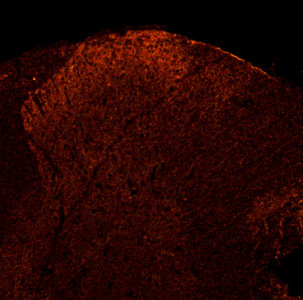
P2X3 in rat Dorsal Horn.
Check out our Purinergic Receptor Antibodies today.


Friday, May 27, 2011

bradykinin B2 or purinergic P2Y receptors and SNs

Our Neurotransmission Research Antibodies are proving to be important tools in studying excitability and firing properties of sympathetic neurons (SNs).

In this study the authors probe the role of a particular nerve cell potassium current, called the M-current, in the control of neurotransmitter release, using the contraction rate of the co-cultured heart cells as a functional read-out of noradrenaline release. Using several drugs and receptor agonists, we manipulated the activity of M-current in the nerve cells, which were stimulated by nicotine, and monitored its effect on heart cell beating. We find that the M-type potassium current has a robust role in the control of noradrenaline release from the nerve cells, and in the response of the heart cells to increased beating frequency as a result:

Oleg Zaika, Jie Zhang, and Mark S. Shapiro. Functional role of M-type (KCNQ) K+ channels in adrenergic control of cardiomyocyte contraction rate by sympathetic neurons. J. Physiol., May 2011; 589: 2559 - 2568.
Abstract: M-type (KCNQ) K+ channels are known to regulate excitability and firing properties of sympathetic neurons (SNs), but their role in regulating neurotransmitter release is unclear, requiring further study. We sought to use a physiological preparation in which SNs innervate primary cardiomyocytes to evaluate the direct role of M-channels in the release of noradrenaline (NA) from SNs. Co-cultures of rat SNs and mouse cardiomyocytes were prepared, and the contraction rate (CR) of the cardiomyocyte syncytium monitored by video microscopy. We excited the SNs with nicotine, acting on nicotinic acetylcholine receptors, and monitored the increase in CR in the presence or absence of the specific M-channel opener retigabine, or agonists of bradykinin B2 or purinergic P2Y receptors on the SNs. The maximal adrenergic effect on the CR was determined by application of isoproterenol (isoprenaline). To isolate the actions of B2 or P2Y receptor stimulation to the neurons, we prepared cardiomyocytes from B2 receptor or P2Y2 receptor knock-out mice, respectively. We found that co-application of retigabine strongly decreased the nicotine-induced increase in CR. Conversely, co-application of bradykinin or the P2Y-receptor agonist UTP augmented the nicotine-induced increase in CR to about half of the level produced by isoproterenol. All effects on the CR were wholly blocked by propranolol. Our data support the role of M-type K+ channels in the control of NA release by SNs at functional adrenergic synapses on cardiomyocytes. We conclude that physiological receptor agonists control the heart rate via the regulation of M-current in SNs.


This research has implications for forwarding the understanding of heart pacing.