Saturday, September 30, 2017

Neuromics' FGF-2 for Expanding Human Cells

FGF-2 and Fibrochondrocytes

FGF-2 of FGF-basic is an important a growth factor for many cell-based assays. Neuromics' has rock solid FGFs for supplementing media used to grow cells. Check out publications referencing use of FGF-2.

Here's the latest publication: Yan Liang, Enaam Idrees, Stephen H. J. Andrews, Kirollos Labib, Alexander Szojka, Melanie Kunze, Andrea D. Burbank, Aillette Mulet-Sierra, Nadr M. Jomha & Adetola B. Adesida. Plasticity of Human Meniscus Fibrochondrocytes: A Study on Effects of Mitotic Divisions and Oxygen Tension. Scientific Reports 7, Article number: 12148 (2017) doi:10.1038/s41598-017-12096-x. ...Thereafter the number of viable MFCs were counted using a haemacytometer after trypan blue staining. MFCs were plated at 104 cells/cm2 and cultured in the standard medium described above supplemented with FGF-2 (5 ng/mL; Neuromics, MN, USA, Catalog#: PR80001) and TGFβ1 (1 ng/mL; ProSpec, NJ, USA, Catalog#: cyt-716) under normal oxygen tension (21% O2) at 37 °C in a humidified incubator...
Images: Immunofluorescence analysis of collagen I and collagen II in pellets derived from T1F2-expanded MFCs of four passages after 21 days chondrogenic stimulation under NRX or HYP from one representative donor (male, 20 years old). Blue (DAPI): cells, Red (Texas Red): collagen I, Green (FITC): collagen II. (A) Pellets cultured under NRX, (B) Pellets cultured under HYP from four passages. Scale bar: 100 µm
Our ISO-Kine FGF-2 is especially potent as it virtually endotoxin free.

Thursday, September 21, 2017

i-Fect Delivers Plasmids!

Important for Gene Expression Studies.
I have posted many examples of how our customers use i-FectTM  and other Transfection Solutions for Gene Manipulation Studies. There are also many publications.

Here we feature how i-Fect was used to delivery plasmids to the CNS: Sara Elramah, María José López-González, Matthieu Bastide, Florence Dixmérias, Olivier Roca-Lapirot, Anne-Cécile Wielanek-Bachelet, Anne Vital, Thierry Leste-Lasserre, Alexandre Brochard, Marc Landry & Alexandre Favereaux. Spinal miRNA-124 regulates synaptopodin and nociception in an animal model of bone cancer pain. Scientific Reports 7, Article number: 10949 (2017) doi:10.1038/s41598-017-10224-1...Intrathecal administration of miRNAs and ShRNA To over-express miR-124, we cloned the pre-miRNA sequence of miR-124 into a plasmid. To determine cells expressing this miR-124 encoding plasmid, we added a GFP-coding sequence to the construct under the control of an IRES. Thus, miR-124 over-expressing cells also express GFP. To inhibit synaptopodin expression, we cloned a ShRNA sequence directed against synaptopodin into a plasmid. To determine cells expressing this ShRNA, we added a GFP-coding sequence to the construct under the control of an IRES. Thus, ShRNA expressing cells also expressed GFP. Two micrograms of these plasmids or the corresponding controls, were solubilized in 10 µl of i-Fect reagent (Neuromics, Edina, USA), and injected intrathecally between the L5 and L6 lumbar vertebrae every two days for a total of 3 injections, according to the manufacturer’s instructions and previously published experiments...
Figures: (C and D) Immunostaining of synpo in spinal cord after miR-124 intrathecal injections: only the dorsal horn which receive nociceptive information was quantified (white dash area). Measurement of synaptopodin stained area reveals ability of miR-124 to inhibit endogenous Synpo expression (20/3 and 17/3 denotes number of sections/animals for control and miR-124-injected mice, respectively.
I am confident there will be many more positive reports regarding our Transfection Reagents.

Saturday, September 16, 2017

Dynamics of Stem Cell Differentiation

Differentiation Markers
Neuromics has excellent markers for determining stem cell differentiation.

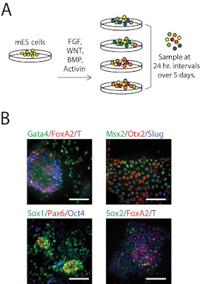
This new publication references use of our OTX2 marker: Sumin Jang, Sandeep Choubey, Leon Furchtgott, Ling-Nan Zou, Adele Doyle, Vilas Menon, Ethan B Loew, Anne-Rachel Krostag, Refugio A Martinez, Linda Madisen, Boaz P Levi, Sharad Ramanathan. Dynamics of embryonic stem cell differentiation inferred from single-cell transcriptomics show a series of transitions through discrete cell states. eLife 2017;6:e20487. DOI: http://dx.doi.org/10.7554/eLife.20487


Figures: Single-Cell Gene Expression Profiling of mESCs during early germ layer differentiation. (A) Mouse embryonic stem cells (mESCs) were exposed to various differentiation conditions to perturb FGF, WNT, and TGF-beta signaling for up to five days of differentiation. (B) Images of immunostained mESCs undergoing differentiation show cell-to-cell variability in their expression of known germ layer marker genes. (Scale bar = 100 μm).
We will continue to post new developments.

Friday, September 01, 2017

Neuronal Markers

New Pub References 3 Markers
We are recognized for our large catalog of neuronal markers. Our strength, in this area, includes markers designed for pain researchers.

They are widely used and frequently published. This new publication references use of our Guinea Pig Substance P, Guinea Pig PGP9.5 and Chicken NF200 of NF-Heavy. andla, Jagadeesha, Lomada, Santosh Kumara, Jianninga; Kuner, Rohinia, Bali, Kiran Kumar. miR-34c-5p functions as pronociceptive microRNA in cancer pain by targeting Cav2.3 containing calcium channels. Pain: September 2017 - Volume 158 - Issue 9 - p 1765–1779 doi: 10.1097/j.pain.0000000000000971.
Neuromics' PGP9.5 Staining of  Mouse DRGs.
.Neuromics SP and NF200 Staining of Mouse DRGSs