Showing posts with label Immunhistochemistry. Show all posts

Tuesday, March 03, 2015

The Roots of Anxiety Induced Pain

Corticotropin-Releasing Factor and ERK1/2 Pathway

This study crossed my radar scope because the investigators referenced use of our phosphoERK1/2 for Western Blotting and Immunohistochemistry: Gisela Patrícia da Silva Borges , Juan Antonio Micó Segura , Fani Lourença Moreira Neto , Esther Berrocoso. Corticotropin-Releasing Factor Mediates Pain-Induced Anxiety through the ERK1/2 Signaling Cascade in Locus Coeruleus Neurons. DOI: http://dx.doi.org/10.1093/ijnp/pyv019 First published online: 25 February 2015

Conclusion: pain-induced anxiety is mediated by CRF neurotransmission in the LC through ERK1/2 signaling cascade.

Figure: a) Schematic representation of the anatomical pathways implicated. Briefly, the contralateral LC indirectly receives inputs from the inflamed paw (red dashed line; ascending pathways) and, subsequently, the information is sent to corticolimbic areas. Additionally, the LC sends direct projections to the spinal cord (blue straight line; descending pathways). b) Body weight of the control and MA rats. c) Body rectal temperature of control and MA rats. d) Mechanical hyperalgesia represented by a significant decrease in the paw withdrawal threshold of the ipsilateral paw of MA rats. e) Mechanical allodynia represented by a significant decrease in the force threshold of the ipsilateral paw of MA rats. Graph depicting the expression of pERK1/2 in the LC after intra-LC administration of the αCRF receptor antagonist, showing that the significant increase of pERK1/2 in MA4W animals was no longer observed when this antagonist was administered.  g) Graph showing that the local administration of the αCRF antagonist had no significant effect on mechanical hyperalgesia in MA4W rats. h) Graph showing that local administration of h) Graph showing that local administration of the α-helical CRF antagonist had no significant effect on mechanical allodynia in the ipsilateral paw of MA rats. i) Graph showing that the time spent in the open arms decreased in MA4W rats receiving the vehicle alone but this effect was successfully reversed by administration of the αCRF antagonist. j) Graph showing that local administration of the α-helical CRF antagonist had no significant effect on the total distance traveled in the elevated zero maze. k) Graph showing that local administration of the α-helical CRF antagonist reversed the decrease in the number of entries into the open arms observed in MA4W rats receiving the vehicle alone. B=Baseline; LC=Locus Coeruleus; αCRF=antagonist of the corticotropin-releasing factor receptor I and II; W=Week; MA=Monoarthritis.
 

Western Blotting: The membranes were blocked with 5% Bovine Serum Albumin (BSA; Sigma, Spain) in TBST and probed overnight at 4 ºC with a rabbit anti-phospho-ERK1/2 (1:5,000; Neuromics). Immunohistochemistry: Brains were removed and processed for free-floating immunohistochemistry. One in five sequential transverse brain sections (30 µm) containing the PVN from each rat were washed, blocked and incubated with a rabbit antiserum against the phosphorylated ERK1 and ERK2 isoforms (pERK1/2; 1:1000; 48 hours at 4-8ºC: Neuromics, USA). Immunodetection was achieved with a biotinylated donkey anti-rabbit antiserum (1:500; 1 hour; Jackson ImmunoResearch, USA), followed by an ABC solution (1:200, 1 hour; ABC Elite kit, Vector Laboratories, UK) and a colorimetric reaction with 3,3-diaminobenzidine tetrahydrochloride (DAB; 10 min) in 0.05M Tris-HCl buffer containing 0.003% hydrogen peroxide (Cruz et al., 2005). Sections were then washed in PBS, mounted on gelatin-coated glass slides, cleared in xylene, cover-slipped with DPX and analyzed by light microscopy.

We have a broad range of pain and inflammatory response research markers. Check us out today.


Saturday, May 18, 2013

The Role of Obesity in Obstructive Sleep Apnea (OSA)

Intermittent Hypoxia and Leptins

Scott Mesenger recently published a Master's Thesis that implicates compelling links between Leptin Signaling and OSA: http://ir.lib.uwo.ca/cgi/viewcontent.cgi?article=2487&context=etd. This work was done in the John Cirello Lab at University of Western Ontario. The study extensively uses our Ob-Rb Antibody to generate data.

There is a strong link between obesity and OSA. "Perhaps the single most important factor affecting OSA risk is body weight. Weight gain of 10% increases the risk of developing OSA by six-fold (Peppard et al., 2000a) and a 10% loss of body weight is estimated to decrease the apnea/hypopnea index (events/hour) by 26%  Within the obese population (BMI ≥ 30) (Wolk et al., 2003), approximately 40% experience significant OSA and approximately 70% of OSA patients are obese (Vgontzas et al., 1994), daunting numbers considering the obesity epidemic occurring in North America and estimates suggesting 41% of Americans will be obese by 2015 (Nejat et al., 2009)."

"Leptin can exert cardiovascular effects by acting centrally, as has been shown by studies introducing leptin directly into the CNS. Central administration serves to increase plasma catecholamines epinephrine and norepinephrine (Satoh et al., 1999). In other studies, leptin has been found to increase SNA, arterial pressure as well as heart rate and to inhibit the baroreflex (Arnold et al., 2009; Mark et al., 2009). Leptin may also have a role in modulating the peripheral chemoreflex at the level of the CNS as microinjection of leptin into caudal pressor areas of the NTS has been found to potentiate the sympathetic and blood pressure responses to chemoreflex activation (Ciriello & Moreau, 2012). These cardiovascular effects may not be debilitating under normal homeostatic conditions, however in conditions of increased circulating leptin, such as in OSA and obesity, the cardiovascular responses may be detrimental."

This study shows the increase in the expression of leptins like Ob-Rb in Carotid .

Images: Fluorescent (a-c) photomicrographs showing the effect of IH on Ob-Rb (a) and ERK 1/2 (b) expression in carotid body glomus cells. Note that Ob-Rb and ERK 1/2 are co-expressed in the same cells (c). Calibration mark in (a) represents 100 μm and applies to (a-c).
Images: Western blots showing the presence of Ob-Rb (a) and Ob-R100 (b) in carotid bodies after IH. Note that IH significantly (*) decreases the protein level of Ob-Rb (a), while significantly (*) increasing the protein level of Ob-R100 (b) compared to normoxic controls (b). p values are indicated. n=5-6.

This study will give the reader great insights on the molecular biology of OSA. Check it out: http://ir.lib.uwo.ca/cgi/viewcontent.cgi?article=2487&context=etd.

Sunday, April 14, 2013

P2X3 Receptors and Migraine

P2X3 Receptors of Trigeminal Sensory Neurons and Familial Hemiplegic Migraine Type 1 (FHM-1).

Our P2X Receptor Markers continue to be used in interesting and novel ways. Here researchers use our P2X3 Receptor Antibody to study expression using primary rat ganlia cultures: Swathi K. Hullugundi,Michel D. Ferrari, Arn M. J. M. van den Maagdenberg, Andrea Nistri. Andrea Nistri. The Mechanism of Functional Up-Regulation of P2X3 Receptors of Trigeminal Sensory Neurons in a Genetic Mouse Model of Familial Hemiplegic Migraine Type 1 (FHM-1). PLoS ONE 8(4): e60677. doi:10.1371/journal.pone.0060677

Abstract: A knock-in (KI) mouse model of FHM-1 expressing the R192Q missense mutation of the Cacna1a gene coding for the α1 subunit of CaV2.1 channels shows, at the level of the trigeminal ganglion, selective functional up-regulation of ATP -gated P2X3 receptors of sensory neurons that convey nociceptive signals to the brainstem. Why P2X3 receptors are constitutively more responsive, however, remains unclear as their membrane expression and TRPV1 nociceptor activity are the same as in wildtype (WT) neurons. Using primary cultures of WT or KI trigeminal ganglia, we investigated whether soluble compounds that may contribute to initiating (or maintaining) migraine attacks, such as TNFα, CGRP, and BDNF, might be responsible for increasing P2X3 receptor responses. Exogenous application of TNFα potentiated P2X3 receptor-mediated currents of WT but not of KI neurons, most of which expressed both the P2X3 receptor and the TNFα receptor TNFR2. However, sustained TNFα neutralization failed to change WT or KI P2X3 receptor currents. This suggests that endogenous TNFα does not regulate P2X3 receptor responses. Nonetheless, on cultures made from both genotypes, exogenous TNFα enhanced TRPV1 receptor-mediated currents expressed by a few neurons, suggesting transient amplification of TRPV1 nociceptor responses. CGRP increased P2X3 receptor currents only in WT cultures, although prolonged CGRP receptor antagonism or BDNF neutralization reduced KI currents to WT levels. Our data suggest that, in KI trigeminal ganglion cultures, constitutive up-regulation of P2X3 receptors probably is already maximal and is apparently contributed by basal CGRP and BDNF levels, thereby rendering these neurons more responsive to extracellular ATP.

Images: Examples of TNFR2 and P2X3 co-exexpression in (wildtype) WT and R192Q (knockin) KI neurons. Left panel shows P2X3 expression (green), and right panel shows TNFR2 staining (red). B, Histograms quantifying % of cells co-expressing TNFR2 and P2X3: both WT and KI cultures show similar TNFR2 and P2X3 co-expression. N = 3 independent experiments (6 mice). C, Representative traces of currents induced by application of α,β-meATP (10 µM, 2 s) to WT or R192Q KI neurons in control conditions or after 4 h TNFα application. D, Histograms show average peak amplitudes of P2X3 receptor-mediated currents: WT control (open bar), n = 30; WT TNFα (stippled bar), n = 38; KI control (grey bar), n = 34; KI TNFα (stippled gray bar), n = 34; ** = p<0 .006="" i="" nbsp="" p="">doi:10.1371/journal.pone.0060677.g001.

Understanding the interplay between TNFR2 and P2X3 could lead to a better understanding of the root causes of migraines. This could open up yet more potential drug targets for this insidious condition.

Check out these related reagent categories:
All Purinergic Receptor Antibodies
Pain and Inflammation Research Antibodies 
Neurotransmission Research Antibodies
Primary Neurons and Astrocytes-Primary human, rat and mouse neurons and astrocytes

Thursday, April 04, 2013

Science Behind Elecroacupuncture for Treating Shingles

Postherpetic neuralgia (PHN) or shingles, caused by caused by herpes zoster, causes nerve damage in the skin and results in abnormal electrical signals to the brain,  and may persist or recur for months, years or for life.

Electroacupuncture (EA) is effective in relieving pain in patients with PHN. Researchers in this study determined the beneficial effect of EA and the potential mechanisms in a rat model of PHN. They use our TRPV1 antibody to track expression in the Dorsal Root Ganglia (DRG) and Dorsal Horn (DH): Cai-hua Wu, Zheng-tao Lv, Yin Zhao, Yan Gao, Jia-qing Li, Fang Gao, Xian-fang Meng, Bo Tian, Jing Shi, Hui-lin Pan and Man Li. Electroacupuncture improves thermal and mechanical sensitivities in a rat model of postherpetic neuralgia. Molecular Pain 2013, 9:18 doi:10.1186/1744-8069-9-18


Conclusions: EA treatment improves thermal perception by recovering TRPV1-positive sensory neurons
and nerve terminals damaged by RTX. EA Also reduces RTX-induced tac tile allodynia by attenuating the damage of myelinated afferent nerves and their abnormal sprouting into the spinal lamina II. Our study provides new information about the mechanisms of the therapeutic actions of EA in the treatment of PHN.
Figure 2:  Effect of EA on RTX-induced deletion of TRPV1-immunoreactive neuron s in the DRG. A, Representative images showing TRPV1-immunoreactive neurons in the lumbar DRG of vehicle ( a ) , RTX ( b ), RTX plus 2 Hz EA ( c ), RTX plus 15 Hz EA ( d ), RTX plus 100 Hz EA ( e ), and RTX plus sham EA ( f ) groups. Scale bar, 50 μ m. B, Summary data show the number of TRPV1 immunoreactive neurons in different groups. Data are expresse d as means ± SEM (n = 6 rats in each group). *P < 0.05, compared with the vehicle group; # P< 0.05, compared with the sham EA group.
Figure 3: Effect of EA on RTX-induced deletion of TRPV1 immunoreactive central terminals in the spinal dorsal horn. A, Representative images showing TRPV1 immunoreactive central terminals of afferent fibers in the spinal dorsal horn of vehicle (a) ,RTX (b), RTX plus 2 Hz EA (c), RTX plus 15 Hz EA (d), RTX plus 100 Hz EA (e), and RTX plus sham EA (f) groups. Scale bar, 50 μm. B, Summary data show the area of TRPV1 immunoreactive central terminals in different groups. Data are expressed as means ± SEM (n= 6 rats in each group). *P < 0.05, compared with the vehicle group; # P < 0.05, compared with the sham EA group. 
I am always interested in how our Pain and Inflammation Research Markers are used to help understand the science behind pain therapies and also discovery of new therapies. There are multiple postings on these subjects with many more to come.

Sunday, January 27, 2013

Epidermal Nerve Fibers in Neuropathic Pain Model

Re-innervation in spared nerve injury (SNI) vs normal rats
Neuromics' Pain and Inflammation Research Antibodies are frequently used to help researchers find root causes of neuropathic pain. This is a fascinating study of re-innervation patterns of epidermal and dermal nerve fibers in a rat neuropathic pain model (Note: our Guinea Pig Polyclonal P2X3 Antibody was used in this study): Liron S. Durakua, Mehdi Hossaini, Barthold N. Schüttenhelm, b, Joan C. Holstege, Martijn Baas, Tom J.H. Ruigrok, Erik T. Walbeehm. Re-innervation patterns by peptidergic Substance-P, non-peptidergic P2X3, and myelinated NF-200 nerve fibers in epidermis and dermis of rats with neuropathic pain. Experimental Neurology Volume 241, March 2013, Pages 13–24. doi.org/10.1016/j.expneurol.2012.11.029.
Non-footpad area vs. footpad.The distribution pattern between the center non-footpad area and footpad is compared for the different subgroups of sensory skin fibers. For epidermal Sub P-IR and upper dermal NF-200-IR fibers there is an equal density of fibers in the center and footpad area suggesting a homogenous distribution over the foot sole of a rat. Whereas for epidermal P2X3-IR fibers there is a significant lower number of P2X3-IR fibers in the footpads as compared to the center non-footpad area, suggesting a heterogeneous distribution in the foot sole for this type of sensory fibers. N = 5 per group. Unpaired t-test. ***: p < 0,001. Scale bar: 250 μm. doi.org/10.1016/j.expneurol.2012.11.029

Illustration of the skin innervation in normal and SNI situation.Illustration showing the innervation pattern of subgroups of sensory skin fibers in the normal situation and in the SNI model 10 weeks PO. In the naïve animals the peptidergic CGRP and non-peptidergic P2X3 fibers have a lower density in the footpads as compared to the non-footpad areas, while the Substance P and NF-200 fibers have an equal distribution over the complete foot sole. In the SNI model there is a significant increase of CGRP epidermal fibers in the medial and lateral area of the foot sole and there is a complete re-innervation of the center area. In addition the footpads in the SNI model are hyper-innervated with CGRP fibers. Substance P and P2X3 and NF-200 fibers show no increased density in the uninjured medial and lateral area after 10 weeks PO. In addition, Substance P and P2X3 fibers hardly re-innervate the center area; however, the NF-200 fibers have the same density of fibers in the denervated area as in the normal situation. In the SNI model the medial, center and lateral area have all an increase in LC's, with the center area being the most prominent one. In addition the epidermis thickness of the SNI model has decreased significantly in all the three areas after 10 weeks PO.
Related Postings: http://neuromics.blogspot.com/search/label/Neuropathic%20Pain.

Sunday, October 21, 2012

P2X3 Receptor and CGRP Antibodies Immunostaining

Dr. Alfredo Ribeiro-da-Silva, McGill University, is a serial publisher of studies using our pain and inflammation research antibodies.

Here, use of our rabbit anti-CGRP and guinea pig anti-P2X3 is referenced. Please note the high titer of these antibodies (dilution is 1:25,000): Abeer W Saeed, Alfredo Ribeiro-da-Silva. Non-peptidergic primary afferents are presynaptic to neurokinin-1 receptor immunoreactive lamina I projection neurons in rat spinal cord. Molecular Pain 2012, 8:64 doi:10.1186/1744-8069-8-64.


Images: CGRP, IB4 and P2X3 staining in transverse spinal cord sections. A and B show low magnification confocal images of CGRP-IR and IB4 positive (A) or P2X3-IR (B) fibers. C and D represent high magnification confocal images from the middle third of the lateromedial extent of the superficial dorsal horn. In C, note that there is limited co-localization of IB4 and CGRP (in yellow). Arrowheads show axonal varicosities (boutons) from nonpeptidergic fibers in lamina I, which do not co-localize CGRP immunoreactivity. The framed regions in A and B indicate the approximate regions from where C and D, respectively, were obtained (the latter originate from other sections). CGRP (in green); IB4 (in red); P2X3 (in red). Scale bar (A, B) = 200 μm; scale bar (C, D) = 20 μm

Tissue processing: The injection site at the level of the parabrachial nucleus was examined by cutting serial, 100 μm-thick coronal sections of the relevant brain region. The dorsal aspect of the L4-L5 spinal cord segment was cut into serial, 50 μm-thick horizontal sections (n = 10), 50 μm-thick parasagittal sections (n = 4) or 50 μm-thick transverse sections (n = 4). All sections were cut using a freezing sledge microtome (Leica, Richmond Hill, Ontario) and collected as freefloating in phosphate-buffered saline (PBS) with 0.2% Triton-X 100 (PBS + T). To block unspecific staining, all spinal cord sections were incubated, for one hour, in 10% normal donkey serum (NDS) (Jackson, West Grove, PA) in PBS + T at room temperature. Subsequently, the sections were placed in primary antibodies (or conjugated lectin IB4 - see below) for 48 hours at 4 °C. We used a mixture of 2 or 4 primary antibodies (each raised in a different species), or IB4, in PBS + T containing 5% NDS. Next, the sections were washed in PBS + T and then incubated in species-specific secondary antibodies that were raised in donkey and conjugated to either AlexaFluor 488, AlexaFluor 405, Rhodamine RedX or biotin. The sections were incubated in 3 different cocktails: #1) rabbit anti-CGRP at a 1:200 dilution (Sigma, St Louis, MO) and lectin IB4 conjugated to AlexaFluor 568 at a 1:200 dilution (Molecular Probes); #2) rabbit anti-CGRP and guinea pig anti-P2X3 at a 1:25,000 dilution (Neuromics, Edina, MN); #3) goat anti-CTb at a 1:5000 dilution (List Biological), rabbit anti-NK-1r at a 1:10000 dilution (Sigma, St Louis, MO), guinea pig anti-CGRP at a 1:8000 dilution (Peninsula, San Carlos, CA) and lectin IB4 conjugated to AlexaFluor 647 at a 1:200 dilution (Molecular Probes). All the sections were washed with PBS + T and then (for #1) incubated for 2 hours at room temperature with donkey anti-rabbit AlexaFluor 488; (for #2) incubated for 90 minutes in a biotin conjugated donkey anti-guinea pig IgG (Jackson Immunoresearch, West Grove, PA, 1:200). Further signal amplification was achieved by treating the sections with 1 hour incubation in an avidin-biotin (A + B) complex (Vectastain Elite ABC kit, Vector Laboratories) followed by tyramide (Perkin-Elmer, Norwalk, CT, 1:75) for 7 minutes. Sections were then incubated in streptavidin conjugated to AlexaFluor 568 (Molecular Probes, Eugene, OR, 1:200) and donkey anti-rabbit AlexaFluor 488; or (for #3) incubated for 2 hours at room temperature with secondary antibodies: donkey anti-goat Rhodamine Red X, donkey anti-rabbit AlexaFluor 488, and donkey anti-guinea pig AlexaFluor 405. Finally, sections were washed with PBS, mounted on gelatin-subbed slides and coverslipped with an anti-fading mounting medium (Aqua Polymount; Polysciences, Warrington, PA). Slides were stored at −4 °C pending further processing.

I will continue to publish outstanding customer data/images using our natibodies/markers.

Saturday, September 22, 2012

Neuron-specific class III beta-tubulin (TuJ1) Staining in hNT2.19 Neurons

Our Tuj-1 antibody is widely used and frequently published. It is proving a potent marker for confirming the differentiation of human neural progenitors to neurons.

In this study the authors use the marker for staining hNT2.19 Neurons: Mary J. Eaton, Yerko Berrocal, and Stacey Q. Wolfe. Potential for Cell-Transplant Therapy with Human Neuronal Precursors to Treat Neuropathic Pain in Models of PNS and CNS Injury: Comparison of hNT2.17 and hNT2.19 Cell Lines. Pain Research and Treatment. Volume 2012 (2012), Article ID 356412, 31 pages. doi:10.1155/2012/356412. 
The results show great promise. They show hNT2 or hNT2-derived cell lines, such as hNT2.17 and hNT2.19, have great potential to permanently reverse symptoms of neuropathic pain following PNS and CNS injuries and can offer new hope to treat these intractable conditions to significantly improve human health. This includes neuropathic pain resulting from diabetic neuropathy and Spinal Cord Injury.


Images: Comparison of graft sites of hNT2.17 and hNT2.19 in the QUIS and severe contusive-SCI models, respectively, at 6 weeks after cell transplant. (a) Sagittal section of anti-GABA-immunostained QUIS + hNT2.17 transplant lumbar spinal cord 6 weeks after grafting. Easily detectible hNT2.17 cells stain for GABA (arrows) on the pial membranes. (b) Sagittal section of anti-NuMA-immunostained QUIS + hNT2.17 transplant lumbar spinal cord 6 weeks after grafting. Easily detectible hNT2.17 cells stain for NuMA (arrows) on the pial membranes in adjacent sections. The hNT2.19 were alternately injected into the subarachnoid space two weeks after severe contusive SCI. Cell graft sites were co-localized with 5HT (c) and the human-specific marker TuJ1(d) (neuron-specific class III β-tubulin). There are many surviving hNT2.19 (d) grafted cells visible on the pial surface, which stain for TuJ1 (arrows) at the end of the experiment, 56 days after SCI and about 6 weeks after cell transplant. Adjacent sections with the same grafted hNT2.19 (c) are labeled for 5HT (arrows). Magnification bar = 20 μm.

Protocol: Modified methods for staining spinal cord sections for the human neuron-specific class III beta-tubulin (TuJ1) to identify grafted hNT2.19 neurons after grafting have previously been described [51]. The sections were washed with 0.1 M PBS pH 7.4 and permeabilized with 0.4% Triton-X-100 in 0.1 M PBS, 10% normal goat serum (NGS) for one hour. The sections were then incubated overnight at 4°C in the primary anti-TuJ1 antibody (1 : 100 DPBS), and the permeabilizing solution, followed by a one-hour incubation at room temperature with the secondary antibody solution, biotinylated mouse IgG raised in goat (Vector; 1 : 200), a Peroxidase ABC reporter in 0.1 M PBS (Vector) and “VIP” substrate (Vector). Some sections were stained in the absence of primary antibody and served as the negative controls.

I will continue to post new applications for our stem cell research reagents.

Thursday, May 17, 2012

Pinpointing Neuropathic Pain

I am pleased to feature our Guinea Pig P2X3 Antibody as a tool for studying the root causes of Neuropathic Pain.

In this study, the authors showed ablation of nonpeptidergic fibers in a chronic constriction injury model caused significant sympathetic and parasympathetic P2X3 fiber sprouting, and led to an exacerbated pain response. This was an unexpected finding, as it has been suggested that nonpeptidergic fibers play a major role in mechanical pain, and suggests that these fibers play a complex role in the development of neuropathic pain: Anna M.W. Taylora, Maria Osikowicza, Alfredo Ribeiro-da-Silva. Consequences of the ablation of nonpeptidergic afferents in an animal model of trigeminal neuropathic pain. PAIN. Volume 153, Issue 6, June 2012, Pages 1311–1319. doi.org/10.1016/j.pain.2012.03.023....Sections were then incubated for 48 hours at 4°C with a guinea pig polyclonal anti-P2X 3 antibody (1:25,000; Neuromics, Edina, MN, USA), diluted in PBS-T. Following primary antibody incubation, sections were treated with a biotin-conjugated...

Image: Example of  Non-petidergic fibers expressing P2X3 receptor for ATP are present in lamina II of the contralateral dorsal horn (arrows). The P2X3-positive fibers are eliminated from the rat dorsal horn ipsilateral to the rhizotomy (arrow heads). Spinal cord segment C6. Scale bar 400 um. P2X3 antibody dilution 1:25,000. Biomédica vol.24 no.2.

Check out our related reagents categories:
 I will continue to post important findings related to the root causes of Neuropathic Pain.


Thursday, April 26, 2012

Primary Hippocampal Neurons Performing!

I advertise our Primary Neurons and Astrocytes as being easy to culture, grow and maintain. We confirm this via the data/images our customers generously share and the many publications referencing use of the cells.

I would like to thank George Kenneth Todd (Patterson Lab at UC Davis) for these wonderful images of our e18 Primary Rat Hippocampal Neurons. These were taken at day 67!


Prior to staining, the cells were treated with Wnt5 for 2 min, then Wnt5 + Wnt3 for an additional 2 min during Calcium Imaging experiments. The cells were then fixed and stained for IP3R (green), Frizzled2 (blue), and B-Catenin (red), and the confocal images were captured at 10x. Notice the parallel neurites formation in image 3.


For staining culture options, check out our neuron-glial-astrocyte markers.

Wednesday, February 15, 2012

Guinea Pig P2X3 Antibody-It's Back!

Our Guinea Pig P2X3 Antibody has historically been one of our most popular Purinergic Receptor Antibodies. It has been frequently cited in publications.

We exhausted our supply of the antibody in the spring of 2011 and it took us many rounds with multiple partners and a degree of disappointing results. Well our efforts have finally yielded the positive testing results required to again provide users this important antibody. Here're related images from our internal testing:
P2X3 staining in rat Dorsal Root Ganglia.
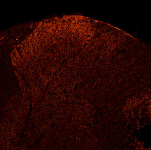
P2X3 in rat Dorsal Horn.
Check out our Purinergic Receptor Antibodies today.


Tuesday, November 08, 2011

Opioid Induced Itch

Our widely used and frequently published Opioid Receptors Antibodies are used for a spectrum of pain research. Here is an interesting study on the root causes of opioid induced itch.

Xian-Yu Liu, Zhong-Chun Liu, Yan-Gang Sun, Michael Ross1, Seungil Kim, Feng-Fang Tsai, Qi-Fang Li, Joseph Jeffry, Ji-Young Kim, Horace H. Loh, Zhou-Feng Chen. Unidirectional Cross-Activation of GRPR by MOR1D Uncouples Itch and Analgesia Induced by Opioids. Cell, Volume 147, Issue 2, 14 October 2011, Pages 261-262.


Root Causes: Spinal opioid-induced itch, a prevalent side effect of pain management, has been proposed to result from pain inhibition. We now report that the μ-opioid receptor (MOR) isoform MOR1D is essential for morphine-induced scratching (MIS), whereas the isoform MOR1 is required only for morphine-induced analgesia (MIA). MOR1D heterodimerizes with gastrin-releasing peptide receptor (GRPR) in the spinal cord, relaying itch information. We show that morphine triggers internalization of both GRPR and MOR1D, whereas GRP specifically triggers GRPR internalization and morphine-independent scratching. Providing potential insight into opioid-induced itch prevention, we demonstrate that molecular and pharmacologic inhibition of PLCβ3 and IP3R3, downstream effectors of GRPR, specifically block MIS but not MIA. In addition, blocking MOR1D-GRPR association attenuates MIS but not MIA. Together, these data suggest that opioid-induced itch is an active process concomitant with but independent of opioid analgesia, occurring via the unidirectional cross-activation of GRPR signaling by MOR1D heterodimerization.

Monday, August 22, 2011

MOR and NMDAR Interplay-Implications in Pain Control

Our Opioid Receptor Antibodies continue to be referenced in publications by Pain Researchers. Many of these studies provide a greater understanding of how opioids alleviate pain and what modulates this ability.

For example, the capacity of opioids to alleviate inflammatory pain is negatively regulated by the glutamate-binding N-methyl-D-aspartate receptor (NMDAR). This study drills down into the specifics of this regulation and references use of Neuromics' MOR1C Antibody: María Rodríguez-Muñoz, Pilar Sánchez-Blázquez, Ana Vicente-Sánchez, Esther Berrocoso and Javier Garzón. María Rodríguez-Muñoz, Pilar Sánchez-Blázquez, Ana Vicente-Sánchez, Esther Berrocoso and Javier Garzón. The Mu-Opioid Receptor and the NMDA Receptor Associate in PAG Neurons: Implications in Pain Control. Neuropsychopharmacology , (3 August 2011) | doi:10.1038/npp.2011.155.

Abstract: The capacity of opioids to alleviate inflammatory pain is negatively regulated by the glutamate-binding N-methyl-D-aspartate receptor (NMDAR). Increased activity of this receptor complicates the clinical use of opioids to treat persistent neuropathic pain. Immunohistochemical and ultrastructural studies have demonstrated the coexistence of both receptors within single neurons of the CNS, including those in the mesencephalic periaqueductal gray (PAG), a region that is implicated in the opioid control of nociception. We now report that mu-opioid receptors (MOR) and NMDAR NR1 subunits associate in the postsynaptic structures of PAG neurons. Morphine disrupts this complex by protein kinase-C (PKC)-mediated phosphorylation of the NR1 C1 segment and potentiates the NMDAR–CaMKII, pathway that is implicated in morphine tolerance. Inhibition of PKC, but not PKA or GRK2, restored the MOR–NR1 association and rescued the analgesic effect of morphine as well. The administration of N-methyl-D-aspartic acid separated the MOR–NR1 complex, increased MOR Ser phosphorylation, reduced the association of the MOR with G-proteins, and diminished the antinociceptive capacity of morphine. Inhibition of PKA, but not PKC, CaMKII, or GRK2, blocked these effects and preserved morphine antinociception. Thus, the opposing activities of the MOR and NMDAR in pain control affect their relation within neurons of structures such as the PAG. This finding could be exploited in developing bifunctional drugs that would act exclusively on those NMDARs associated with MORs.

I will continue to post these studies. They give hope for pain sufferers as many propose potential new druggable targets.

Friday, June 17, 2011

Chronic Type II Diabetes Mellitus and Cardiovascular Disease

Our Neuropeptide and Neuropeptide Receptors and Leptin and Leptin Receptor Antibdodies are frequently used to study pathologies and biology specific to Obesity and Diabetes.

Here's a new publication studying the relationship between diabetic neuropathy and altered neuropeptide Y and its receptor expression levels in myocardium and plasma.

Robina Matyal, Feroze Mahmood, Michael Robich, Hiliary Glazera, Kamal Khabbaza, Philip Hessa, Cesario Bianchia, Robert Hagberga, Shu-Xu Hua, and Frank W. Sellkea. Chronic type II diabetes mellitus leads to changes in neuropeptide Y receptor expression and distribution in human myocardial tissue. European Journal of Pharmacology. Volume 665, Issues 1-3, 31 August 2011, Pages 19-28.

Abstract: Neuropeptide Y is one of the most abundant neurotransmitters in the myocardium, and is known to influence cardiovascular remodeling. We hypothesized that diabetic neuropathy could possibly be associated with altered neuropeptide Y and its receptor expression levels in myocardium and plasma. Plasma neuropeptide Y levels in diabetic (n = 24, HgbA1c 7.9 ± 1.1%) and non-diabetic (n = 27, HgbA1c 5.8 ± 0.5%) patients undergoing cardiac surgery utilizing cardiopulmonary bypass were analyzed. Right atrial tissue of these patients was used to determine the expression of neuropeptide Y, the receptors 1–5, and leptin by immunoblotting, real-time PCR and immunofluorescence. Apoptosis signaling and endostatin and angiostatin were measured to determine the effects of leptin.


Plasma neuropeptide Y levels were significantly increased in patients with Type II diabetes mellitus as compared to non-diabetic patients (P = 0.026). Atrial tissue neuropeptide Y mRNA levels were lower in diabetic patients (P = 0.036). There was a significant up-regulation of myocardial Y2 and Y5 receptors (P = 0.009, P = 0.01 respectively) in the diabetic patients. Leptin, involved with apoptosis and angiogenesis, was down regulated in diabetic patients (P = 0.05). The levels of caspase-3, endostatin and angiostatin were significantly elevated in diabetic patients (P = 0.003, P = 0.008, P = 0.01 respectively). Y1 receptors were more likely to be localized within the nuclei of cardiomyocytes and vascular smooth muscle cells.


Neuropeptide expression is altered differentially in the serum and myocardium by diabetes. Altered regulation of this system in diabetics may be in part responsible for the decreased angiogenesis, increased apoptosis, and increased vascular smooth muscle proliferation leading to coronary artery disease and heart failure in this patient population.