Showing posts with label Oligodendrocyte Precursor Cells. Show all posts

Thursday, April 13, 2017

mGluRs Protect OPCs

Valuable for Remyelination of Damaged Neurons

We add a new publication to our mGluR Markers Category: Arthur M. Butt, Ilaria Vanzulli, Maria Papanikolaou, Irene Chacon De La Rocha, Virginia E. Hawkins. Metabotropic Glutamate Receptors Protect Oligodendrocytes from Acute Ischemia in the Mouse Optic Nerve. Neurochem Res (2017). doi:10.1007/s11064-017-2220-1. Its focus is the protective characteristics of mGluRs.


Images: Expression of mGluR in optic nerve oligodendrocytes. a RT-qPCR of mGluR subtypes in the postnatal (P8–12) and young adult (P30–35) optic nerve, compared to cortex at the same ages (inset); data are expressed as mean ± SEM ΔΔCT relative to GAPDH, ***p < 0.001 determined by ANOVA and post hoc Bonferroni’s test. b, c Oligodendrocytes in optic nerve explant cultures from P8 PLP-DsRed reporter mice after 10 DIV were immunolabelled for mGluR2/3 (d) and mGluR5 (e), illustrating single channels (Di, Dii, Ei, Eii) and the merged channel in which mGluR colocalization with PLP appears white (Diii, Eiii); scale bars 10 µm

Saturday, January 21, 2017

Leptin and Remyelination

Leptin Promotes Proliferation of OPCs

Demyelination occurs in many diseases of the Central Nervous System (CNS) including Multiple Sclerosis and Parkinson's Disease. Here researchers show that Leptin plays a role in the proliferation of Oligodendrocyte Precursor Cells (OPCs): These cells are critical for keeping the myelin sheath on Neurons of the CNS healthy and happy.  Ken Matoba, Rieko Muramatsu & Toshihide Yamashita. Leptin sustains spontaneous remyelination in the adult central nervous system. Scientific Reports 7, Article number: 40397 (2017) doi:10.1038/srep40397.

Our LepRB Antibody is used in this study to stain OPCs.


Figures: (a) Representative image of cultured OPCs stained with antibodies against LepRb (green) and PDGFRα (red). Scale bar: 25 μm. (b) Relative BrdU incorporation into the OPC obtained from the brain (left graph) and spinal cord (right graph). Cells were treated with recombinant leptin for 48 h (n = 4). (Left graph) P = 0.005993 (control vs 10 ng/mL), 0.045616 (control vs 100 ng/mL), (Right graph) P = 0.004456 (control vs 10 ng/mL), 0.017859 (control vs 100 ng/mL). (c) Relative BrdU incorporation into the OPC after leptin stimulation (10 ng/ml) with U0126 (20 μM), a MEK inhibitor (n = 4 for brain OPCs, n = 3 for spinal cord OPCs). (Left graph) P = 0.019753 (control vs leptin), 0.039433 (leptin vs leptin + U0126), (Right graph) P = 0.045545 (control vs leptin), 0.04486 (leptin vs leptin + U0126). (d) Representative images of western blotting (upper panels) and quantitative analysis of ERK phosphorylation (lower graph) are shown. OPCs were treated with leptin (10 ng/ml) under indicated periods (n = 3). P = 0.006352 (2 min), 0.016571 (5 min), 0.017675 (10 min), 0.024100 (15 min), 0.081342 (30 min).
We are in the process of looking for Labs to sponsor Neuromics' to isolate and purify adult human OPCs in return for receiving 2,000,000 cells. Stay tuned.

Tuesday, April 14, 2015

Markers for Oligodendrocyte Progenitor Cells (OPCs)

Improving OPC Expansion

Researchers reference use of Neuromics' GFAP and PDGF R Alpha/CD140A for OPC Selection

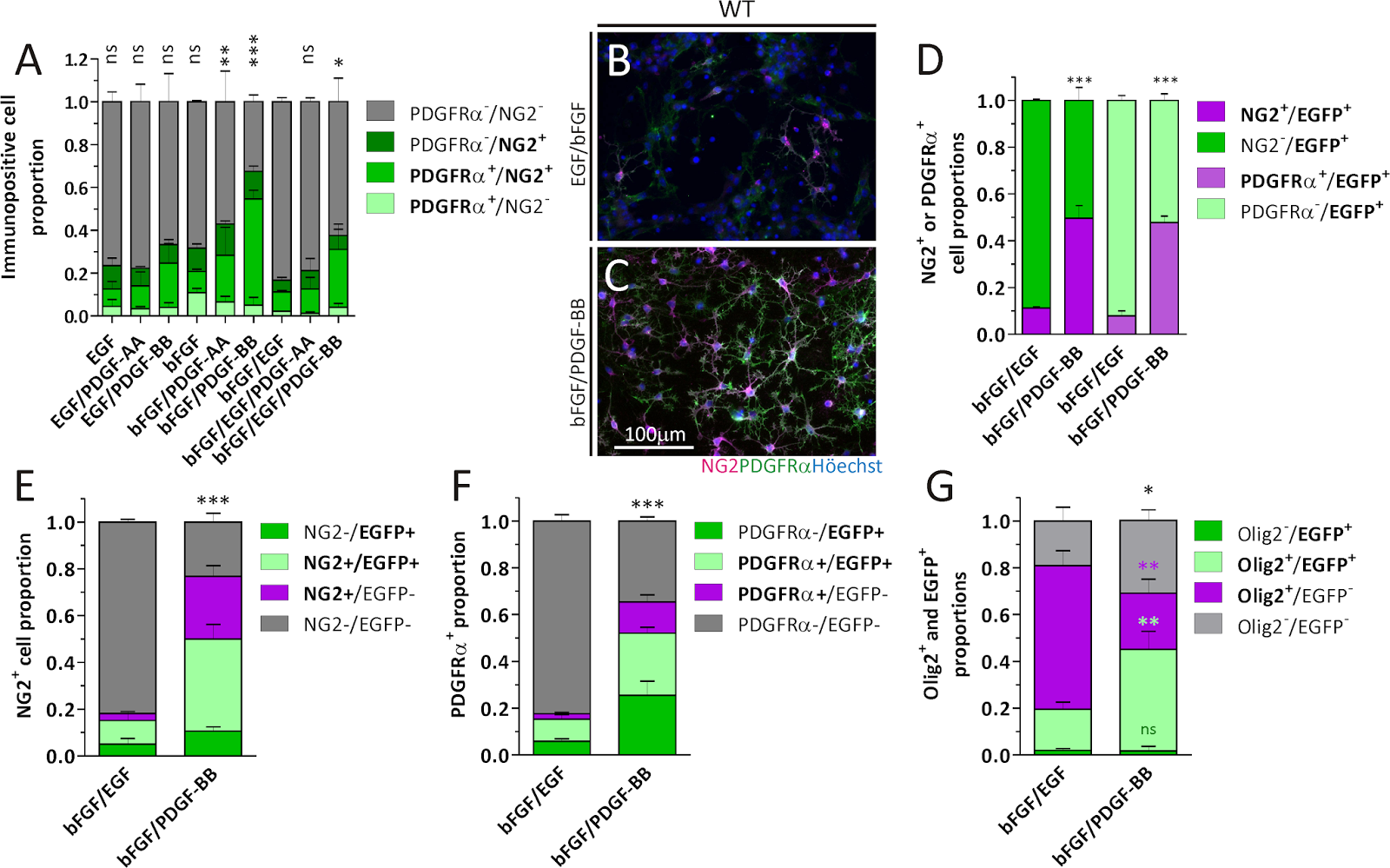
The data provided in this publication demonstrates that the OPC yield from SVZ-derived cell cultures can be improved with the PDGF-BB isoform in comparison to classical bFGF-EGF, or PDGF-AA-based protocols. Additionally, it would be expected that the OPC-enriched cultures obtained from NSC/NPC exposure to PDGF-BB and heparin generate cells suitable for cell transplantation for treating demyelinating diseases: Paula G. Franco, Juana M. Pasquini, Lucas Silvestroff. Optimizing Culture Medium Composition to Improve Oligodendrocyte Progenitor Cell Yields In Vitro from Subventricular Zone-Derived Neural Progenitor Cell Neurospheres. Published: April 2, 2015DOI: 10.1371/journal.pone.0121774:

Images: Detection of OPC markers by ICC on NS cells. A) Quantitation of NG2+ and/or PDGFRα+ cell proportions in WT mice NS generated in the presence of different growth factor combinations. Data belong to three independent experiments for each condition. Data for NG2-/PDGFRα- was analyzed with a One-way ANOVA plus Dunnett´s post test, where bFGF/EGF was set as the control. B, C) Representative images of NG2+ and PDGFRα+ cells generated from WT mice cultures in the presence of either bFGF/EGF or bFGF/PDGF-BB. D) Comparison of NG2+ or PDGFRα+ cell proportions in NS cultures generated from Act::EGFP mice in the presence of bFGF/EGF or bFGF/PDGF-BB. Data for NG2+ (dark magenta) or PDGFRα+ (light magenta) cells was analyzed separately with Student´s t test. E, F) NG2 and PDGFRα immunodetection in CNP::EGFP derived cultures. Gray bars in each graph were analyzed with Student´s t test. G) Olig2 expression in WT mice NS cultures. Asterisks are colour coded to indicate the pairs of bars compared and analyzed with Student´s t test. Cell proportions in A, and D-G are expressed as a fraction of the total cell nuclei counted for each condition. Error bars represent the SD for all bar graphs.. doi:10.1371/journal.pone.0121774.g001.

Neuromics has an excellent catalog of Stem Cell Selection Markers.

Monday, October 29, 2012

OPC Markers!

Effective Oligodendrocyte, Oligodendroglial Oligodendrocyte Lineage Markers are important for determining the differentiate state of Oligodendrocyte Precursor Cells. This is important for the study of de and re-myelination of neurons and the discovery of potential therapeutic targets for diseases like MS and ALS.

Here researchers use our Olig2 antibody to study the differentiation state of Fetal Human Oligodendrocyte Progenitor Cells: Crystal R. McClain, Fraser J. Sim and Steven A. Goldman. Pleiotrophin Suppression of Receptor Protein Tyrosine Phosphatase-β/ζ Maintains the Self-Renewal Competence of Fetal Human Oligodendrocyte Progenitor Cells. The Journal of Neuroscience, 24 October 2012, 32(43): 15066-15075; doi: 10.1523/​JNEUROSCI.1320-12.2012.
Abstract: Oligodendrocyte progenitor cells (OPCs) persist in human white matter, yet the mechanisms by which they are maintained in an undifferentiated state are unknown. Human OPCs differentially express protein tyrosine phosphatase receptor β/ζ (PTPRZ1) and its inhibitory ligand, pleiotrophin, suggesting the maintenance of an autocrine loop by which PTPRZ1 activity is tonically suppressed. PTPRZ1 constitutively promotes the tyrosine dephosphorylation of β-catenin and, thus, β-catenin participation in T cell factor (TCF)-mediated transcription. Using CD140a/PDGFRα-based fluorescence-activated cell sorting to isolate fetal OPCs from the fetal brain at gestational ages 16–22 weeks, we asked whether pleiotrophin modulated the expansion of OPCs and, if so, whether this was effected through the serial engagement of PTPRZ1 and β-catenin-dependent signals, such as TCF-mediated transcription. Lentiviral shRNAi knockdown of PTPRZ1 induced TCF-mediated transcription and substantially augmented GSK3β inhibition-induced TCF-reporter luciferase expression, suggesting dual regulation of β-catenin and the importance of PTPRZ1 as a tonic brake upon TCF-dependent transcription. Pharmacological inhibition of GSK3β triggered substrate detachment and initiated sphere formation, yet had no effect on either proliferation or net cell number. In contrast, pleiotrophin strongly potentiated the proliferation of CD140a+-sorted OPCs, as did PTPRZ1 knockdown, which significantly increased the total number of population doublings exhibited by OPCs before mitotic senescence. These observations suggest that pleiotrophin inhibition of PTPRZ1 contributes to the homeostatic self-renewal of OPCs and that this process is mediated by the tonic activation of β-catenin/TCF-dependent transcription.


Images: To verify that GSK3β inhibition was effecting TCF activation through altering localization of β-catenin, the Wnt signaling intermediate, β-catenin, was localized by confocal imaging in OPCs, validated as such by their coexpression of Olig2.

Marker Options:
NameCatalog #TypeSpeciesApplicationsSizePrice
CNPaseCH23013Chicken IgYH; MICC; IHC100 ul$89
Caspr2SP15104Sheep IgGH; MIHC; WB; E100 ug$365
HSP105MO20028Mouse IgGH; M; RIHC; WB100 ul$155
MAG/Siglec 4aGT15152Goat IgGRIHC; WB; E100 ug$365
MOGGT15141Goat IgGHIHC; WB; E100 ug$365
Mash1GT15216Goat IgGMIHC; WB; E100 ug$365
Mash1MO15048Rat IgGH; MICC; WB; E100 ug$255
NOGO ReceptorGT15154Goat IgGHIHC; WB; E100 ug$365
OMgpGT15200Goat IgGHWB; E100 ug$365
Olig1RA14141Rabbit IgGRIHC100 ul
100 ul @ 1mg/ml
$350
$95
Olig1,2,3MO15059Mouse IgGH; RIHC100 ug$305
Olig2GT15132Goat IgGH; MIHC; WB; E100 ug$365
Olig2RA25081Rabbit IgGH; M; RICC; IHC; WB; IP100 ul$395
Oligodendrocyte Marker O1MO15001Mouse IgMH; M; RIHC; FC50 ug$215
Oligodendrocyte Marker O4MO15002Mouse IgMC; H; M; RIHC50 ug$215
Oligodendrocyte Marker O4-Phycoerythrin LabeledFC15013Mouse IgMHFC100 Tests$305
PDGF R Alpha/CD140AGT15150Goat IgGMIHC; WB; E100 ug$365

Sunday, April 08, 2012

ApoTransferrin and the fate of Neural Stem Cell/Progenitors

Implications for De-Myelinating Diseases Like MS and ALS

Dr. Juana María Pasquini and her team at the University of Buenos Aires are ongoing users of our Neural Stem Cell-Progenitor (NSC-NP) Markers. In this study, they use these markers to determine the states and fates of NSCs and NPs as they proliferate and differentiate and the related role of ApoTransferrin (aTF). Here we learn aTf exposure during differentiating conditions favours OL maturation from OPCs by promoting OL morphological development. This evidence supports a key role of Tf on the generation of OL from NSC/NPCs and highlights its potential in demyelinating disorder treatment: Silvestroff L , Franco PG , Pasquini JM (2012) ApoTransferrin: Dual Role on Adult Subventricular Zone-Derived Neurospheres. PLoS ONE 7(3): e33937. doi:10.1371/journal.pone.0033937.

Proliferation rates under different conditions are shown in A–C. BrdU incorporation (red) during proliferation (CTLP, A) or differentiation (CTLPCTLD, B). BrdU+ cells are expressed as a percentage of total nuclei for either condition in C. Free floating NS during proliferation express Nestin (D, green) and GFAP (E, green). After dissociation, NS-derived cells continue to express Nestin (F, green). PDGFRα+ (G, green) and NG2+ cells (H, green). Few MBP+ (I, green) cells were found under proliferative conditions. A large proportion of BrdU incorporating cells (J, red) co-expressed with NG2 (J, green). Some BLBP+ cells (K, green) incorporated BrdU (K, red). After differentiation (L–O), MBP+ cells were found with a highly branched and complex morphology (L, green). Cells expressed GFAP (M, green), as well as the neuronal NF200 marker (N, green). BrdU incorporating (O, red) cells were mostly NG2+ (O, green) during differentiation conditions. BrdU+ cells co-expressing NG2, as a proportion of total BrdU+ cells, are shown in P for either culture condition. A representative Western Blot membrane in Q shows how MBP levels increase in whole cell protein extracts as cells differentiate. The densitometric analysis of the MBP isoforms/GAPDH ratio of 5 independent experiments was semi-quantitated in R. All 4 MBP isoforms were pooled and considered as a single value before normalizing to GAPDH values. Blue colour in images indicates Höechst nuclear dye. Scale bar in A represents 250 µm for A and B. Scale bar in D equals 100 µm in D–I and L–N, scale bar in J equals 250 µm in J and O, and scale bar in K represents 50 µm. Bars in P represent mean values of 2 independent experiments. Bars in C and R represent Mean + SD of 4 and 5 individual cultures, respectively. Student's t Test was used to analyze data in C, while a One Way ANOVA with an SNK Post-test was used to analyze data in R. * p<0.05, ** p<0.01, *** p<0.001

Note:  PDGFRα+ is a marker for oligodendrocytes (OLs).

Here's the pathway model that sumarizes authors' findings
I will keep you posted on research that could implications for the discovery of de-myelinating disorder therapies.

Friday, November 11, 2011

Diabetic retinopathy blindness-root causes

Diabetic retinopathy is a leading cause of acquired blindness. This publication from our friends at University of Buenos Aires touches on potential root causes: Diego C. Fernandez, Laura A. Pasquini, Damián Dorfman, Hernán J. Aldana Marcos, Ruth E. Rosenstein. Early Distal Axonopathy of the Visual Pathway in Experimental Diabetes. doi:10.1016/j.ajpath.2011.09.018
" In animals that had been diabetic for 6 weeks, a large increase in astrocyte reactivity occurred in the distal (but not the intraorbital) portion, which coincided with significant axon loss. Moreover, profound myelin alterations and altered morphologic features of oligodendrocyte lineage were observed at the distal (but not the proximal) optic nerve portion. The present results suggest that axoglial alterations at the distal portion of the optic nerve could be the first structural change in the diabetic visual pathway."
The authors used our PDGFR Alpha/CD140A Marker to Study the change in Oligodendrocyte Lineage precursor cells. Expression of the protein was increased in these cells with the presence of disorganized and hypertrophic cells. This could disrupt formation of myelin resulting the pathological alteration at the distal portion.