Showing posts with label Neurotransmission. Show all posts

Monday, April 22, 2013

Small Molecules-Peptides for Neuroscience Research

Agonists, Antagonists, Inhibitors and Ligands for Studying Neuromodulation

Our friends at R and D Systems/Tocris Bioscience have made available to us select Small Molecules/Peptides. Our focus will be on providing agonists, antagonists, inhibitors and ligands that complement our Neuroscience and Pain Research products and expertise.


We will be adding about 10 new molecules/peptides per month. Here's a sampling our our most recent additions:
NameTypeBioactivity
(±)-trans-ACPDAgonistPotent NMDA agonist. Also group II mGluR agonist
(S)-(-)-5-FluorowillardiineAgonistVery potent AMPA agonist
(S)-4-CarboxyphenylglycineAntagonistCompetitive group I mGluR antagonist/weak group II agonist
2-APBModulatorTRP channel modulator. Also IP3 receptor antagonist
2-Methylthioadenosine triphosphate tetrasodium saltAgonistP2 purinergic agonist
AM 404ModulatorVanilloid receptor agonist. Also anandamide transport inhibitor
BRL 52537 hydrochlorideLigandPotent and selective κ opioid receptor agonist
CNQXAntagonistPotent AMPA/kainate antagonist
Clocinnamox mesylateAntagonistIrreversible μ-opioid receptor antagonist
Endomorphin-1AgonistPotent and selective μ opioid receptor agonist
Endomorphin-2AgonistPotent and selective μ opioid receptor agonist
FITAgonistIrreversible δ opioid receptor agonist
GBR 13069 dihydrochlorideAgonistPotent dopamine uptake inhibitor
L-NIO dihydrochlorideInhibitorPotent eNOS inhibitor
L-Quisqualic acidAgonistVery potent group I mGluR agonist
N-Benzylnaltrindole hydrochlorideAgonistOpioid receptor selective non-peptide antagonist
NociceptinInhibitorEndogenous NOP agonist
O-Phospho-L-serineAntagonistGroup III mGluR agonist; enhances neuronal differentiation
Ro 51AntagonistPotent P2X3, P2X2/3 antagonist
cis-ACPDAgonistPotent NMDA agonist. Also group II mGluR agonist
We will be posting new additions and related data and publications

Tuesday, July 17, 2012

mGluR Antibodies in Action

Metabotropic glutamate receptors (mGluRs) are involved in normal brain function and can be perturbed in many neuropathologic conditions. This makes mGluR Markers important tools for Neuroscience Research.

Neuromics has a comphrensive catalog of these markers. They are widely used and frequently reference in pubs-Neuromics' mGluR customer publications. I get excited when a mGluR we recently manufactured is referenced in a publication: Hoon Shim, Chih-Ting Wang, Yen-Lin Chen, Viet Q. Chau, Kevin G. Fu, Jianqi Yang, A. Rory McQuiston, Rory A. Fisher, and Ching-Kang Chen. Defective Retinal Depolarizing Bipolar Cells (DBCs) in Regulators of G-protein Signaling (RGS) 7 and 11 Double Null Mice. JBC Papers in Press. Published on February 27, 2012 as Manuscript M112.345751. The latest version is at http://www.jbc.org/cgi/doi/10.1074/jbc.M112.345751...Animals were sacrificed by CO2 inhalation and the eyeballs were immediately enucleated. After removal of cornea and lens the resulting eyecups were immersionfixed in 4% paraformaldehyde in 1X PBS at room temperature for 15 minutes. This short fixation time ensured good mGluR6 and Gb5 signals at the OPL. After cryoprotection in 30% sucrose in 1X PBS, the eyecups were embedded in TBS (Richard Allan Scientific, Kalamazoo, MI), sectioned at 20μm thickness, and stained...


Images: Absence of Gb5 immunoreactivity in mGluR6-containing puncta in retinas of RGS7 and RGS11 double knockout (711dKO) mice. Retinal sections from wild-type control (WT) and 711dKO (DKO) mice stained for metabotropic glutamate receptor 6 (mGluR6, 1:200) and Gb5 (CT215, 1:250) demonstrates superimposed signals in the WT but absence of Gb5 signal in the DKO. Scale bar equals 10μm.

I will continue to post new developments reagrding this important category of Neurotransmission Research Antibodies.

Saturday, November 27, 2010

Fragile-X, Astrocytes and BMC Image of the Month

Dr. Laurie Doering and his team at McMaster University are discovering root causes of Fragile X Syndrome. A disease manifested by cognitive impairment, attention deficit and autistic behaviours.

I wanted to share highlights and links to a recent publication as it contains interesting conclusions and some of the best multiple label staining of combined embryonic rat and mouse neurons-astrocytes cultures I have seen. No wonder that this is a highly accessed Biomed Central Article and includes the image of the month. The featured  image references use of our MAP-2 antibody.

Shelley Jacobs , Meera Nathwani and Laurie C Doering. Fragile X astrocytes induce developmental delays in dendrite maturation and synaptic protein expression. BMC Neuroscience 2010, 11:132doi:10.1186/1471-2202-11-132.

Conclusions: These experiments are the first to establish a role for astrocytes in the delayed growth characteristics and abnormal morphological features in dendrites and synapses that characterize the Fragile X syndrome.

Image: Co-culture of embryonic mouse hippocampal neurons and astrocytes. Primary embryonic hippocampal neurons at 7 days in vitro, were stained with Microtubule Associated Protein-2 (MAP, green) to enable the visualization of the dendritic arbors. These neurons were cultured on top of a monolayer of primary cortical astrocytes, stained with an antibody directed against Glial Fibrillary Acidic Protein (GFAP, red). The cell nuclei were visualized by staining with 4',6-diamidino-2-phenylindole (DAPI, blue).

Related Links:
Neuronal-Glial Markers-Astrocytes, Glia, Microglia, Olidogodendrocytes, Progenitors and Schwann Cell Markers
Neurofilament or NF Antibodies
Stem Cell Research Antibodies
Stem Cell Research Reagents
Primary Neurons and Astrocytes-Primary human, rat and mouse neurons and astrocytes.

Wednesday, November 10, 2010

Our TRPV1 Antibodies Rock

We now have 40 publications referencing use of our TRP Antibodies in multiple applications. Here are the October-November 2010 publications referencing use of these antibodies:

VR1 N-Terminus (TRPV1)-Rabbit

T. Wu, L. Song, X. Shi, Z. Jiang, J. Santos-Sacchi and A.L. Nuttal. Effect of capsaicin on potassium conductance and electromotility of guinea pig outer hair cell. doi:10.1016/j.heares.2010.10.010
...anti-TRPV1 (rabbit polyclonal, RA10110, Neuromics, Edina, MN, USA) diluted to 1:500 with 1% BSA-PBS...antibody (TRPV-1) and its blocking peptide (104 M) (Neuromics, Edina, MN, USA)...

(TRPV1) - mouse specific
Julie A. Christianson, Klaus Bielefeldt, Sacha A. Malin and Brian M. Davis. Neonatal colon insult alters growth factor expression and TRPA1 responses in adult mice. Pain Volume 151, Issue 2, November 2010, Pages 540-549.
...primary antiserum to TRPV1 (1:4000; Neuromics, Minneapolis, MN; cat# RA14113)...

VR1 C-terminus (TRPV1)-Guinea Pig
N. Schuelert, C. Zhang, A.J. Mogg, L.M. Broad, D.L. Hepburn, E.S. Nisenbaum, M.P. Johnson and J.J. McDougal. Paradoxical effects of the cannabinoid CB2 receptor agonist GW405833 on rat osteoarthritic knee joint pain. Osteoarthritis and Cartilage Volume 18, Issue 11, November 2010, Pages 1536-1543.
...primary antiserum to TRPV1 (1:4000; Neuromics, Minneapolis, MN; cat# RA14113)...

Mariusz Mucha, Lezanne Ooi, John E. Linley, Pawel Mordaka, Carine Dalle, Brian Robertson, Nikita Gamper, and Ian C. Wood Transcriptional Control of KCNQ Channel Genes and the Regulation of Neuronal Excitability.
J. Neurosci., Oct 2010; 30: 13235 - 13245 ; doi:10.1523/JNEUROSCI.1981-10.2010
...1:1000 guinea pig anti-TRPV1 (Neuromics)...

Related Reagents:
All TRP Antibodies
Pain and Inflammation Research Antibodies
Neurotransmission -Neurotransmission Research Antibody Categories

Monday, July 26, 2010

δ- and μ-opioid receptors co-expression and Nociceptive Pain

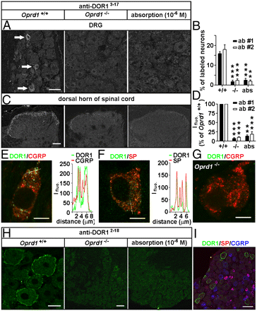
Dr. Tomas Hokfelt and his team at Karolinska Institute recently published use of our Opioid Receptor Antibodies and Substance P Antibody.They show the interplay of DOR and MOR in modulation of nociceptive afferent transmission and opioid analgesia.

Hai-Bo Wanga, Bo Zhaoa, Yan-Qing Zhonga, Kai-Cheng Li, Zi-Yan Li, Qiong Wang, Yin-Jing Lua, Zhen-Ning Zhang, Shao-Qiu He, Han-Cheng Zheng, Sheng-Xi Wu, Tomas G. M. Hökfelt, Lan Baob, and Xu Zhanga. Coexpression of δ- and μ-opioid receptors in nociceptive sensory neurons. PNAS July 20, 2010 vol. 107 no. 29 13117-13122.

Immunostaining. Adult rats, mice, and Oprd1 exon 1-deleted mice were fixed. Cryostat sections of L4 and L5 DRGs and spinal cord segments were processed for immunofluorescence staining (13) with Rb anti-DOR13–17 (1:2,000–1:60,000; DiaSorin and 1:4,000–1:60,000; Neuromics), Rb anti-DOR12–18 (1:30,000–1:120,000; Alomone), Rb anti-DOR1358–372 (1:1,000–1:2,000; Lifespan Biosciences), Rb anti-MOR (1:1,000; Neuromics); guinea pig anti-SP (1:500; Neuromics), and mouse anti-CGRP (1:1,000; Biogenesis) antibodies. IB4-labeling was carried out with fluorescein-labeled GSL I-IB4 (1:200). The Myc-DOR1–transfected HEK293 cells and neurons were fixed and processed with mouse anti-Myc antibodies (1:500; DSHB). Nuclear DAPI staining was used to indicate HEK293 cells in control experiments.

Images: Distinct distribution patterns of DORs in subsets of DRG neurons of mice. Immunostaining with antibodies against DOR13–17 [A: 1:30,000, antibody 1 (ab #1); DiaSorin and C: antibody 2 (ab #2); Neuromics] shows DORs in small DRG neurons and afferent fibers in spinal laminae I–II. This immunostaining pattern is abolished by the antiserum preabsorption or the deletion of Oprd1 exon 1. Reduction in immunostaining is quantitatively assayed by determining the percentage of positive DRG neurons (B; n = 6) and fluorescence intensity (Ifluo.) in the laminae I–II (D; n = 5). **P < 0.01; ***P < 0.001. (Scale bars: A and C, 40 μm.). DOR labeling (anti-DOR13–17, 1:30,000; DiaSorin) associated with vesicles in peptidergic small DRG neurons (E and F) is absent in Oprd1 exon 1-deleted mice (G). Colocalization of DORs and neuropeptides is shown by correlated peaks of Ifluo. measured along lines. (Scale bar: 8 μm.) (H) Immunostaining with antibodies against DOR12–18 (1:60,000; Alomone) shows the presence of DORs on the cell surface of large DRG neurons of mice. (Scale bar: 25 μm.) This staining pattern is abolished by preabsorption and is absent in Oprd1 exon 1-deleted mice. (Scale bar: 80 μm.) (I) Triple-immunostaining shows that DOR+ large DRG neurons contain neither SP nor CGRP. (Scale bar: 80 μm.)

Immunoblotting.The samples were processed for SDS/PAGE, transferred, probed with Rb antibodies against MOR (1:500; Neuromics), phospho-DOR1 (1:1,000; Neuromics), phospho-MOR (1:1,000; Neuromics), Myc (1:500; DSHB), Flag (1:1,000; Sigma), or actin (1:50,000; Chemicon) and visualized with enhanced chemiluminescence (19).

Featured and Related Reagents:

Mu Opioid Receptor-Rabbit


Mu Opioid Receptor-Guinea Pig


Delta Opioid Receptor 3-17


Delta Opioid Receptor 358-372


Delta Opioid Receptor 358-372


Kappa Opioid Receptor


phospho-Mu Opioid Receptor (Ser375)


MOR-1C
 

ORL 1-Pure


ORL1-Whole Serum


All Opioid Receptor Antibodies


Pain and Inflammation


Neurotransmission Research Antibodies
-GPCRs, Ligand Gated Ion
Channels, Biogenic Amines and more

i-Fect Transfection Kit
-gene silencing
of DOR, NaV1.8 tetrodotoxin-resistant sodium channel, NTS2 and more
in-vitro and in vivo

Primary Neurons and Astrocytes
-Primary
human, rat and mouse neurons and astrocytes

Tuesday, May 25, 2010

Dr. Steve Stice to Present the Power of StemEZ Neural Cells

Dr. Steve Stice to Present the Power of StemEZ Neural Cells

Posted using ShareThis

I have profiled Steve Stice's research here. The focus has been the excellent research results he and his team at ArunA Biomedical have generated with STEMEZ(TM) hN2 Human Neurons and hNP1 Human Neural Progenitors.

The story continues. He will be presenting the latest at the 9th Annual World Pharmaceutical Congress in Philadelphia, June 14. Topics include: using these neural cell lines to study neurotoxicity in cell-based assays and disease modeling. Recent work conducted in outside laboratories demonstrates that these lines are more sensitive to environmental toxicants than traditional cellular models.

Sample high throughput assay applications:

  • Cell morphology and neurite outgrowth

  • Cell signaling and transcription factor expression

  • Receptor and ion channel function

  • Cytotoxicity
  • Apoptosis, genotoxicity and DNA damage

These capabilities has been confirmed by our customers. I look for the use of the STEMEZ cell lines to continue to grow as researchers discover their value in Drug Discovery and Basic Neuroscience capabilities.

Saturday, May 08, 2010

TRPV1 Expression and Temporal Lobe Epilepsy

Dr Bret. Smith and his lab at Tulane have demontrated a link between Temporal Lobe Epilepsy (TLE) and VR1 N-Terminus (TRPV1) expression.

In this excellent study they showed an increase in expression in TLE mice vs controls.

Muthu D. Bhaskaran and Bret N. Smith. Effects of TRPV1 activation on synaptic excitation in the dentate gyrus of a mouse model of temporal lobe epilepsy. doi:10.1016/j.expneurol.2010.01.021
...with polyclonal VR1 N-terminus (1:2500) (Neuromics, Edina)..


Abstract: Temporal lobe epilepsy (TLE) is a condition characterized by an imbalance between excitation and inhibition in the temporal lobe. Hallmarks of this change are axon sprouting and accompanying synaptic reorganization in the temporal lobe. Synthetic and endogenous cannabinoids have variable therapeutic potential in treating intractable temporal lobe epilepsy, in part because cannabinoid ligands can bind multiple receptor types. This study utilized in vitro electrophysiological methods to examine the effect of transient receptor potential vanilloid type 1 (TRPV1) activation in dentate gyrus granule cells in a murine model of TLE. Capsaicin, a selective TRPV1 agonist had no measurable effect on overall synaptic input to granule cells in control animals, but significantly enhanced spontaneous and miniature EPSC frequency in mice with TLE. Exogenous application of anandamide, an endogenous cannabinoid that acts at both TRPV1 and cannabinoid type 1 receptors (CB1R), also enhanced glutamate release in the presence of a CB1R antagonist. Anandamide reduced the EPSC frequency when TRPV1 were blocked with capsazepine. Western blot analysis of TRPV1 receptor indicated protein expression was significantly greater in the dentate gyrus of mice with TLE compared with control mice. This study indicates that a prominent cannabinoid agonist can increase excitatory circuit activity in the synaptically reorganized dentate gyrus of mice with TLE by activating TRPV1 receptors, and suggests caution in designing anticonvulsant therapy based on modulating the endocannabinoid system.

Images: Western blot detection of TRPV1 receptor expression in the dentate gyrus. A. Diagram of dentate gyrus showing the microdissected area (box). B. Western blot showing TRPV1 receptor expression in two untreated mice and in two pilocarpine-treated mice that survived SE. Actin was used as the loading control which did not change significantly. C. Graph showing a significant (p less than 0.05; n=4) TRPV1 expression in epileptic mice.

Related Reagents:

TRPV (Vanilloid); TRPM; TRPA and TRPCs


Neurotransmission Research Antibodies
-GPCRs, Ligand Gated Ion Channels, Biogenic Amines and more

Primary Neurons and Astrocytes
- Primary human, rat and mouse neurons and astrocytes by Category.

Saturday, March 13, 2010

Neuropeptides and Large Dense Core Vesicles

We continue to be recognized by for the quantity and quality of our Neuropeptide and Neuropeptide Receptor Antibodies for studying Neurotransmission and Pain.

We wanted to feature an new article referencing use of our Guinea Pig Substance Antibody. Dr. Richard Mains and his team shed light on the function and behavior of large dense core
vesicles (LDCVs) concluding that under basal conditions, LDCVs move faster away from the soma than toward the soma, but fewer LDCVs travel anterograde than retrograde. Stimulation decreased average anterograde velocity and increases granule pausing. Data from antibody uptake, quantification of enzyme secretion and appearance of pHluorin fluorescence demonstrate distributed release of peptides all along the axon, not just at terminals.
Jacqueline A Sobota , William A Mohler , Ann E Cowan , Betty A Eipper and Richard E Mains.
Dynamics of peptidergic secretory granule transport are regulated by neuronal stimulation. BMC Neuroscience 2010, 11:32doi:10.1186/1471-2202-11-32.

Image: Sustance P staining of trigeminal ganglia from P3-P5 rat pups-note this pub features some of the best IHC staining we have seen.

Related Products and Product Categories:
Substance P-Pure

Substance-P-Mouse Monoclonal

Neurokinin-1 (NK 1) Receptor

Neurokinin-1 (NK 1) Human Receptor

Neurokinin-3 (NK 3) Receptor

proNeurokinin B (proNKB or P2)

Neurotransmission Research Antibodies

Neuropeptide and Neuropeptide Receptor Antibodies

Pain and Inflammation Antibodies

Diabetes and Obesity Antibodies

Primary Neurons and Astrocytes
- Primary human, rat and mouse neurons and astrocytes.

Wednesday, September 16, 2009

Thursday, October 16, 2008

Purinergic Receptor Pubs

Our Purinergic Receptor Antibodies have recently been referenced in several publications.

In the first study 5 of our antibodies are used. It investigates the expression of purinergic P2 receptors in oxygen-induced retinal neovascularization.

Sylvia Sarman; Jorge Mancini; Ingeborg van der Ploeg; J. Oscar Croxatto; Anders Kvanta; Juan E. Gallo. Involvement of Purinergic P2 Receptors in Experimental Retinal Neovascularization. Current Eye Research. 10.1080/02713680701885470 .

...Five eyes (5 animals) from either hyperoxa- or non-hyperoxia-treated mice were enucleated and fixed for 48 hr at 4°C in paraformaldehyde (Sigma-Aldrich, St Louis, MO, USA). Afterward, eyes were immersed for cryoprotection in graded sucrose solution (5% overnight, 10%, 15%, and 20%) and interlocked with resin. Fourteen micron sections were obtained (Shandon AS325 Retraction Microtome, Thermo Scientific, Waltman, MA, USA) and fixed on polylysine-treated glass slides. After overnight incubation in primary antibody (P2X1 1:1000, P2X2 1:1000,P2X3 1:1000, and P2Y2 1:800) (Neuromics, Minneapolis, MN, USA) at 4°C, sections were treated with biotinylated secondary antibodies followed by an avidin peroxidase complex step (Vectastin Elite ABC, Vector Laboratories, Burlingame, CA, USA). Finally, a color reaction was obtained using 3,3'-diaminobenzidine (DAB)/nickel-enhanced solution for staining (Sigma-Aldrich, St Louis, MO, USA). At least 6 sections per retina were analyzed. For immunofluorescence, the sections were labeled with lissamine rhodamine-conjugated goat anti-rabbit Ig-G or with fluorescein-5-isothiocyanate-conjugated goat anti-guinea pig Ig-G (Jackson Immunoresearch Laboratories, West Grove, PA, USA). At least 6 sections per retina were analyzed. Visualization was done using a Nikon Fluorescence Eclipse Microscope (Tokyo, Japan), and photographs were taken with a Nikon DN 100 Digital Camera (Tokyo, Japan)...

Images: Expression of P2X and P2Y receptors in normal mice and in a model of ischemia-induced pathological retinal angiogenesis as assessed by immunohistochemistry. (A) P2X2 receptor expression was found in the outer plexiform layer of control mice. (B) In mice
treated with oxygen, the expression was similarly found in the outer plexiform layer. In addition, a strong signal was also found in the inner plexiform layer. This induction was noted in all slides analyzed. (C) Retina without primary antibody for P2X2. (D) In control mice,
weak P2Y2 receptor expression was seen in the ganglion cell and in the nerve fiber layer. (E) In mice treated with oxygen, P2Y2 expression
showed a similar overall distribution as control mice. The signal was, however, strongly up-regulated. This increase was noted in all slides analyzed. (F) Retina without primary antibody for P2Y2. Black bar = 20 μm.

The second studies P2Xs' role in neurotransmision.

Elsa Fabbretti*, Elena Sokolova*, Lara Masten, Marianna D’Arco, Alessandra Fabbro, Andrea Nistri and Rashid Giniatullin. Identification of negative residues in the P2X3 ATP receptor ectodomain as structural determinants for desensitization and the Ca2+ sensing modulatory sites. JBC Papers in Press. Published on October 8, 2004 as Manuscript M409772200.

...Western immunoblots of transfected or untransfected HEK cells were performed as recently reported (19). In brief, these were lysed using a buffer containing 100 mM Tris HCl (pH 6.8), 200 mM dithiothreitol, 4 % SDS, 20 % glycerol and a cocktail of protease inhibitors (Sigma), and separated on 10 % polyacrylamide gel. After blocking with Tris saline buffer (TBS) containing milk, Tween 20 and preimmune serum, proteins were incubated overnight at 4°C with an anti-P2X3 antibody (Neuromics; 1:2000). Immunocomplexes were incubated for 1 h with a peroxidase-conjugated secondary antibody (Sigma; 1:4000) and detected with a chemiluminescence ECL kit (Amersham). Negative controls were obtained by mock transfection of HEK cells with the pEGFP-N1 plasmid (CLONTECH). Controls for efficient loading of different lysate materials were carried out by using an anti-β actin antibody (mouse monoclonal, 1:2000, Sigma)...

Related Reagents:

P2X1-Rabbit Antibody
P2X2-Guinea Pig Antibody
P2X2-Rabbit Antibody
P2X3-Rabbit Antibody
P2X3-Guinea Pig Antibody
Related Reagents:
All Purinergic Receptor Antibodies
All Pain and Inflammation Antibodies
Vision and Retina Research Antibodies