Thursday, December 27, 2018

Primary Human Brain Pericytes

Pure and Potent
The authors of a chapter on pericytes published in SpringerLink referred to our human primary pericytes as "pericyte-like", but chose not to characterize them because of the "exorbitant cost". Dore-Duffy P., Esen N. (2018) The Microvascular Pericyte: Approaches to Isolation, Characterization, and Cultivation. In: Birbrair A. (eds) Pericyte Biology - Novel Concepts. Advances in Experimental Medicine and Biology, vol 1109. Springer, Cham

Given that these pericytes are also part of our hot selling 3-D Human BBB Model, we are in the process of challenging these claims. We will always take inaccurate claims regarding our solutions seriously, and we take immediate action.
  • Characterization of cells-we validated several key markers by immunofluorescence.


Staining of Desmin (dilution 1:100). Secondary antibody conjugated to Alexa 594 (red) and counterstained with DAPI (blue). Cells were mounted using iBrite mounting media.

Staining of Actin (dilution 25 ug/mL). Secondary antibody conjugated to Alexa 594 (red) and counterstained with DAPI (blue). Cells were mounted using iBrite mounting media.
We also plan on doing a phenotypic analysis of these cells and will post here when completed.
  • Cost of cells-isolating pericytes from human donors, and expanding to the required number of cells involves much time and effort. We know! Further differentiating stem cells into pericytes is hard and time-consuming. Against this backdrop, we consider our pricing to be inexpensive. Though they are more difficult to derive, they are priced equivalently with our other human primary cells ($789/500,000 USD Cells).
As always, we will post new data here.

Saturday, December 15, 2018

Neuromics FBS at 259 USD/500 ml.

Only 2 Weeks Left
We wanted to get our FBS in the hands of more cell culturing experts. Our low pricing has accelerated purchasing of this culture supplement.

More importantly, we are pleased with the positive feedback. Here are some recent reviews.
on BirdEye
 ★
9 days ago
My experience with Neuromics was very positive. I asked for a sample of FBS and after to use the FBS I received. 
on BirdEye
 
month ago
I purchased FBS from Neuromics. It was shipped to Canada without any problems. It arrived safely and still frozen. Cells grow well in the product. Customer service was great and I will definitely shop here again.

Here are the most recent publications:
  1. Fei Cao, Li-Xue Yin. (2018). miR-122 enhances sensitivity of hepatocellular carcinoma to oxaliplatin via inhibiting MDR1 by targeting Wnt/β-catenin pathway. Experimental and Molecular Pathology. https://doi.org/10.1016/j.yexmp.2018.10.009 
  2. Amélie Robert, Peirun Tian, Stephen A. Adam, Mark Kittisopikul, Khuloud Jaqaman, Robert D. Goldman, and Vladimir I. Gelfand. Kinesin-dependent transport of keratin filaments: a unified mechanism for intermediate filament transport. 26 Jun 2018https://doi.org/10.1096/fj.201800604R 
  3. Mayuri Manoj Vaidya (2018). Verification of Apoptosis in MDA-MB-231 Triple Negative Breast Cancer Cells Post NBA Photodynamic Therapy Using DNA Fragmentation Assay and Cell Death Dyes. (Doctoral Dissertation). University of Texas San Antonio
The pricing is good through December 31, 2018.

Tuesday, November 27, 2018

Pain Receptors and Diabetic Neuropathy

We Got the Markers
Our Markers for Pain Research are widely used and frequently published. They are often referenced in publications on Diabetic nerve pain.

Here are some of my past blog postings:
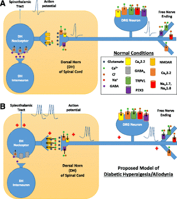
Here's a model of the actual nerve receptors behind both intractable pain and loss of sensation.
A: Simplified model of nociception under normal conditions. Free nerve endings transduce a painful stimulus into a neural signal, which propagates to DRG centrally and eventually synapses on a nociceptive neuron within the DH of the spinal cord.B: Proposed model of nociception under conditions of diabetic hyperalgesia and allodynia. A pain signal augmented by upregulated pronociceptive ion channels in sensory neurons is carried toward the DH, where it is further augmented by a hypoactive GABAergic system and subsequently diminished inhibition from an inhibitory interneuron. NMDAR, N-Methyl-D-aspartate receptors. https://doi.org/10.2337/dbi15-0006
We will continue to post findings on the root causes and potential treatment for Neuropathy.

Saturday, November 17, 2018

Primary Human Schwann Cells

Research Ready

Our Schwann Cells have proven "rock solid" in the hands of our users.

Image: Human Primary Schwann Cells in Culture

Human Schwann Cells (HSwC) are isolated from the human spinal nerve. HSwC are cryopreserved at passage one and delivered frozen. Each vial contains >5 x 10^5 cells in 1 ml volume. HSwC are characterized by immunofluorescence with antibodies specific to S100, GFAP, and CD90

Saturday, October 27, 2018

Cardiomyocytes

Pure, Potent and Research Ready
We are recognized for our growing catalog of human primary and stem cells.

These include hMSC Derived Cardiomyocytes.

Human Cardiomyocytes stained with Cardiac Tryponin T (green) and counter stained with DAPI (blue).

These cells are optimized to provide additional options for in-vitro testing of drug to drug candidate toxicities allowing researchers to rule out the ineffective and potentially toxic small molecules/compounds early in the process.

Thursday, October 18, 2018

TRP Antibodies

Some of the Best Available
Our TRP Channel Antibodies are widely used and frequently published.

Here's some data from researchers using on of our TRPV1 Antibodies.

Immunohistochemical expression of serum response factor in rat dorsal root ganglia. Dorsal root ganglia dissected from male Sprague-Dawley rat and imaged by confocal microscopy for (A) TRPV1 (blue), (B) AKAP (AKAP150, green), and (C) SRF (red), with colocalization represented in panel (D). White arrows indicate cells expressing all 3 proteins. Yellow line represents 50 μM in length; images are representative of 4 individual sections taken from 6 L4 to L6 DRG. (E) Venn diagram of coexpression between SRF (red circle), AKAP (AKAP150, green circle), and TRPV1 (blue circle). (F) Percentages of SRF coexpression with AKAP (AKAP150) or TRPV1 calculated from 8 randomly selected coverslips. DRG, dorsal root ganglia; SRF, serum response factor. Source Serum response factor mediates nociceptor inflammatory pain plasticity PAIN Reports3(3):e658, May/June 2018.
We offer 100% refund or free replacements should you not be delighted with your results.

Sunday, October 07, 2018

Neuromics New Neuronal Markers

Best of the Best
We are recognized for having some of the best Neuronal-Glial Antibodies in the business. We are pleased to announce the addition of these:

CH22122 – Tyrosine Hydroxylase
GT22101 - GFP
GT22102 – MAP2
GT22103 - Vimentin
MO22186 – Tyrosine Hydroxylase
RA22135 – Tyrosine Hydroxylase

Immunofluorescent analysis of rat brainstem section stained with GT22102, MAP2 dilution 1:2,000 in red, and costained with mouse mAb to MO22121, MBP dilution 1:5,000, in green. Following transcardial perfusion of rat with 4% paraformaldehyde, brain was postfixed for 24 hours, cut to 45μM, and free-floating sections were stained with the above antibodies.
If you have questions on these or any of our solutions, not hesitate to contact me directly-pshuster@neuromics.com or 612-801-1007,

Sunday, September 30, 2018

Quality FBS-Only 259 USD/500 ml.

First Come; First Serve
We have an inventory of FBS of approximately 2000 bottles.  This inventory includes the lots we've been selling and using internally all year.  These have proven to be of the highest quality.
Here’s a sampling of user feedback: 
  • After a referral from an ex-colleague, we tested a sample from a single batch and found that the product is great for cell cloning & hybridoma work, and generally for all other cell culture. We subsequently ordered several bottles of FBS. The sales person was very friendly and worked with me to make the purchase possible. The order arrived promptly. - Peter Dias, The Biomedical Research Institute of SC
  • We ordered several bottles of Fetal Bovine Serum on 2 occasions and are pleased with its performance in our cultures. The FBS arrived frozen in enough dry ice and was neatly and carefully packed. The FBS was very reasonably priced. Thank you. - Ken Patrene, University of Pittsburgh
Referenced in Publications:
 FBS
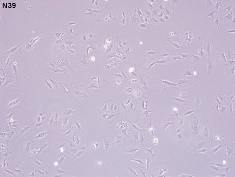
N39 Cell Line Cells grown in Media supplemented with Neuromics' FBS, Courtesy of Deng Guo, University of Iowa.
FBS
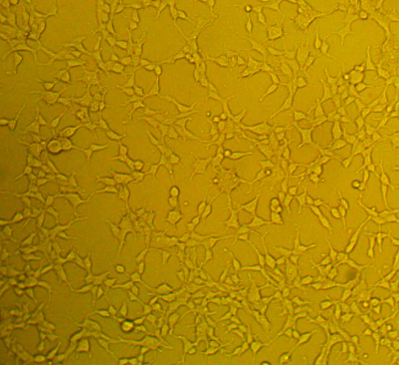
HEK Cells cultured in Media Supplemented with Neuromics' FBS Courtesy of Kavita Shah, Purdue University

The reason for this great offer is we need to clear inventory so that we can place a volume order of raw FBS stock yet this year and lock in 2019 pricing.  Our goal is to provide the best FBS at the lowest price.  This offer of $259/500ml is valid until the current stock is exhausted.
Like a sample to try?  Contact rose@neuromics.com for a 50 ml sample.  Rose Ludescher-Manager of Customer Satisfaction-

Thursday, September 20, 2018

Human Brain Microvascular Endothelial Cell in Action

Recent Pubs
Our Human Brain Microvascular Endothelial Cells (HBMECs) are known for the performance in drug discovery and tox assays. These 21-CFR compliant cells are used in the "blood side" of our 3-D BBB Models.

  1. Shavali Shaik, Bridget Kennis, Shinji Maegawa, Keri Schadler, Yang Yanwen, Keri Callegari, Rishi R. Lulla, Stewart Goldman, Javad Nazarian, Veena Rajaram, Jason Fangusaro, and Vidya Gopalakrishnan. (2018). REST upregulates gremlin to modulate diffuse intrinsic pontine glioma vasculature. Oncotarget. 2018 Jan 12; 9(4): 5233–5250. doi: 10.18632/oncotarget.23750 
  2. Hu et al. (2016). Epigenetic Activation of WNT5A Drives Glioblastoma Stem Cell Differentiation and Invasive Growth. Cell. 167, 1281–1295. http://dx.doi.org/10.1016/j.cell.2016.10.039,
GREM-1 is required for tube formation in vitro (A) Q-RT-PCR analysis of GREM-1 gene expression in SU-DIPG-IV cells stably expressing either control shRNA or GREM-1-specific shRNA. Lentiviral constructs expressing two different GREM-1 shRNAs (GREM-1.1 and GREM-1.2) were used to knockdown GREM-1. Efficiency of GREM-1 knockdown was determined by Q-RT-PCR and expression was normalized to 18s RNA. Significance is as shown (**less than.01). (B-C) HUVEC or human brain microvascular endothelial cells (HBMEC) were cultured in endothelial cell medium and or conditioned medium from either control shRNA or shGREM-1.2 transfected SU-DIPG-IV cells. Tube formation in matrigel was measured after 16h and images were obtained. The ability of GREM-1 to rescue loss of tube formation upon REST knockdown was determined by addition of human-recombinant GREM-1 (rGREM-1) to conditioned media-endothelial media mix. Scale bars, 100μm. (C) Quantification of tubes in matrigel shown in Figure B (right panels). Data shown is mean +/- SD, ***p less than .001, n=3. (D) Western blot analysis to assess VEGFR2 levels in SU-DIPG-IV, -VI and –XIII cells, HUVECs and HBMECs was done using anti-VEGFR2 antibodies. Tubulin served as a loading control. (E) Western blot analysis was performed to assess AKT signaling downstream of GREM-1 interaction with its potential receptor VEGFR2 in HUVEC and HBMEC. Anti-pAKT (S473), anti-pAKT (T308), total AKT, and anti-actin were employed.
If these cells fit your assay requirements and need more data/info, do not hesitate to contact me, Pete Shuster, CEO at pshuster@neuromics.com or cell: 612-801-1007.

Friday, September 14, 2018

Neuromics' BBB Model and Permeability Assays

Crossing the Blood-Brain Barrier
Our BBB Model is being increasingly used by Bio-Pharma for drug permeability studies. Customers include Amgen, Genentech, Boehringer Ingelheim, and Merck.

These assays are key for making sure molecules/compounds of interest will cross the barrier into the brain and at what rate. This data, in part, add clarity on best candidates to move into in-vivo testing.
Human Blood Brain Barrier Model 3D45002 12 well
Human Blood Brain Barrier Model 3D45002 24 well
Human Blood Brain Barrier Model 3D45002 6 wells
I you have interest in using our models, Rose Ludescher, Manager of Customer Satisfaction, can provide information aligned with your assay requiremenst. rose@neuromics.com or 1-866-350-1500.

Monday, August 27, 2018

Energize your Cell-Based Assays

Check out our AlphaBioCoat 
Collagen is a fibrous protein found in the extracellular matrix and connective tissue. The most common form of collagen is type I and is most prevalent in bone, tendon and skin. It consists of 3 intertwined coiled subunits: 2 x α1 (I) chains and 1 x α2 (I) chain. Each chain contains 1050 amino acids wound tightly around one another in a characteristic right-handed triple helix. The triple-helical structure of collagen arises from unique abundance of the amino acids in collagen appear in a characteristic repeating motif Gly-X-Y, where X is usually proline and Y is usually hydroxyproline.

AlphaBioCoat Solution (AC001) is a biocompatible complex of extracellular matrix binding solution that is supplemented with growth factors. It helps accelerate cell attachment and cell growth. AC001 is the premium version of our Smooth Coat Solution (SC300).

It is ideal for plate coating due to its unique viscosity. Its coating greatly enables cell migration on cultured plate surfaces. Perfect for establishing primary cell lines, it can increase endothelial cell attachment, survival in culture, and cell growth. AlphaBioCoat Solution is great for both coating plates and T-flasks.

Interested? Please contact rose@neuromics.com.

Thursday, August 16, 2018

Human Brain Cells

Our Expertise
When researchers are considering using our human brain cells, they often voice concerns about growth. In order to address these concerns, we explain that we have optimized our media and coatings in order to meet their assay needs.

Here's an example of how our human brain pericytes grow over time.
We have primary human neurons, astrocytes, microglia and Schwann cells.

If you have interest you can contact us at 866-350-1500 or rose@neuromics.com. We will provide the information required to help you make an informed decision.

Tuesday, August 07, 2018

New Neuronal Markers

Check Them Out!
We continue to add new antibodies. Many are additions to our frequently used and widely published Neuron-Glial Markers.

Image: Immunofluorescent analysis of cortical neuron-glial cell culture from E20 rat stained with mouse mAb to GAP43, MO22170, dilution 1:1,000, in red, and costained with chicken pAb to MAP2, dilution 1:10,000, in green. The blue is DAPI staining of nuclear DNA. GAP43 antibody labels protein expressed in the axonal membrane of the neuronal cells, while the MAP2 antibody stains dendrites and perikarya of neurons.

Name Species Applications
Aurora A/B Kinase Human, Mouse, Rat ICC, WB, IHC, IF
Aurora B Kinase Human, Mouse, Rat ICC, WB, IHC, IF
Beta-Tubulin Human, Mouse, Rat, Primate ICC, WB, IHC, IF
DJ1/Park7 Polyclonal Rabbit Antibody Human, Mouse, Rat ICC, WB, IF
Fibrillarin Human, Mouse, Rat ICC, WB, IHC, IF
Galectin-3 Rabbit Polyclonal Antibody Human, Mouse, Rat ICC, WB, IF
GAP43 Human, Mouse, Rat ICC, WB, IHC, IF
GAP43 (IgG) Human, Mouse, Rat ICC, WB, IHC, IF
HSP60 (Heat Shock Protein 60) Human, Mouse, Rat ICC, WB, IHC, IF
IBA1 Polyclonal Rabbit Antibody Human, Mouse, Rat ICC, WB, IHC, IF
Ki67 Human ICC, WB, IF
Nestin Human, Mouse, Rat ICC, WB, IHC, IF
Nsp1p ICC, WB, IHC, IF
OPA1 Human, Mouse, Rat ICC, WB, IHC, IF
Pdi1p WB, IP, IF
Secretagogin Polyclonal Chicken Antibody Human, Mouse, Rat, Bovine ICC, WB, IF
Vimentin Human, Rat ICC, WB, IHC, IF
All our products are tested, characterized and research ready.

Tuesday, July 31, 2018

Easy Immunostaining Staining

10X the Signal at a Fraction of the Cost
Time is money. This is especially true of routine immunostaining assays.

We are pleased to introduce you to EZ-HRPTM Polymer Detection. Features include:
  • 10-15 times more sensitive than conventional avidin-biotin detection 
  • Allows for entire staining procedure to be completed in just under 2 hours Requires less primary antibody (3-4 times less) 
  • Does not produce non-specific background staining Can be used on unfixed or chemical fixed (formalin, formaldehyde, alcohol, etc), paraffin-embedded and frozen tissue sections.
Comparison of IHC images using EZ-HRP™ Polymer vs. a conventional avidin-biotin antibody
We are continuously adding new products to improve your research results.

Tuesday, July 24, 2018

Our Hearing Adapts in Space

Synaptic Plasticity in the Ear
Our mission to Mars is driving a need to better understand the physiological impact on the human body. Deep space travel places unique challenges for humans. It is important that there is minimal impact on space travelers' senses. This includes hearing.

This study uses our Shank 1a Antibody to determine changes in the neuronal structure of the ear in microgravity.

Image: Verification of antibodies to CtBP2 and Shank1a. A and B: serial 14-µm cryosections were obtained from a postnatal day 71 (P71) mouse utricle, and maximum-intensity projections are shown. Hair cell (hc) and support cell (sc) nuclei are illuminated by the DAPI stain (blue). Numerous closely associated CtBP2-and Shank1a-positive puncta can be observed in the positive immunostained section represented in A (block arrowheads). The CtBP2-positive puncta highlighted by the flared arrowhead in A may represent an undocked synaptic ribbon. No primary antibodies were included in the processing represented in the micrograph in B. The scale bar in B represents 5 µm and also applies to A. C and D: maximum-intensity projection micrographs from right and left whole mount utricles from a P65 mouse. Importantly, fixative administration into the temporal bones yielding the specimens represented in C and D was delayed 7 min to replicate the conditions associated with specimens derived from the microgravity and control specimens. The positive-immunostained specimen of the pair is shown in C, where numerous closely associated CtBP2-positive and Shank1a-positive puncta can be observed. Though faint, CtBP2-immunostained nuclei are highlighted by the flared arrowheads. The micrograph in D illustrates the results of withholding primary antibodies from the processing. Immunolabeled puncta are not observed. The scale bar in D represents 5 µm and also applies to C. https://www.physiology.org/doi/full/10.1152/jn.00240.2016 

These results demonstrate that structural plasticity was topographically localized to the utricular region that encodes very low frequency and static changes in linear acceleration, and illuminates the remarkable capabilities of utricular hair cells for synaptic plasticity in adapting to novel gravitational environments.

Wednesday, July 18, 2018

Staining Cells and Tissue

The background is Bad!
Yes, it is. It compromises data. We have a solution.
FluoMuteTM

FluoMute™ ready-to-use reagent to reduce autofluorescence in cells and tissue. Just incubate fixed cells of tissue sections with FluoMute™ for 30-60 min at room temperature, rinse with PBS and continue with immunofluorescence ICC or/and IHC protocols. Treatment with FluoMute™ does not affect cell morphology and the integrity of tissue antigens to be detected with primary antibodies (see bottom image). FluoMute™ is compatible with paraffin-embedded and frozen tissue sections, stem cells, lymphocytes and mammalian cell lines of different origin.

We plan on adding products that will provide stronger signals with fewer protocols steps vs traditional solutions. Stay tuned.

Wednesday, July 11, 2018

Human Cells in Action

Human Microvascular Retinal Endothelial Cells (HMRECS)
We have built the foundation of Neuromics on satisfied customers. We make a practice of following up with each user to make sure our solutions are working as expected. If not, we offer "no question asked" refunds or replacements.

This is especially important for our human cells, media, and supplements. Success with these is easy to measure as either the cells are healthy and happy or they are not.

We also use reviews and publications as another measure of satisfaction. Here I would l highlight the latest publication using our HREMCS. A. P. Da Cunha, Q. Zhang, M. Prentiss, X. Q. Wu, V. Kainz, Y. Y. Xu, J. Vrouvlianis, H. Li, N. Rangaswamy, B. Leehy, T. L. McGee, C. L. Bell, C. E. Bigelow, V. Kansara, Q. Medley, Q. Huang & H. Y. Wu. The Hierarchy of Proinflammatory Cytokines in Ocular Inflammation. Current Eye Research, Volume 43, 2018 - Issue 4 Published Online: 04 Dec 2017.

Images: Effect of HG (high glucose) and pro-inflammatory cytokines on connexin43 expression in HRMECs. Immunohistochemical data showing connexin43 expression in (A) normal medium; (B) HG (25 mM); (C) pro-inflammatory cytokines (IL-1β and TNF-α 10 ng/mL each); and (D) a combination of HG and pro-inflammatory cytokines inducing a change in cell morphology with signs of cell swelling, possibly owing to hemichannel opening (indicated by white arrows).

If you have questions or interest in any of these, please contact rose@neuromics.com or 866-350-1500.

Monday, July 02, 2018

Neuromics' Fetal Bovine Serum (FBS) Strikes Again

Potent FBS at Pricing You'll Like
Neuromics started providing FBS to researchers in early 2017. Our goal was to provide thorough tested and 9-CFR compliant FBS with the lowest pricing anywhere.

In order to ensure our initial claims are trustworthy, we follow up with all our users and ask that they provide feedback. Here's the latest review-Ordered Fetal bovine serum. Best price and quality" - Juwen D, Albert Einstein College of Medicine. Product: Heat-inactivated FBS, cat no. FBS001-HI.

We are now starting to see the use of our FBS reference in publications: Amélie Robert, Peirun Tian, Stephen A. Adam, Mark Kittisopikul, Khuloud Jaqaman, Robert D. Goldman, and Vladimir I. Gelfand. Kinesin-dependent transport of keratin filaments: a unified mechanism for intermediate filament transport. 26 Jun 2018https://doi.org/10.1096/fj.201800604R.

Images: Keratin filaments are associated with microtubules. A) 3D-SIM imaging of keratin and tubulin immunostaining of RPE cells. The enlargements show the alignment of keratin filaments with microtubules. Scale bar, 5 mm. B) Confocal imaging of keratin and tubulin immunostaining. In control cells, keratin filaments extend to the cell periphery, and the filaments retract into the perinuclear region after 3-h treatment with 10 mM nocodazole to depolymerize microtubules. Scale bar, 10 mm.

Give our serum a try today! Neuromics US-Origin Fetal Bovine Serum is only 349USD/500ml. Heat-inactivated FBS only 364USD/500ml. Need a large quantity? Bulk discounts are available. Like a sample to try? Contact rose@neuromics.com for a 50 ml sample.

Thursday, June 28, 2018

You'll Love our FBS!

Check out Testimonials from Users
Neuromics is proud to offer you the high-quality Fetal Bovine Serum, the same serum we use internally, without the high cost. Our customers, researchers like you, agree.

Ordered Fetal bovine serum. Best price and quality" - Juwen D, Albert Einstein College of Medicine. Product: Heat-inactivated FBS, cat no. FBS001-HI

Outstanding service. Fetal bovine serum was of highest quality, easy to order and packaging and shipping were best possible." - Rosemary, University of Buffalo Product: Heat-inactivated FBS, cat no. FBS001-HI (see all of Neuromics reviews here).
Give our serum a try today! Neuromics US-Origin Fetal Bovine Serum is only 349USD/500ml. Heat-inactivated FBS only 364USD/500ml. Need a large quantity? Bulk discounts are available. Like a sample to try? Contact rose@neuromics.com for a 50 ml sample.

Tuesday, June 26, 2018

Autophagy Assay Kits

Detects Autophagy in Living Cells
Autophagy is a conserved lysosomal recycling process by which cells break down their own components such as proteins, lipids, and carbohydrates. The process plays an important role in maintaining homeostasis and destroying intracellular pathogens. In addition, autophagy can be upregulated in times of starvation or stress to provide additional nutrients for the cell. Dysregulation of autophagy has implications for cancer, infection, and degenerative diseases.

Autophagy is a three-stage process. First, cytoplasmic components targeted for degradation are sequestered, resulting in the formation of the autophagosome. Next, the autophagosome fuses with the lysosome to form the autophagolysosome or autolysosome. Finally, degradation of the autophagosomal contents occurs.

Our Autophagy Assay, Red (cat# KF17373) enables researchers to detect and monitor the in vitro development of autophagy in living cells. The Autophagy Probe is cell-permeant and fluoresces red when inserted in the lipid membranes of autophagosomes and autolysosomes. Results can be read using a flow cytometer.
Figure. Flow Cytometry Results. Autophagy Assay Kit, Red was used to assess the induction of autophagy in Jurkat cells. Cells were either untreated (Black) or treated with 0.5 μM Rapamycin (Orange), 10 μM Chloroquine (Blue), or both 0.5 μM Rapamycin and 10 μM Chloroquine (Red) for 18 hours. After staining with Autophagy Probe, Red for 60 minutes, cells were washed and analyzed by flow cytometry (BD LSRFortessa Special Order flow cytometer equipped with a green/yellow laser (561 nm excitation) and a 610/20 emission filter). An overlay of the histograms is shown on the right. A table displaying the median fluorescence signal, % negative, and % positive cells is shown below. Treatment with Rapamycin or chloroquine increased the fluorescence signal detected compared to the untreated control. Combined treatment of rapamycin and chloroquine further increased the fluorescence signal detected. Data courtesy of Dr. Kristi Strandberg (ICT 228:37-40).

We have many options for detecting apoptosis, necrosis, autophagy, and cytotoxicity in living cells. 

Thursday, June 14, 2018

Markers for Tyrosine Hydroxylase-TH

Proven and Published
Tyrosine hydroxylase TH is a necessary enzyme to create neurotransmitters and protect the body against oxidative stress.

Dysregulation of this enzyme results in manic-depression and schizophrenia. Parkinson's disease is also considered a TH deficiency as low dopamine levels are a consistent neurochemical abnormality.

Markers to TH are important for studying its role in neurotransmission and how it is altered in disorders and diseases.

We have many options and this enables you to choose the best markers for your research. All are proven and published.
TH staining of mouse salivary gland tissue. https://doi.org/10.1016/j.omtm.2018.02.008
Check out all our markers for neuroscience research.

Wednesday, June 06, 2018

Put our Antibodies to Work for You!

Tested and Characterized for Results You Can Trust
When you select a vendor for antibodies, there is a measure of trust. We know you are asking, "will it work in the applications as advertised".

Since the inception of Neuromics, our guiding principle is providing well-characterized solutions for results you can trust and understand. In order to provide as much comfort in the purchase of our solutions, we encourage access to publications, data, and testimonials.

Here're some recent testimonials:
  • Good antibody! Product Name: Tuj 1, Chicken – (Cat# CH23005) Good for neuronal staining. Bright and specific.Liang Shi - May 03, 2018 - Rating: 5.0 
  • Neuromics has a conjugated antibody for oligodendrocytes that is unavailable anywhere else and works really well. Very pleased with the product and have already ordered it a few times. Edit: This antibody is available from Miltenyi now. They both behave comparably. Matthew Smith - Apr 24, 2018 - Rating: 5.0 
  • I bought the secondary antibodies (488-Goat anti mouse IgG and 546-goat anti rabbit IgG) from Neuromics and used them for IF assay. Both antibodies worked well. I highly recommend them.Bonnie Dai - Apr 21, 2018 - Rating: 5.0 
Expression of TH (green) and TRPV1 (blue) relatively to CGRP-cre+ neurons (red) in DRG from CGRPcre-ER/+;Rosa26LSL-tDTomato/+ mice. Some TRPV1+ DRG neurons are not expressed in CGRP-cre+ sensory neurons. These CGRP-cre-/TRPV1+ neurons are marked with yellow arrows (a and a1). b. Expression of calbindin-28K (Calb; green) relatively to CGRP-cre+ DRG neurons (red). Yellow arrows mark CGRP-cre-/Calb+ neurons (b and b1). c. Expression of trkB (green) relatively to CGRP-cre+ DRG neurons (red). Yellow arrows mark CGRP-cre-/trkB+ neurons; and a blue arrow shows a rare example of the CGRP-cre+/trkB+ neuron (c and c1). d. Expression of trkC (green) relatively to CGRP-cre+ DRG neurons (red). Yellow arrows mark CGRP-cre-/trkC+ neurons; and; and a blue arrow shows an example of the CGRP-cre+/trkC+ neuron (d and d1). White horizontal bar shows 20μm scale for each panel.

We offer an easy, "no question asked" replacement or refund policy. Give us a try. You will be satisfied.

Monday, June 04, 2018

New Fibrillarin Antibody

Potent Nucleoli Marker
Given the importance of Nucleus and Related Markers to all branches of cell/molecular biology, we are dedicated to having the best.

We have just added a new Fibrillarin Antibody.
Images: Immunofluorescent analysis of (A) C6 rat glioma cells and (B) Human embryonic kidney cells stained with mouse mAb to fibrillarin, MO22169, dilution 1:1,000 in red, in both cases costained with chicken pAb to vimentin, CPCA-Vim, dilution 1:10,000 in green. The blue is DAPI staining of nuclear DNA.

Nop1p/Fibrillarin was originally identified as a nucleolar protein of bakers yeast, Saccharomyces cerevisiae (accession P15646). The Nop1p protein is 327 amino acids in size (34.5kDa), is essential for yeast viability, and is localized in the nucleoli. Nop1p is the yeast homolog of a protein apparently found in all eukaryotes and archaea generally called fibrillarin. Fibrillarin/Nop1p is extraordinarily conserved so that the yeast and human proteins are 67% identical, and the human protein can functionally replace the yeast protein. This antibody is becoming widely used as a convenient marker for nucleoli in a wide variety of species (e.g. 4-6). The HGNC name for this protein is FBL. To raise the MCA-4H4 antibody, mice were injected with full length recombinant human fibrillarin.

Wednesday, May 30, 2018

New 3-D Eye Model

More in-vivo like Model
We see our world in 3-D. Diseases of the eye compromise this ability.

Neuromics' is pleased to announce that we have a 3-D model aimed at accelerating drug discovery for these diseases. Sight is a terrible thing to lose and the faster new drugs can be discovered, fewer people will have to suffer the loss of sight.


Our 3D Human Retinal Microvascular Angiogenesis model is constructed using GFP‐Tagged human Retinal Microvascular Endothelial cells. They are co-cultured with RFP-Tagged human supporting cells. GFP positive human retinal capillary-like tubule formation can be monitored in real time under fluorescence microscope throughout the whole process of the experiment.

Sunday, May 20, 2018

Antibodies Against Neuropeptides

Another Nod From Nature Scientific Reports
Publications referencing use of our antibodies continue to build. We are recognized for having potent Neuropeptide Antibodies.

This most recent publication references our Guinea Pig Substance-P Antibody: Khaled Abdallah, Francis Nadeau, Francis Bergeron, Sylvie Blouin, Véronique Blais, Kelly M. Bradbury, Christine L. Lavoie, Jean-Luc Parent;, Louis Gendron. (2018). Adeno-associated virus 2/9 delivery of Cre recombinase in mouse primary afferents. Scientific Reports volume 8, Article number: 7321 (2018) doi:10.1038/s41598-018-25626-y
Figure: Distribution of the Cre-GFP in the neuronal subpopulations within the lumbar dorsal root ganglia. Representative photomicrographs showing co-localization of GFP with markers of peptidergic (Substance P), non-peptidergic (Isolectine B4) and large diameter myelinated (NF200) neurons are shown for mice (n = 6) injected in the plantar surface of both hindpaws with the AAV2/9-CBA-Cre-GFP virus. Arrows indicate neurons co-labeled for GFP and the indicated marker. Scale bars = 50 µm.
We stand behind all our products. If you are not 100% satisfied with one of our antibodies, we offer full replacements or your money back,

Thursday, May 10, 2018

Researchers Love our FBS

Potent Serum at a Great Price!
We are pleased by the 5-star ratings on our Fetal Bovine Serum (FBS). Check them out.
  • Outstanding service. Fetal bovine serum was of highest quality, easy to order and packaging and shipping were best possible.Rosemary Dziak - May 08, 2018 - Rating: 5.0
  • This is first time to buy FBS from them. Rose is very nice person. they make you easy and comfortable to purchase.jie wei - Feb 16, 2018 - Rating: 5.0
  • I enjoyed working with the people at Neuromics. The sale person was very friend and willing to take extra time to solve our problems. Product Name: FBS - (Cat#FBS001) https://www.neuromics.com/FBS001 Organization: MayoWenqian Hu - Oct 15, 2017 - Rating: 5.0
All Testimonials
FBS-500 ml-only 349 USD


Our FBS is 9CFR-tested, meeting FDA and USDA requirements. Fetal bovine serum products can also be tested to meet EMEA requirements. Note: We test each lot of FBS on our primary human cell cultures enabling us to choose lots yielding the best results...
Testimonial: "We have used the FBS from Neuromics in feeding media for primary mouse astrocytes as well as for some cell lines. Your product is good and we plan to continue using it.” - Svetlana Vidensky, MS, Senior Research Specialist in Dr. Jeffrey Rothstein lab, Department of Neurology, Johns Hopkins University Contact Rose at rose@neuromics.com or 952-374-6161 for bulk pricing.

Tuesday, May 01, 2018

Fibroblast Compression and Tumor Cells Migration

Role of Compression in Metastasis
Our Human Pancreatic Fibroblasts play a key role in this study.

Pancreatic fibroblasts are continuously gaining ground as an important component of tumor microenvironment that dynamically interact with cancer cells to promote tumor progression. In addition, these tumor-infiltrated fibroblasts can acquire an activated phenotype and produce excessive amounts of extracellular matrix creating a highly dense stroma, a situation known as desmoplasia. Maria Kalli, Panagiotis Papageorgis, Vasiliki Gkretsi, Triantafyllos Stylianopoulos. (2018). Solid Stress Facilitates Fibroblasts Activation to Promote Pancreatic Cancer Cell Migration. Annals of Biomedical Engineering. https://doi.org/10.1007/s10439-018-1997-7.

FIGURE 1. A schematic of the in vitro transmembrane pressure device. (a) Fibroblasts were grown as a monolayer on the transmembrane of a 0.4 lm transwell insert and a piston of adjustable weight was applying a compressive stress. Control cells were covered with an agarose cushion only. (b) The experimental set-up of the co-culture system consisted of fibroblasts and pancreatic cancer cells (MIA PaCa-2 or CFPAC-1) in the upper and lower chamber of a transwell insert, respectively. A piston with adjustable weight, applying 4.0 mmHg of compressive stress on fibroblasts for 48 h is shown. A co-culture system consisting of fibroblasts and cancer cells without a compressive load was used as a control.
FIGURE 2. Neuromics'  Human Pancreatic Fibroblast in culture-Controls and Compressed.


FIGURE 1. A schematic of the in vitro transmembrane pressure device. (a) Fibroblasts were grown as a monolayer on the transmembrane of a 0.4 lm transwell insert and a piston of adjustable weight was applying a compressive stress. Control cells were covered with an agarose cushion only. (b) The experimental set-up of the co-culture system consisted of fibroblasts and pancreatic cancer cells (MIA PaCa-2 or CFPAC-1) in the upper and lower chamber of a transwell insert, respectively. A piston with adjustable weight, applying 4.0 mmHg of compressive stress on fibroblasts for 48 h is shown. A co-culture system consisting of fibroblasts and cancer cells without a compressive load was used as a control.

Solid stress developed within tumors is able by itself to activate normal fibroblasts, which in turn produce excessive amounts of ECM proteins leading to desmoplasia.

Thursday, April 26, 2018

Pure and Potent Human Primary Retinal Pigment Endothelial Cells (HRPES)

Culture Ready
Neuromics is a recognized leader in providing researchers 21-CFR Compliant Primary Human Cells.

Here we feature our new Retinal Pigment Epithelial Cell (HRPES). These hard to find cells are reasonably priced and culture ready,
HRPES in Culture

Cells are provided at passage 3. HRPEs growth medium (contains 10% serum and growth supplements, Alpha-33) is recommended for cell culture and these cells have a minimum average population doubling capacity of 8 when cultured following the detailed protocol.

Thursday, April 19, 2018

ATP and Pain

P2XRs Play a Key Role
In dorsal root ganglion (DRG) neurons, ATP is an important neurotransmitter in nociceptive signaling through P2 receptors (P2Rs) such as P2X2/3R, and adenosine is also involved in anti-nociceptive signaling through adenosine A1R.

ENNPs interact with P2XRs to metabolize ATP to AMP in DRGs. Kentaro Nishida, Yuka Nomura, Kanako Kawamori, Akihiro Ohishi, Kazuki Nagasawa. ATP metabolizing enzymes ENPP1, 2 and 3 are localized in sensory neurons of rat dorsal root ganglion. European Journal of Histochemistry 2018; 62:2877.
Images: Rat DRG stained with Neuromics P2X2R and ENPP1.

Modulating ATP Processing in DRGs could prove a target for pain therapies.

Saturday, April 14, 2018

Long Term Culturing of Cells

Non-Perfusion Model
Researchers, for some studies, are demanding the ability to maintain vibrant cultures for long periods of time.

Perfusion models afford this, but are expensive, sensitive and require specialized expertise. Given this, I believed it a good time to represent a protocol that can be used for our primary neurons and can be extrapolated to many of our other primary and stem cells.

Rose Ludescher, Manager of Customer Satisfaction, is an expert in helping our customers successfully culturing cells. So if you need help with any of your cell-based assays do not hesitate to contact her. Just email your request to rose@neuromics.com.


Saturday, April 07, 2018

National Eye Institute's 3-D ROC Challenge

Are you Ready?
Neuromics is a proud Sponsor of this challenge. It enables us to further leverage our potent, proven and published 3-D Cell-Based Assay Solutions into drug discovery for eye-related diseases.

Our solution set includes 21-CFR Compliant human primary and stem cells and research ready custom and off the shelf 3-D Models. We also provide defined media and supplements.
Neuromics 3-D Blood-Brain Barrier Model
In addition to models, we offer: ECMS
•Engineered hydrogels optimized for cell types
•Coming soon: Bio-Inks for 3-D Printing-Engineered for Cells
•Nanofibers

Neuromics’ HUVECS in an engineered ECM
For all participants we offer We offer a 10% discount on all cells, 3-D models, media and supplements Contacts: Pete Shuster, pshuster@neuromics.com, 612-801-1007; Rose Ludescher, rose@neuromics.com, 866-350-1500

Thursday, March 29, 2018

Unmasking Root Causes of Stress/Anxiety

i-Fect Knocks Down Suspected Stress/Anxiety Receptor
The molecular pathogenesis underlying anxiety disorders is still unclear. Here, the authors demonstrate that myristoylated alanine-rich C-kinase substrate like 1 (MARCKSL1) overexpression in mice increases spine formation in the amygdala and induces stress hormone upregulation and anxiety-like behaviors. Suppression of MARCKSL1 in the amygdala ameliorates both the increase in stress hormones and the elevated anxiety-like behaviors. Our results indicate that MARCKSL1 expression in the amygdala plays an important role in anxiety-like behaviors.

This was proved, in part, by the knockdown of MARCKSL1 in vivo in mice using our i-FectTM. Tanaka, Takashi; Shimizu, Shoko; Ueno, Masaki; Fujihara, Yoshitaka; Ikawa, Masahito; Miyata, Shingo. MARCKSL1 Regulates Spine Formation in the Amygdala and Controls the Hypothalamic-Pituitary-Adrenal Axis and Anxiety-Like Behaviors. https://doi.org/10.1016/j.ebiom.2018.03.018

Figure: Knockdown of MARCKSL1 ameliorates anxiety-like behavior in MARCKSL1 Tg mice. (A and B) For the in vivo experiment, siRNA (blue) was injected into the CeA (total 4 sites) with i-Fect siRNA transfection reagents 5 days prior to behavioral tests. (C) In situ hybridization for Marcksl1 mRNA (blue) in the amygdala after injection of Marcksl1 siRNA into the CeA of Tg/Tg mice. Scale bar, 200 μm. (D and E) Light/dark transition test and elevated plus maze performance in 
MARCKSL1 knockdown mice (WT + control siRNA, n = 7; Tg/Tg + control siRNA, n = 7; Tg/Tg + Marcksl1 siRNA, n = 8).

We will continue to post new i-Fect results here.