Showing posts with label i-Fect. Show all posts

Monday, May 13, 2019

i-Fect Scores Again

Knock-down of HIF-1a Attenuates Chemo Induced Pain
i-Fect TM is one of our original products. It has enjoyed 14 years of upping transfection percentages both in-vitro and in-vivo.

Here is a new study showing successful use of i-Fect to knock down HIF-1a in-vivo-Taylor Ludman and Ohannes K. Melemedjian. (2019). Bortezomib-induced aerobic glycolysis contributes to chemotherapy-induced painful peripheral neuropathy. Molecular Pain. https://doi.org/10.1177/1744806919837429.


Figure 1. (a) Treatment of mice with bortezomib (Bor) for five days augmented HIF1A expression in L4-6 DRGs (*P ¼ 0.0412, five mice/ group) relative to the vehicle-treated group. (b) A schematic depicting the site of the intrathecal (IT) siRNA injection. The siRNA was administered between the L4 and L5 vertebrae which is around 17 mm rostral to the spinal cord (SC) section innervated by the L4-6 DRGs. (c) IT injection of siRNA (1 mg in 5 ml) that targets HIF1A (siRNA) but not control siRNA (Cont), for two consecutive days, significantly reduced the levels of HIF1A in L4-6 DRGs. (***P=0.0006, five mice/group). (d) IT siRNA did not affect HIF1A levels in L4-6 spinal cord (five mice/group). (e) After determining baseline withdrawal thresholds using von Frey filaments, male ICR mice received IP injection of vehicle or bortezomib (black arrows) and IT siRNA (blue arrows). The withdrawal thresholds were measured on days 7 to 14. IT HIF1A siRNA prevented the development of bortezomib-induced neuropathic pain. (****P less than 0.0001, five mice/group). DRG: dorsal rootganglia; HIF1A: hypoxia-inducible factor 1 alpha; IT: intrathecal; IP: intraperitoneal; siRNA: small interfering RNA.


This study is the first to demonstrate that the stabilization of HIF1A expression underpins the development of bortezomib-induced neuropathic pain. Crucially, these findings reveal that the initiation and maintenance of bortezomib-induced neuropathic pain are regulated by distinct mechanisms.

Thursday, March 29, 2018

Unmasking Root Causes of Stress/Anxiety

i-Fect Knocks Down Suspected Stress/Anxiety Receptor
The molecular pathogenesis underlying anxiety disorders is still unclear. Here, the authors demonstrate that myristoylated alanine-rich C-kinase substrate like 1 (MARCKSL1) overexpression in mice increases spine formation in the amygdala and induces stress hormone upregulation and anxiety-like behaviors. Suppression of MARCKSL1 in the amygdala ameliorates both the increase in stress hormones and the elevated anxiety-like behaviors. Our results indicate that MARCKSL1 expression in the amygdala plays an important role in anxiety-like behaviors.

This was proved, in part, by the knockdown of MARCKSL1 in vivo in mice using our i-FectTM. Tanaka, Takashi; Shimizu, Shoko; Ueno, Masaki; Fujihara, Yoshitaka; Ikawa, Masahito; Miyata, Shingo. MARCKSL1 Regulates Spine Formation in the Amygdala and Controls the Hypothalamic-Pituitary-Adrenal Axis and Anxiety-Like Behaviors. https://doi.org/10.1016/j.ebiom.2018.03.018

Figure: Knockdown of MARCKSL1 ameliorates anxiety-like behavior in MARCKSL1 Tg mice. (A and B) For the in vivo experiment, siRNA (blue) was injected into the CeA (total 4 sites) with i-Fect siRNA transfection reagents 5 days prior to behavioral tests. (C) In situ hybridization for Marcksl1 mRNA (blue) in the amygdala after injection of Marcksl1 siRNA into the CeA of Tg/Tg mice. Scale bar, 200 μm. (D and E) Light/dark transition test and elevated plus maze performance in 
MARCKSL1 knockdown mice (WT + control siRNA, n = 7; Tg/Tg + control siRNA, n = 7; Tg/Tg + Marcksl1 siRNA, n = 8).

We will continue to post new i-Fect results here.

Monday, June 20, 2016

MeCP2 and Nerve Injury

i-Fect used In Vivo for Study

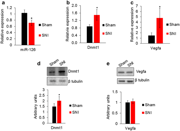
Researchers use our i-FectTM to effectively deliver miR-126 in vivo to modulate Methyl-CpG-binding protein 2 (MeCP2).

MeCP2 regulates gene expression through activation, repression and chromatin remodeling. Mutations in MeCP2 cause Rett syndrome, and these patients display impaired nociception. The researchers observed an increase in MeCP2 expression in mouse dorsal root ganglia (DRG) after peripheral nerve injury: Melissa T. Manners, Adam Ertel, Yuzhen Tian and Seena K. Ajit. Genome-wide redistribution of MeCP2 in dorsal root ganglia after peripheral nerve injury. Epigenetics & Chromatin 20169:23. DOI: 10.1186/s13072-016-0073-5© The Author(s) 2016 Received: 11 March 2016. Accepted: 27 May 2016Published: 7 June 2016...miRNA administration protocol was adapted from previous report of intrathecal miRNA delivery. To administer miRNA mimics, a polyurethane catheter (25G, 5.5 cm long, SAI infusion) was placed into the intrathecal space of the lumber L4–L5 vertebrae under isoflurane anesthesia. The catheter was stereotactically secured under the skin and occluded between injections. A custom miRCURY (Exiqon) miR-126 mimic containing a 5′ cholesterol tag and 3′ fluorescein label was injected at 2 nmol concentration with 4 µl iFECT transfection reagent (Neuromics). A total of 6 µl was delivered into the catheter connection juncture using a 25G blunt end needle on a Hamilton syringe. The catheter was then flushed with 7 µl sterile PBS to ensure miRNA reached the intrathecal space...

Figure: Expression of miR-126 and its target genes Dnmt1 and Vegfa in the DRG after nerve injury. a Relative expression of miR-126 determined by qPCR shows a reduction in miR-126 in SNI model compared to DRG from sham control. U6 was used for normalization (n = 8 sham, n = 7 SNI). b Relative expression of Dnmt1 mRNA and c Vegfa transcripts showed an increase in the DRG after nerve injury compared to control (n = 3). Gapdh was used as a normalizer. d Representative Western blot and quantification showed an increase of Dnmt1 protein in the DRG after nerve injury. e Western blot and quantification showed Vegfa protein was not significantly different in DRG after nerve injury (n = 3 from pooled samples, three DRG were pooled for each sample).


Conclusions: The study shows a regulatory role for MeCP2 in that changes in global redistribution can result in direct and indirect modulation of gene expression in the DRG. Alterations in genome-wide binding of MeCP2 therefore provide a molecular basis for a better understanding of epigenetic regulation-induced molecular changes underlying nerve injury.

Thursday, March 24, 2016

Epigenetics and Pain Research

i-Fect Used to Study Impacts

Our i-Fect siRNA, miRNA and shRNA Trasfection Kit was recently used to study the impact of G9a-specific siRNA (AGUAACGGGCAUCAAUGC) on Mu Opioid Receptors: Yuhao Zhang, Shao-Rui Chen, Geoffroy Laumet, Hong Chen and Hui-Lin Pan. Nerve Injury Diminishes Opioid Analgesia through Lysine Methyltransferase-Mediated Transcriptional Repression of µ-Opioid Receptors in Primary Sensory Neurons. First Published on February 25, 2016, doi: 10.1074/jbc.M115.711812... In some SNL rats, G9a-specific siRNA (4 µg) or the negative control siRNA was administered intrathecally. G9a-specific siRNA(AGUAACGGGCAUCAAUGC) or universal negative control siRNA (#SIC001, Sigma-Aldrich) was mixed with i-Fect (Neuromics, Edina, MN) to a final concentration of 400 mg/L for the intrathecal injections...

Figures: G9a knockdown with siRNA reverses the MOR expression in the DRG and the morphine analgesic effect diminished by nerve injury. (A,B) Quantitative PCR (A) and Western blotting (B) analyses show the mRNA and protein levels of MORs in the DRGs of sham and SNL rats treated with control or G9a-specific siRNA (n = 10 rats in each group). The ipsilateral L5 and L6 DRG tissues were removed 24 h after the last siRNA injection. The amount of MOR mRNA and protein was normalized to GAPDH in the same samples, and the mean value of MOR levels in sham control rats was considered to be 1. (C) Time course of the intrathecal morphine effects on the tactile and pressure withdrawal thresholds in sham and SNL rats treated with G9a-specific siRNA or negative control siRNA (n = 9 rats in each group). The withdrawal thresholds after the last siRNA injection were plotted as the baseline control (BL).

Summary: The findings provide new insight into the epigenetic mechanism regulating MOR expression in primary sensory neurons in neuropathic pain. This multidisciplinary approach provides conclusive evidence for G9a as a key chromatin regulator responsible for MOR downregulation in the DRG and the analgesic efficacy of opioids reduced by nerve injury. A better understanding of the epigenetic mechanisms underlying nerve injury-induced downregulation of MORs in primary sensory neurons could help improve the analgesic efficacy of opioids for treating chronic neuropathic pain. G9a inhibitors could be used to enhance the opioid analgesic effect and reduce opioid consumption in patients with chronic neuropathic pain.

Wednesday, December 09, 2015

Kv Channels and Pain Transmissiom

i-Fect TMis a Proven Tool for Gene Manipulation in Studying All Types of Pain

I previously posted on use of our i-Fect Transfection Kit to silence Kv Channels Receptors. This has enabled researchers to study the role of these receptors in vitro and in vivo (see:i-Fect™ Delivers Your siRNA Payload).

Sample Data

Figure: Figures. siRNA-mediated knockdown of Kv1.1 expression in thoracic DRG significantly increased gastric sensitivity in naive adult rats. (A) Western blots showed a significant decrease in Kv1.1 protein in thoracic DRG (T8–T12) after intrathecal treatment with Kv1.1 siRNA but not with control siRNA. siRNA treatment did not alter TrpV1 expression (n = 5 rats each; *P < .01 vs control siRNA). (B) Naive rats treated with Kv1.1 siRNA showed a significant increase in VMR to gastric distention (n = 5 rats each, compared with pretreatment baseline; *P < .05). (C) Treatment with control siRNA had no significant effect on gastric hypersensitivity. (D) Patch clamp recordings from freshly dissociated gastric DRG neurons from FD-like and PND 10 saline-treated littermate controls showed a significant decrease in rheobase in FD-like rats (*P < .05), and (E) a significant increase in the number of action potentials elicited by current injection at 3× the rheobase in gastric DRG neurons from FD-like rats (*P < .05). (F) Sample voltage vs time traces showing action potentials evoked at ×1, ×2, and ×3 rheobase. The patch clamp data were obtained from 16 cells from 5 PND 10 saline control rats and 19 cells from 5 FD-like rats

I am pleased to share with you a new reference detailing how research use i-Fect to optimize and deliver euchromatic histone-lysine N-methyltransferase-2 (G9a) siRNA. This brings the number of publications referencing use of our Transfection Kits to over 45: Geoffroy Laumet, Judit Garriga, Shao-Rui Chen, Yuhao Zhang, De-Pei Li, Trevor M Smith, Yingchun Dong, Jaroslav Jelinek, Matteo Cesaroni, Jean-Pierre Issa & Hui-Lin Pan G9a is essential for epigenetic silencing of K+channel genes in acute-to-chronic pain transition. Nature Neuroscience (2015) doi:10.1038/nn.4165.

The authors report: "Selective knockout of the gene encoding G9a in DRG neurons completely blocked K+ channel silencing and chronic pain development after nerve injury. Remarkably, RNA sequencing analysis revealed that G9a inhibition not only reactivated 40 of 42 silenced genes associated with K+ channels but also normalized 638 genes down- or upregulated by nerve injury."

I will continue to post here new and unique solutions and related referencing for our Gene Expression Analysis Tools.

Saturday, February 21, 2015

i-Fect Delivers siRNA in vitro and in vivo

Gene Expression Analysis for Studying Stroke

We have posted over 35 publications that reference use of our i-Fect Transfection Kit to deliver siRNA, miRNA and shRNA in vitro and in vivo. Results documented in these publications prove that this kit is both non-toxic and delivers ultra-high transfection efficiency.

i-Fect Data Example


Figure. The effects of HDAC2 siRNA Infusion into the CeA of P rats on voluntary ethanol consumption as measured by the two-bottle free choice paradigm. Monitoring the voluntary ethanol consumption of alcohol-preferring (P) rats via the two bottle free choice paradigm following infusion of vehicle or histone deacetylase isoform 2 (HDAC2) siRNA into the central amygdala (CeA) demonstrates that high HDAC2 levels may mediate the high alcohol drinking behaviors of P rats. P rats were given access to water and 7% ethanol followed by water and 9% ethanol. On the sixth day of ethanol access P rats received infusion of vehicle or HDAC2 siRNA and consumption of water and 9% ethanol were monitored for sevnfusion. Total fluid intake did not significantly differ between the groups. Values are represented as the mean ± SEM of the ethanol consumption (g / kg / day) and total fluid intake (mL) plotted daily for n=6 rats per treatment group. *Significantly different between the groups.SACHIN MOONAT. The Role of Amygdaloid Chromatin and Synaptic Remodeling in Anxiety and Alcoholism. THESIS Submitted as partial fulfillment of the requirements for the degree of Doctor of Philosophy in Neuroscience in the Graduate College of the University of Illinois at Chicago, 2014.

Here's yet another recent publication highlighting use of i-Fect:

In this study data suggest the protective effects of EPO on NUV injuries are highly associated with the increase of p-Cx43, which improves GJIC to reduce neurotoxic substances: Ziyi Zhoua, Xiaobai Weib, Jun Xiang, Junpeng Gao, Lixin Wang, Jinsong You, Yefeng Cai , Dingfang Caid. Protection of erythropoietin against ischemic neurovascular unit injuries through the effects of connexin43. Biochemical and Biophysical Research Communications. doi:10.1016/j.bbrc.2015.02...The strands were incubated at 90°C for 5 min and then at 37°C for 1 h. SiRNA was prepared immediately before administration by mixing the RNA solution (1 μg/μl in annealing buffer) with the transfection reagent i-Fect (v/v: 1/3; Neuromics, Edina, MN, USA) to a final siRNA ...

Highlights
•EPO has protective effects on ischemic NVU injuries.
•EPO up-regulates phosphorylation of Cx43, not total Cx43.
•EPO's protective effects on NUV injuries are p-Cx43-GJIC dependent.

Thursday, August 07, 2014

Pain Research Pubs

2014 Is Already a Record Year

I am pleased with the acceleration of publications by Pain Researchers using our Pain Research Markers and Gene Expression Analysis Tools.

Here's a sampling of the most recent: Transfection Regent Publications: Lili Hou, Yanfeng Zhang, Yong Yang, Kai Xiang, Qindong Tan, Qulian Guo. Intrathecal siRNA Against GPNMB Attenuates Nociception in a Rat Model of Neuropathic Pain. Journal of Molecular Neuroscience. July 2014...Ten micrograms of siRNA1- GPNMB dissolved in 30 μl i-Fect transfection reagent (Neuromics, Edina, MN, USA) was administered intrathecally once daily for 7 days, starting from 1 day before CCI surgery...

TRPV1 PublicationsCapsaicin-responsive corneal afferents do not contain TRPV1 at their central terminals in trigeminal nucleus caudalis in ratsOriginal Research ArticleJournal of Chemical Neuroanatomy, Volumes 61–62, November 2014, Pages 1-12 Deborah M. Hegarty, Sam M. Hermes, Tally M. Largent-Milnes, Sue A. Aicher

New insights into mechanisms of opioid inhibitory effects on capsaicin-induced TRPV1 activity during painful diabetic neuropathy. Neuropharmacology, Volume 85, October 2014, Pages 142-150 Mohammed Shaqura, Baled.I. Khalefa, Mehdi Shakibaei, Christian Zöllner, Mahmoud Al-Khrasani, Susanna Fürst, Michael Schäfer, Shaaban A. Mousa
TRPV1 IF in Mouse DRGs


Mohammed Shaqura, Baled.I. Khalefa, Mehdi Shakibaei, Christian Zöllner, Mahmoud Al-Khrasani, Desipramine and citalopram attenuate pretest swim-induced increases in prodynorphin immunoreactivity in the dorsal bed nucleus of the stria terminalis and the lateral division of the central nucleus of the amygdala in the forced swimming test. DOI: http://dx.doi.org/10.1016/j.npep.2014.07.001...... After rinsing with PBS, sections were incubated in PBS containing 0.3% Triton X-100 and 5% normal goat serum at room temperature for 30 min and then incubated with polyclonal guineapig anti-prodynorphin antibody (1:1000, Neuromics, Edina, MN, USA) or polyclonal rabbit ...

All Neuromics' Publications

I will continue to post Pain Research Updates!

Wednesday, July 23, 2014

Pain Research Gene Expression Analysis

Potent and Proven Transfection Kits


Pain Researchers have successfully modulated 25+ genes involved in pain pathways using our Transfection Kits. Highlights include: DOR,The β3 subunit of  Na+,K+-ATPase, NTS1, NAV1.8, Kv 1.1, Kv 9.1, TROY, NOV, β-arrestin, TRPV1, CAV1.2, TLR4 and ASIC and more!  To learn more, check out our Transfection Kit Publications and Blog.

Figures: Intrathecal Kv9.1 siRNA treatment induces pain behaviors in naive rats. A, qRT-PCR quantification of Kv9.1 mRNA in rat PASMC cultures transfected with one of three Kv9.1 siRNA sequences or control siRNA. B, qRT-PCR showing Kv9.1 in vivo knock-down in L5 DRG, 4 d after intrathecal delivery of siRNA #1 compared with vehicle or matched scrambled control.  C, IHC for Kv9.1 in scrambled- and siRNA-treated DRG to determine protein knockdown. Graphs illustrate quantification of number of positive myelinated neurons and mean Kv9.1 signal intensity. D, Kv9.1 siRNA infusion inflicts a reduction in mechanical pain withdrawal thresholds. E, There was no change in heat pain thresholds after siRNA treatment. Vertical arrows on x-axis denote siRNA injections. doi: 10.1523/​JNEUROSCI.3561-12.2012.


We can now add the GPNMB gene to the list of those anaylyzed: Lili Hou, Yanfeng Zhang, Yong Yang, Kai Xiang, Qindong Tan, Qulian Guo. Intrathecal siRNA Against GPNMB Attenuates Nociception in a Rat Model of Neuropathic Pain. Journal of Molecular Neuroscience. July 2014...Ten micrograms of siRNA1- GPNMB dissolved in 30 μl i-Fect transfection reagent (Neuromics, Edina, MN, USA) was administered intrathecally once daily for 7 days, starting from 1 day before CCI surgery...
Abstract: Neuropathic pain is characterized by hyperalgesia, allodynia, and spontaneous pain. Recent studies have shown that glycoprotein nonmetastatic melanoma B (GPNMB) plays a pivotal role in neuronal survival and neuroprotection. However, the role of GPNMB in neuropathic pain remains unknown. The aim of the present study was to assess the role of GPNMB in neuropathic pain. In cultured spinal cord neurons, we used two small interfering RNAs (siRNAs) targeting the complementary DNA (cDNA) sequence of rat GPNMB that had potent inhibitory effects on GPNMB, and siRNA1-GPNMB was selected for further in vivo study as it had the higher inhibitory effect. After sciatic nerve injury in rats, the endogenous level of GPNMB was increased in a time-dependent manner in the spinal cord. Furthermore, the intrathecal injection of siRNA1-GPNMB inhibited the expression of GPNMB and pro-inflammatory factors (TNF-α, IL-1β, and IL-6) and alleviated mechanical allodynia and thermal hyperalgesia in the chronic constriction injury (CCI) model of rats. Taken together, our findings suggest that siRNA against GPNMB can alleviate the chronic neuropathic pain caused by CCI, and this effect may be mediated by attenuated expression of TNF-α, IL-1β, and IL-6 in the spinal cord of CCI rats. Therefore, inhibition of GPNMB may provide a novel strategy for the treatment of neuropathic pain.

If you would like to learn how you can optimize your gene expression analysis studies, do not hesitate to e-mail: pshuster@neuromics.com or direct line: 612-801-1007.

Thursday, March 20, 2014

Irritable Bowel Syndrome (IBS) and BDNF

Gene Expression Analysis Determines BDNF's Role in IBS

In this study, Researchers used Neuromics'  i-FectTM Transfection Kit to deliver BDNF to determine effect on visceral hypersensitivity (VHS): J. H. Winston1 Q. Li1, S. K. Sarna1. Chronic prenatal stress epigenetically modifies spinal cord BDNF expression to induce sex-specific visceral hypersensitivity in offspring. Article first published online: 4 MAR 2014. Neurogastroenterology & Motility. DOI: 10.1111/nmo.12326. The Journal of Pain, Volume 14, Issue 11, November 2013, Pages 1485–1491 http://dx.doi.org/10.1016/j.jpain.2013.07.007

Intrathecal treatment with brain-derived neurotrophic factor (BDNF) antagonists reduced VMR to colorectal distension (CRD) in female chronic prenatal stress (CPS)+HeICS rats. (A) Graph shows that intrathecal administration of BDNF antagonist trkBFc in female CPS rats significantly decreased VMR to CRD, 24 h following adult 29 HeICS (two-way repeated measures ANOVA found a significant main effect of treatment, F1,53 = 10.4, p = 0.015; post hoc tests found significant differences at 30, 40, 50, and 60 mmHg, n = 4). (B) Graph shows that intrathecal administration of BDNF siRNA in female CPS rats significantly decreased VMR to CRD, 24 h following adult 29 HeICS (two-way repeated measures ANOVA: treatment 9 pressure interaction, F1,77 = 3.49, p = 0.008, tukey post hoc tests found significance at 30 mmHg, p = 0.013 and at 40, 50 50, 80 mmHg, p < 0.001, n = 7 Ctr., n = 6 BDNF siRNA). (C) Western blot shows a significant decrease in spinal cord BDNF protein expression in rats treated with BDNF siRNA (*p < 0.05).

Conclusion: Chronic prenatal stress followed by a second exposure to HeICS in adult offspring exacerbated visceral hypersensitivity (VHS) greater in female offspring that persisted longer than in male offspring. Chronic prenatal stress up-regulated BDNF expression in the lumbar-sacral dorsal horn that correlated with the exacerbation of VHS in female, but not in male offspring by increasing RNA Pol II binding and histone H3 acetylation, and decreasing histone deacetylase 1 association with the core promoter of BDNF in female offspring.

Monday, December 10, 2012

Pain Research and Gene Expression Analysis

There have been multiple publications referenced here on using Neuromics' i-Fect siRNA Delivery Kit to study the effect of silencing genes known to play a role in Pain Signaling. These include: DOR,The β3 subunit of the Na+,K+-ATPase, NTS1, NAV1.8, TRPV1 NOV, β-arrestin, TRPV1, CAV1.2 and ASIC.

I would like to highlight an exciting new study referencing how knocking down Kv9.1 Potassium Channel Subunit in vivo mediates neuropathic pain: Christoforos Tsantoulas, Lan Zhu, Yasin Shaifta, John Grist, Jeremy P. T. Ward, Ramin Raouf, Gregory J. Michael, and Stephen B. McMahon. Sensory Neuron Downregulation of the Kv9.1 Potassium Channel Subunit Mediates Neuropathic Pain following Nerve Injury. The Journal of Neuroscience, 28 November 2012, 32(48): 17502-17513; doi: 10.1523/​JNEUROSCI.3561-12.2012.

Highlights: Here, we report that the potassium channel subunit Kv9.1 is expressed in myelinated sensory neurons, but is absent from small unmyelinated neurons. Kv9.1 expression was strongly and rapidly downregulated following axotomy, with a time course that matches the development of spontaneous activity and pain hypersensitivity in animal models. Interestingly, siRNA-mediated knock-down of Kv9.1 in naive rats led to neuropathic pain behaviors. Diminished Kv9.1 function also augmented myelinated sensory neuron excitability, manifested as spontaneous firing, hyper-responsiveness to stimulation, and persistent after-discharge. Intracellular recordings from ex vivo dorsal root ganglion preparations revealed that Kv9.1 knock-down was linked to lowered firing thresholds and increased firing rates under physiologically relevant conditions of extracellular potassium accumulation during prolonged activity. Similar neurophysiological changes were detected in animals subjected to traumatic nerve injury and provide an explanation for neuropathic pain symptoms, including poorly understood conditions such as hyperpathia and paresthesias. In summary, our results demonstrate that Kv9.1 dysfunction leads to spontaneous and evoked neuronal hyperexcitability in myelinated fibers, coupled with development of neuropathic pain behaviors.

n vivo RNA interference: Anesthetized rats were subjected to a thoracic laminectomy and a silastic tube was inserted subdurally to lie just rostral to L3 DRG and externalized to deliver bolus injections (one injection per day for 4 consecutive days). Animals were allowed to recover for 5 d before treatment commenced. On the day of injection, siRNA was mixed with i-Fect (Neuromics) to a final concentration of 0.2 μg μl−1, according to published protocols (Luo et al., 2005). For each treatment, 10–20 μl of Kv9.1 siRNA or scrambled control mixture was injected, followed by a 10 μl saline flush. Twenty-four hours after the fourth injection animals were killed and L5 DRGs fresh dissected for qRT-PCR analysis. A separate set of animals were PFA perfused and DRGs retrieved for IHC. Passenger strand sequences for Kv9.1 and scrambled control siRNAs were cuuggaaucuguaggauca and gaggcctaatcgatatgtt, respectively (Dharmacon; “in vivo processing” option).
Intrathecal Kv9.1 siRNA treatment induces pain behaviors in naive rats. A, qRT-PCR quantification of Kv9.1 mRNA in rat PASMC cultures transfected with one of three Kv9.1 siRNA sequences or control siRNA (control, n = 6; siRNA, n = 3 per group; *p < 0.05 vs control, one-way ANOVA with Tukey's). B, qRT-PCR showing Kv9.1 in vivo knock-down in L5 DRG, 4 d after intrathecal delivery of siRNA #1 compared with vehicle or matched scrambled control (vehicle, n = 4; scrambled, n = 5; Kv9.1, n = 7; *p < 0.05, t test). C, IHC for Kv9.1 in scrambled- and siRNA-treated DRG to determine protein knockdown. Graphs illustrate quantification of number of positive myelinated neurons and mean Kv9.1 signal intensity (scrambled, n = 4; siRNA, n = 6; **p < 0.01, ***p < 0.001, t test). D, Kv9.1 siRNA infusion inflicts a reduction in mechanical pain withdrawal thresholds (Kv9.1, n = 7; control, n = 6; *p < 0.05, **p < 0.01, ***p < 0.001 vs scrambled control or baseline, two-way repeated measurements ANOVA with Tukey's). E, There was no change in heat pain thresholds after siRNA treatment. Vertical arrows on x-axis denote siRNA injections. All data represent mean ± SEM.
Kv9.1 knock-down triggers ectopic activity and a form of peripheral wind-up in response to stimulation. A, Schematic illustrating the positions of stimulating and recording electrodes. B, Example recordings from centrally disconnected L4/L5 strands demonstrating SA in Kv9.1 siRNA-treated or nerve-injured rats, but not in control (scrambled siRNA) animals. C, Frequency-dependent SEA (denoted by double arrowheads) in Kv9.1 siRNA-treated (middle) and injured (right), but not control (left) animals. This activity is not locked in time and can be seen in between stimulation events (vertical arrows on top of 5 Hz stimulation traces, only first 5 shown). Also note the prolonged after-discharge (AD) observed in siRNA-treated and injured animals. D, Percentage of units showing SA and SEA in control (n = 269), Kv9.1 siRNA-treated (n = 369) and injured (n = 176) animals (*p < 0.05, **p < 0.01, ***p < 0.001 vs control, χ2 test). E, Firing rate of SEA units at different stimulation frequencies (mean ± SEM; control, n = 4; siRNA, n = 22; injured, n = 17; *p < 0.05 vs control, two-way ANOVA with Tukey's). F, Quantification of AD rate per SEA unit (mean ± SEM; *p < 0.05 vs control, Mann–Whitney test).

Results propose that Kv9.1 downregulation after nerve injury may be the molecular switch controlling myelinated sensory neuron hyperexcitability. Intriguingly, a recent wide-genome association screen in humans identified a Kv9.1 polymorphism associated with susceptibility to develop chronic neuropathic pain after back surgery or leg amputation (Costigan et al., 2010), suggesting that the mechanisms described in our studies will be of direct clinical relevance to human pain. Future efforts to elucidate the precise pathways involved, combined with approaches aiming to compensate loss of Kv9.1 function, may create novel therapeutic opportunities for neuropathic pain management.

Tuesday, October 30, 2007

SfN Presentation-i-Fect and siRNA in vivo

We are excited about this upcoming presentation at Society for Neuroscience comference presesentation. Neuromics' i-Fect ™ reagent was used to deliver siRNA in vivo to silence the NTS2 gene.

Program#/Poster#:509.6/PP9
Title:Small interfering RNA-mediated selective knockdown of NTS2 receptors reverses neurotensin-induced analgesia in rats
Location:San Diego Convention Center: Halls B-H
Presentation Start/End Time:Monday, Nov 05, 2007, 2:00 PM - 3:00 PM
Authors:*L. DORE-SAVARD1,2, G. ROUSSY1, M.-A. DANSEREAU1, K. BELLEVILLE1, N. BEAUDET1, M. BEHLKE2, P. SARRET1; 1
Physiology and Biophysics, Univ. Sherbrooke, Sherbrooke, PQ, Canada; 2Integrated DNA Technologies Inc., Coralville, IA

2007 Copyright by the Society for Neuroscience all rights reserved. Permission to republish any abstract or part of any abstract in any form must be obtained in writing by SfN office prior to publication.