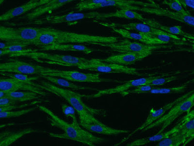
Showing posts with label Human Endothelial Cells. Show all posts

Wednesday, November 24, 2021

Studying Diabetic Retinopathy?

Check out How Neuromic's Tools Are Used

The authors used four of Neuromics to complete this study-

Sulodexide reduces glucose induced senescence in human retinal endothelial cells A. Gericke, K. Suminska-Jasińska & A. Bręborowicz Scientific Reports volume 11, Article number: 11532 (2021) Cite this article.

Material and Methods
Experiments were performed on HREC (#HEC09, Neuromics, Edina, MN, USA) in in vitro culture. Cells were seeded in 75 cm2 culture flasks coated with AlphaBioCoat Solution (#AC00, Neuromics, Edina, MN, USA) and were grown in Endo Growth Medium (EKG001, Neuromics, Edina, MN, USA) supplemented with fetal bovine serum 0.5% until they formed monolayers. Then the cells were harvested with cell detachment solution (#ADF001, Neuromics, Edina, MN, USA) and seeded in quadruplicates into 25 cm2 coated, as described above, culture flasks at a density of 1.5 × 105 cells/flask.

We conclude that Sulodexide may have a beneficial effect in cases of diabetic retinopathy. It slows down hyperglycemia-dependent senescence of endothelial cells, which translates into the lower angiogenic and inflammatory impact of these cells. An important observation was that Sulodexide also has effective antiangiogenic and anti-inflammatory effects in the senescent HREC. That means that Sulodexide may be effective in retinal endothelial cells, which are already senescent. Further studies are required to explain the potential effect of Sulodexide on the endothelial glycocalyx structure and permeability of the retinal endothelial layer composed of the senescent cells to molecules with various size and electrical charge.

We are pleased to see our solutions used in this important  study. It could lead to drugs that slow diabetic retinopathy.

Monday, February 01, 2021

Malaria and the Blood-Brain Barrier

 EPCR and ICAM Receptors

Neuromics Astrocytes and Pericytes were used in this important study-Yvonne Adams, Rebecca W. Olsen, Anja Bengtsson, Nanna Dalgaard, Mykola Zdioruk, Sanghamitra Satpathi, Prativa K. Behera, Praveen K. Sahu, Sean E. Lawler, Klaus Qvortrup, Samuel C. Wassmer, and Anja T.R. Jensen. (2021). Plasmodium Falciparum Erythrocyte Membrane Protein 1 Variants Induce Cell Swelling and Disrupt the Blood–Brain Barrier in Cerebral Malaria. Journal of Experimental Medicine, 218 (3). doi: 10.1084/jem.20201266.

This is the first study showing malaria pericytes interacting with human endothelial cells. Please note we have the complete BBB Model that can be used for studies like these.


Tuesday, March 17, 2020

We Are Open

Providing Cells, Media and Supplements in the time of Covid-19
If you call us, we will answer the phone. If you submit an order, it will ship.

Neuromics stands ready to serve you.
Image: Neuromic's Human Pancreatic CAF-Stellate Cells in Culture
FBS only $299/500 ml. We have plenty of inventory so we can continue with this pricing for the duration of the Pandemic.
Stay safe.

Tuesday, December 03, 2019

Human Cells-utopia!

Great for use a controls vs Differentiated iPSCs

We have a cornucopia of Human Primary Neuron, Astrocytes and Schwann Cells plus CAFS. Featured Assays:
Human Brain Pericytes used to study Guide Axon Guidance.
Human Pancreatic CAFS and Tumor Dynamics.

Axon guidance at the site of a cervical spinal cord injury in a rat model. (Ai) Schematic illustrating transplantation of scaffold into a C-4 hemisection. The injury cavity is shown prior to (ii) and immediately following (iii) transplantation. (Bi) Scaffold conditioned with flow exhibits viable GFP-labeled microvessels (green) (ii) and alignment of host axons (magenta) infiltrating the scaffold in the rostral-caudal direction (grey arrow). (C) Scaffold conditioned in static conditions showing disrupted alignment of both microvessels (ii) and host axons (iii). (D–F) Microvessel and axon plots showing alignment (D,E) and length (F). Scale bars, 1 mm (Aii,Aiii) and 50 μm (B,C). Data are presented as mean ± s.e.m. ***P < 0.001; statistical significance was calculated using Welch Two Sample t-test. White arrows denote proximity of axons with microvessels. Microvessel alignment values (n = 30), axon alignment values (n = 30), microvessel length values (n = 15), and axon length values (n = 15) are from single hydrogel samples per condition.
Representative CAFs spheroids embedded in collagen gels at time 0 and at 6 h post implantation, respectively.
It is imperative that all our cells work as advertised. If your results do not meet expectations, we will run similar tests to make sure they walk and talk as they should.

We wish you and yours a Happy Holiday Season, Pete Shuster, CEO and Owner Direct phone: 612-801-1007 or pshuster@neuromics.com

Wednesday, July 11, 2018

Human Cells in Action

Human Microvascular Retinal Endothelial Cells (HMRECS)
We have built the foundation of Neuromics on satisfied customers. We make a practice of following up with each user to make sure our solutions are working as expected. If not, we offer "no question asked" refunds or replacements.

This is especially important for our human cells, media, and supplements. Success with these is easy to measure as either the cells are healthy and happy or they are not.

We also use reviews and publications as another measure of satisfaction. Here I would l highlight the latest publication using our HREMCS. A. P. Da Cunha, Q. Zhang, M. Prentiss, X. Q. Wu, V. Kainz, Y. Y. Xu, J. Vrouvlianis, H. Li, N. Rangaswamy, B. Leehy, T. L. McGee, C. L. Bell, C. E. Bigelow, V. Kansara, Q. Medley, Q. Huang & H. Y. Wu. The Hierarchy of Proinflammatory Cytokines in Ocular Inflammation. Current Eye Research, Volume 43, 2018 - Issue 4 Published Online: 04 Dec 2017.

Images: Effect of HG (high glucose) and pro-inflammatory cytokines on connexin43 expression in HRMECs. Immunohistochemical data showing connexin43 expression in (A) normal medium; (B) HG (25 mM); (C) pro-inflammatory cytokines (IL-1β and TNF-α 10 ng/mL each); and (D) a combination of HG and pro-inflammatory cytokines inducing a change in cell morphology with signs of cell swelling, possibly owing to hemichannel opening (indicated by white arrows).

If you have questions or interest in any of these, please contact rose@neuromics.com or 866-350-1500.

Friday, October 14, 2016

"Hard to Find" Cells

You can Trust Your Results
Cells form the foundation of your cell based assays. We work hard to provide the cells you need and, more importantly that they are healthy and happy.

We are pleased to announce that we are recognized for having primary human cells that comply with federal and international regulations. This is especially important in drug discovery, because if the cells yield targets that result in future therapies this validation becomes increasingly valuable.
Human Brain Microvascular Endothelial Cells in Culture

Looking for "hard to find cells"? Check us out today.
Cancer-Associated Fibroblasts (CAFs) and Cells
Solutions for studying tumor growth, angiogenesis and metastasis.
Human Brain Astrocytes-New
Derived from Cortex
Immortalized Human Brain Microglia Cells
Isolated from healthy human brain tissue
Human Schwann Cells - New
Human spinal nerve derived primary cells
hN2™ Neuron Discovery Kits
Derived from H9 (WA09) ECSs-Consistent, Easy to Use & Cost Effective
Human alpha Motor Neurons Kit
hMSC Derived-includes 100 ml of media to optimize your cultures
Human Cardiomyocytes-New
Derived from hMSCs
Human Chondrocytes-New
Derived from hMSCs
Human Endothelial Cells-New
Artery, Microvascular, Vein & Umbilical Cord Derived + Culture Media
Human Fibroblasts-New
Pancreas and Neonatal Dermal Derived
NeuroNet Pure Human Neurons Discovery and HTS Kits
Pure Population of hPSC Derived Neurons
Human Osteoblasts-New
Derived from hMSCs
Human Mesenchymal Stem Cells (hMSCs)
hMSCs Derived from Umbilical Cord Blood
Stressed Selected Stem Cells
They Survive to Thrive
Human Pericytes
Brain and Retinal Derived
3-D Cell Based Assay Solutions
Nanofibers, Hydrogels and Extracellular Matrix (ECM) Proteins

Tuesday, October 04, 2016

Your Brain Trust

Trust is hard to gain and easy to lose. My focus over the past 12 years has been to earn the trust of all our clients. This means that we do everything possible to insure our solutions work. When we come up short, we strive to resolve your issues the same day, and we have one of the easiest replacement policies in the market today. Here's proof.
Trust and Testimonials: We use BirdEye, a 3rd party, to collect and manage feedback from our clients.
5on BirdEye, Aug 29, 2016
C. H.
The order arrived quickly. The service was great!
Product Name: Human Brain Microvascular Endothelial Cells, 500,000+ Cells, - (Cat# HEC02)

Human Brain Microvascular Pericytes (HBMPVCs), 500,000+ Cells, - (Cat# HMP104)
http://bit.ly/2bzQlPn
ENDO-Growth Media, - (Cat#MED001)
http://www.neuromics.com/endo-growth-media
Pericyte-Growth Media, - (Cat# PGB001)
http://www.neuromics.com/pericyte-growth-media
Organization: UCSF

HBMPVCs in Culture
Trust and Publications: There are hundreds of publication referencing use of our products. Want to see if a product of interest is published? Just Ask
Need hard to find human cells? Please contact me directly pshuster@neuromics.com or 612-801-1007. We can together determine your needs and desired outcomes and provide a statement of work with pricing, project milestones and delivery.
We wish you a productive October filled with new and exciting discoveries.

Thursday, July 21, 2016

Human Endothelial Cells and Drug Discovery

Pure, Potent and Culture Ready
Alterations of endothelial cells and the vasculature play a central role in the pathogenesis of a broad spectrum of the most dreadful of human diseases, as endothelial cells have the key function of participating in the maintenance of patent and functional capillaries. This makes them a key component for Drug Discovery.

We are proud that Biotech and Biopharma Cos. are increasingly using our cells. We believe we represent the best options. Here're some of the cells currently available:
Microvascular Endothelial Cells in Action

Figures: HRMECs treated with SUZ12 siRNA demonstrate increased miR-200b expression, decreased VEGF expression and decreased endothelial branching. A,B: Gene knockdown of EZH2 and SUZ12 was confirmed by qPCR. C,D: In HRMECs transfected with EZH2 siRNA in HG, miR-200b and VEGF were not significantly different from HG+control siRNA but decreased compared to NG+control siRNA. In HRMECs transfected with SUZ12 siRNA in HG, miR-200b was significantly increased with decreased levels of VEGF compared to HRMECs transfected with control siRNA, with levels similar to NG+control siRNA. E,F: Tube formation assay to measure endothelial branching. HRMECs transfected with HG+control siRNA demonstrated significantly increased branching compared to NG+control siRNA. Transfection of EZH2 siRNA did not reduce endothelial branching significantly compared to HG+control siRNA. However, transfection of SUZ12 siRNA significantly reduced endothelial branching compared to HG+control siRNA. 10.1371/journal.pone.0123987
We will keep you posted as we add more cell options.

Saturday, September 26, 2015

Retinal Microvascular Endothelial Cells & Diabetic Retinopathy

HRMECS in Action

Our Human Retinal Microvascular Endothelial Cells in this publication on Diabetic Retinopathy. In this study, the authors show that human retinal microvascular endothelial cells exposed to high levels of glucose regulate miR-200b repression through histone methylation and that inhibition of PRC2 increases miR-200b while reducing VEGF: Michael Anthony Ruiz, Biao Feng, and Subrata Chakrabarti. Polycomb Repressive Complex 2 Regulates MiR-200b in Retinal Endothelial Cells: Potential Relevance in Diabetic Retinopathy. PLoS One. 2015; 10(4): e0123987. Published online 2015 Apr 17. doi: 10.1371/journal.pone.0123987...To investigate PRC2 and miR-200b regulation in the context of diabetic retinopathy, the major cell type used for investigation was the human retinal microvascular endothelial cell (HRMECs,Neuromics/ Olaf Pharmaceuticals, Worchester, MA, Cat# HEC09)...

Figure: High levels of glucose alter VEGF and miR-200b expression in HRMECs. A: HRMECs exposed to various concentrations of D-glucose for 24 hours exhibited differential mRNA levels of VEGF. Compared to 5mM D-glucose, VEGF expression was significantly increased at 15mM and 25mM D-glucose concentrations, with no change at 20mM L-glucose. B: Measured by WST-1 assay, HRMECs exposed to increasing concentrations of D-glucose for 24 hours exhibited decreased cell viability at 25mM, 50mM and 100mM compared to 5mM. C: HRMECs exposed to 25mM (high glucose; HG) glucose for 24 and 48 hours demonstrated significantly increased VEGF mRNA compared to 5mM (normal glucose; NG). These differences were not observed at time points earlier than 24 hours. D,E: HRMECs exposed to 5mM D-glucose (NG) 25mM D-glucose (HG) and 20mM L-glucose+5mM D-glucose (osmotic control; OSM). HRMECs cultured for 24 hours and 48 hours in HG showed significantly decreased levels of miR-200b with parallel increased levels of VEGF expression compared to NG and OSM. F,G: VEGF is also increased at the protein level in HG compared to NG as measured by Western Blotting. [* p < 0.05 compared to NG; n = 6; data expressed as mean ± SEM, normalized to β-actin or U6 and expressed as a fold change of NG]. doi:10.1371/journal.pone.0123987.

Check out our growing catalog of Human Endothelial Cells-Artery, Microvascular, Vein, Umbilical Cord Derived + Culture Media.