Thursday, October 21, 2010

Stems Cells and SCI

This is fascinating and encouraging to sufferers of Spinal Cord Injury:

ScienceDaily (Oct. 9, 2010) — Researchers at Karolinska Institutet have shown how stem cells, together with other cells, repair damaged tissue in the mouse spinal cord. The results are of potential significance to the development of therapies for spinal cord injury: http://www.sciencedaily.com/releases/2010/10/101008082736.htm#

Saturday, October 09, 2010

Enhancing Neurite Outgrowth

We are pleased to announce the addition of new proteins to our Axon Growth and Guidance category. These are designed to enhance neurite out growth.


Name
Catalog #TypeSpeciesSizePrice
NGF-b, NSO Derived
PR15083
ProteinH; M; R100 ug$285
NGF-b, NSO Derived, CFPR15084CFProteinH; M; R100 ug$285
S100A13, CFPR15085CF-50ProteinH; R50 ug$315
Slit1PR15075-50ProteinCh; H50 ug$315
Slit2, CHO
PR15085-50
ProteinCh; H50 ug$315

Image: Cultured chick dorsal root ganglion neurons were grown in the presence of recombinant human NGF-b with (A) or without (B) recombinant mouse Slit2. The presence of the Slit2 protein signifi cantly enhanced neurite outgrowth.

Wednesday, September 22, 2010

Glutaredoxin 2 prevents aggregation of mutant SOD1

Our PTEN-induced kinase, PINK1 or PARK6 Antibody is an excellent marker for Amyotrophic Lateral Sclerosis (ALS) and Parkinson's Disease (PD) researchers.

Here's  new publication referencing use of this antibody:  Alberto Ferri, Paolo Fiorenzo, Monica Nencini, Mauro Cozzolino, Maria Grazia Pesaresi, Cristiana Valle, Sara Sepe, Sandra Moreno, and Maria Teresa Carrì. Glutaredoxin 2 prevents aggregation of mutant SOD1 in mitochondria and abolishes its toxicity.
Hum. Mol. Genet., first published on Sep 20, 2010 as doi: doi:10.1093/hmg/ddq383

Abstract:
Vulnerability of motoneurons in amyotrophic lateral sclerosis (ALS) arises from a combination of several mechanisms, including protein misfolding and aggregation, mitochondrial dysfunction and oxidative damage. Protein aggregates are found in motoneurons in models for ALS linked to a mutation in the gene coding for Cu,Zn superoxide dismutase (SOD1) and in ALS patients as well. Aggregation of mutant SOD1 in the cytoplasm and/or into mitochondria has been repeatedly proposed as a main culprit for the degeneration of motoneurons. It is, however, still debated whether SOD1 aggregates represent a cause, a correlate or a consequence of processes leading to cell death. We have exploited the ability of glutaredoxins (Grxs) to reduce mixed disulfides to protein thiols either in the cytoplasm and in the IMS (Grx1) or in the mitochondrial matrix (Grx2) as a tool for restoring a correct redox environment and preventing the aggregation of mutant SOD1. Here we show that the overexpression of Grx1 increases the solubility of mutant SOD1 in the cytosol but does not inhibit mitochondrial damage and apoptosis induced by mutant SOD1 in neuronal cells (SH-SY5Y) or in immortalized motoneurons (NSC-34). Conversely, the overexpression of Grx2 increases the solubility of mutant SOD1 in mitochondria, interferes with mitochondrial fragmentation by modifying the expression pattern of proteins involved in mitochondrial dynamics, preserves mitochondrial function and strongly protects neuronal cells from apoptosis. The toxicity of mutant SOD1, therefore, mostly arises from mitochondrial dysfunction and rescue of mitochondrial damage may represent a promising therapeutic strategy.
Related Reagents:
Parkin
Parkin-2
PARK2 Co-regulated (PACRG)
PARK7 (DJ-1)
LRRK2 (PARK8)                        
Neurodegenerative Disease Research Antibodies                          

Neurodegenerative Disease Research Proteins
Neurotransmission -Neurotransmission Research Antibody Categories                         
Neurotrophins and Growth Factor Antibodies
Neuron-Glial Expressed-Includes Neurotrophin Proteins
Apoptosis Research Reagents-Apoptosis Categories-includes: detection kits, antibodies and proteins

Primary Neurons and Astrocytes -Primary human, rat and mouse neurons and astrocytes by Category

Wednesday, September 15, 2010

TRHR1 and LepRb receptors and Thermogenesis

I would like to thank Montina Van Meter, Lab Manager, Autonomic Neuroscience at Pennington Biomedical Research Center, for alerting me to this just published study. Included are excellent images of stained LepRb (OB-Rb) and GAD1 expressing neurons localized in loose clusters of cells in the DMN, NST, and the VLM.

This study focus on identifying loci in the hindbrain where leptin and TRH act synergistically to increase thermogenesis. Since thermogenic processes are at the root of how our bodies regulate energy, understanding the related expression and signaling pathways could be key to finding therapies for obesity.

Maria J. Barnes, Richard C. Rogers, Montina J. Van Meter and Gerlinda E. Hermann. Co-localization of TRHR1 and LepRb receptors on neurons in the hindbrain of the rat. doi:10.1016/j.brainres.2010.07.094


Example images: Distribution of LepRb+ fibers in hindbrain. LepRb-ir (red) fibers and varicosities are seen among TRHR1-ir (green) cells and fibers. These red and green fibers are adjacent and co-mingle but do not show co-localization of receptors. This pattern is seen in (A) fascicles of the solitary tract (ST); (B) raphe pallidus (RP), and (C) raphe obscurrus (RO). (D) Border between the medial solitary nucleus (NST) and the area postrema (AP; white dashed line) showing an abundance of LepRb-ir (red) fibers and
 neurons (white arrows for selected neurons) in the NST but not the AP. (E) LepRb-ir staining is suppressed by pretreatment of tissue with LepRb epitope blocking peptide. (F) TRHR1-ir staining is suppressed by treatment with excess TRHR1. Scale bar A–D=100 microns; E, F=300 microns. cc=central canal.
Abstract: We have reported a highly cooperative interaction between leptin and thyrotropin releasing hormone (TRH) in the hindbrain to generate thermogenic responses (Hermann et al., 2006) (Rogers et al., 2009). Identifying the locus in the hindbrain where leptin and TRH act synergistically to increase thermogenesis will be necessary before we can determine the mechanism(s) by which this interaction occurs. Here, we performed heat-induced epitope recovery techniques and in situ hybridization to determine if neurons or afferent fibers in the hindbrain possess both TRH type 1 receptor and long-form leptin receptor [TRHR1; LepRb, respectively]. LepRb receptors were highly expressed in the solitary nucleus [NST], dorsal motor nucleus of the vagus [DMN] and catecholaminergic neurons of the ventrolateral medulla [VLM]. All neurons that contained LepRb also contained TRHR1. Fibers in the NST and the raphe pallidus [RP] and obscurrus [RO] that possess LepRb receptors were phenotypically identified as glutamatergic type 2 fibers (vglut2). Fibers in the NST and RP that possess TRHR1 receptors were phenotypically identified as serotonergic [i.e., immunopositive for the serotonin transporter; SERT]. Co-localization of LepRb and TRHR1 was not observed on individual fibers in the hindbrain but these two fiber types co-mingle in these nuclei. These anatomical arrangements may provide a basis for the synergy between leptin and TRH to increase thermogenesis.

Related Reagents:
Leptin and Leptin Receptor Antibodies
Leptin Proteins

Tuesday, September 14, 2010

Isolation of Medulloblastoma Stem Cells (Video Protocol)

I am pleased to present this excellent video. It also has excellent images using key stem cell markers as the cells undergo differentiation:


Sep 1, 2010 ... GFAP antibody, Neuromics, CH22102,
Chicken, 1:1000. Tuj1 antibody, Sigma, T5076, Mouse, 1:2000. NeuN
antibody, Millipore, MAB377, Mouse, ...
www.jove.com

Thursday, September 09, 2010

Potential Therapeutic Targets for Bone Cancer Pain-P2X Receptors

Cancer pain is difficult to treat as it appears to be driven simultaneously by inflammatory, neuropathic and tumorigenic mechanisms. I have reported on multiple occasions publication referencing use of our Pain and Inflammation Research Antibodies in studying bone cancer pain.

I would like to alert you to the latest reference:

Timothy K. Y. Kaan, Ping K. Yip, Sital Patel, Meirion Davies, Fabien Marchand, Debra A. Cockayne, Philip A. Nunn, Anthony H. Dickenson, Anthony P. D. W. Ford, Yu Zhong, Marzia Malcangio, and Stephen B. McMahon Systemic blockade of P2X3 and P2X2/3 receptors attenuates bone cancer pain behaviour in rats. Brain, September 2010; 133: 2549 - 2564.

......Slides were then incubated with rabbit anti-P2X3 (1:2000, Neuromics) and sheep anti-calcitonin gene-related peptide (1:1000, Biomol...anti-beta-III-tubulin (1:4000, Promega) and guinea pig anti-P2X3 (1:100, Neuromics). The next day, after three washes with phosphate-buffered......

Summary: Pain remains an area of considerable unmet clinical need, and this is particularly true of pain associated with bone metastases, in part because existing analgesic drugs show only limited efficacy in many patients and in part because of the adverse side effects associated with these agents. An important issue is that the nature and roles of the algogens produced in bone that drive pain-signalling systems remain unknown. Here, we tested the hypothesis that adenosine triphosphate is one such key mediator through actions on P2X3 and P2X2/3 receptors, which are expressed selectively on primary afferent nocioceptors, including those innervating the bone. Using a well-established rat model of bone cancer pain, AF-353, a recently described potent and selective P2X3 and P2X2/3 receptor antagonist, was administered orally to rats and found to produce highly significant prevention and reversal of bone cancer pain behaviour. This attenuation occurred without apparent modification of the disease, since bone destruction induced by rat MRMT-1 carcinoma cells was not significantly altered by AF-353. Using in vivo electrophysiology, evidence for a central site of action was provided by dose-dependent reductions in electrical, mechanical and thermal stimuli-evoked dorsal horn neuronal hyperexcitability following direct AF-353 administration onto the spinal cord of bone cancer animals. A peripheral site of action was also suggested by studies on the extracellular release of adenosine triphosphate from MRMT-1 carcinoma cells. Moreover, elevated phosphorylated-extracellular signal-regulated kinase expression in dorsal root ganglion neurons, induced by co-cultured MRMT-1 carcinoma cells, was significantly reduced in the presence of AF-353. These data suggest that blockade of P2X3 and P2X2/3 receptors on both the peripheral and central terminals of nocioceptors contributes to analgesic efficacy in a model of bone cancer pain. Thus, systemic P2X3 and P2X2/3 receptor antagonists with central nervous system penetration may offer a promising therapeutic tool in treating bone cancer pain.

Related Reagents:

All Purinergic Receptors
Neurotransmission Research Antibodies

Sunday, August 08, 2010

Angiogenesis Research Reagents

The processes of angiogenesis and neurogenesis show striking similarities. Given our strong roots in providing Neuroscience research reagents, it is a natural extension for us to provide potent reagents for the studying the growth of new blood vessels.

Angiogensis processes are important for healing wounds and for restoring blood flow to tissues after injury or insult. In females, it also occurs during the monthly reproductive cycle and during pregnancy(to build the placental circulation between mother and fetus.

Images: Increased arteriogenesis and hyperemia in TSP2-null mice. Smooth muscle actin-positive and ephrin B2-positive vessels in WT (F, H) and TSP2-null (G, I). DOI: 10.2353/ajpath.2008.080128.

Angiogenesis-dependent diseases result when new blood vessels either grow excessively or insufficiently. These disease include: cancers, diabetic blindness, age-related macular degeneration, rheumatoid arthritis, psoriasis, neurodegeneration and many more.

Product Categories

Thursday, July 29, 2010

Let-7 microRNAs and Nociceptive Pain

Our Opioid Receptor Antibodies have set a potent standard for studying Nociceptive and Neuropathic Pain. Related Publications.

We want to recognize Dr. Zaijie Jim Wang and his team for being the first to use our Mu Opioid Receptor for studying the potential role of microRNAs in Nociception.

Ying He, Cheng Yang, Chelsea M. Kirkmire, and Zaijie Jim Wang. Regulation of Opioid Tolerance by let-7 Family MicroRNA Targeting the µ Opioid Receptor. The Journal of Neuroscience, July 28, 2010, 30(30):10251-10258; doi:10.1523/JNEUROSCI.2419-10.2010
Abstract: MicroRNA has emerged as a critical regulator of neuronal functions. This study aimed to test whether let-7 microRNAs can regulate the µ opioid receptor (MOR) and opioid tolerance. Employing bioinformatics, we identified a let-7 binding site in the 3'-untranslated region (UTR) of MOR mRNA, which was experimentally confirmed as a direct target of let-7. The repressive regulation of MOR by let-7 was revealed using a LNA-let-7 inhibitor to knockdown let-7 in SH-SY5Y cells. Conversely, morphine significantly upregulated let-7 expression in SH-SY5Y cells and in a mouse model of opioid tolerance. The LNA-let-7 inhibitor decreased brain let-7 levels and partially attenuated opioid antinociceptive tolerance in mice. Although chronic morphine treatment did not change overall MOR transcript, polysome-associated mRNA declined in a let-7-dependent manner. let-7 was identified as a mediator translocating and sequestering MOR mRNA to P-bodies, leading to translation repression. These results suggest that let-7 plays an integral role in opioid tolerance.

  • Western blot analysis. Western blot analysis was performed as previously described (Tang et al., 2006) using the anti-µ opioid receptor antibody (1:1000; Neuromics). The expression of β-actin was similarly determined from the same blots using a monoclonal antibody (1:10,000; Sigma).
  • For immunofluorescence analysis, the antibody for hDcp1a (Santa Cruz Biotechnology) and MOR were used at 1:500 and 1:5000 dilutions, respectively. Secondary anti-goat and anti-mouse antibodies labeled with Alexa 488 and Alexa 594 fluorochromes (Invitrogen), respectively, were used at 1:500 dilutions.

Related Reagent Links:
All Opioid Receptor Antibodies

Pain and Inflammation
       
Neurotransmission Research Antibodies
-GPCRs, Ligand Gated Ion Channels,
Biogenic Amines and more
i-Fect Transfection Kit
-gene silencing of DOR,
NaV1.8 tetrodotoxin-resistant sodium channel, NTS2 and more in-vitro and in vivo
Primary Neurons and Astrocytes
-Primary human,
rat and mouse neurons and astrocytes 


Monday, July 26, 2010

δ- and μ-opioid receptors co-expression and Nociceptive Pain

Dr. Tomas Hokfelt and his team at Karolinska Institute recently published use of our Opioid Receptor Antibodies and Substance P Antibody.They show the interplay of DOR and MOR in modulation of nociceptive afferent transmission and opioid analgesia.

Hai-Bo Wanga, Bo Zhaoa, Yan-Qing Zhonga, Kai-Cheng Li, Zi-Yan Li, Qiong Wang, Yin-Jing Lua, Zhen-Ning Zhang, Shao-Qiu He, Han-Cheng Zheng, Sheng-Xi Wu, Tomas G. M. Hökfelt, Lan Baob, and Xu Zhanga. Coexpression of δ- and μ-opioid receptors in nociceptive sensory neurons. PNAS July 20, 2010 vol. 107 no. 29 13117-13122.

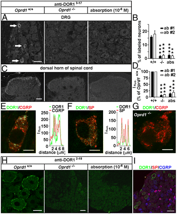
Immunostaining. Adult rats, mice, and Oprd1 exon 1-deleted mice were fixed. Cryostat sections of L4 and L5 DRGs and spinal cord segments were processed for immunofluorescence staining (13) with Rb anti-DOR13–17 (1:2,000–1:60,000; DiaSorin and 1:4,000–1:60,000; Neuromics), Rb anti-DOR12–18 (1:30,000–1:120,000; Alomone), Rb anti-DOR1358–372 (1:1,000–1:2,000; Lifespan Biosciences), Rb anti-MOR (1:1,000; Neuromics); guinea pig anti-SP (1:500; Neuromics), and mouse anti-CGRP (1:1,000; Biogenesis) antibodies. IB4-labeling was carried out with fluorescein-labeled GSL I-IB4 (1:200). The Myc-DOR1–transfected HEK293 cells and neurons were fixed and processed with mouse anti-Myc antibodies (1:500; DSHB). Nuclear DAPI staining was used to indicate HEK293 cells in control experiments.

Images: Distinct distribution patterns of DORs in subsets of DRG neurons of mice. Immunostaining with antibodies against DOR13–17 [A: 1:30,000, antibody 1 (ab #1); DiaSorin and C: antibody 2 (ab #2); Neuromics] shows DORs in small DRG neurons and afferent fibers in spinal laminae I–II. This immunostaining pattern is abolished by the antiserum preabsorption or the deletion of Oprd1 exon 1. Reduction in immunostaining is quantitatively assayed by determining the percentage of positive DRG neurons (B; n = 6) and fluorescence intensity (Ifluo.) in the laminae I–II (D; n = 5). **P < 0.01; ***P < 0.001. (Scale bars: A and C, 40 μm.). DOR labeling (anti-DOR13–17, 1:30,000; DiaSorin) associated with vesicles in peptidergic small DRG neurons (E and F) is absent in Oprd1 exon 1-deleted mice (G). Colocalization of DORs and neuropeptides is shown by correlated peaks of Ifluo. measured along lines. (Scale bar: 8 μm.) (H) Immunostaining with antibodies against DOR12–18 (1:60,000; Alomone) shows the presence of DORs on the cell surface of large DRG neurons of mice. (Scale bar: 25 μm.) This staining pattern is abolished by preabsorption and is absent in Oprd1 exon 1-deleted mice. (Scale bar: 80 μm.) (I) Triple-immunostaining shows that DOR+ large DRG neurons contain neither SP nor CGRP. (Scale bar: 80 μm.)

Immunoblotting.The samples were processed for SDS/PAGE, transferred, probed with Rb antibodies against MOR (1:500; Neuromics), phospho-DOR1 (1:1,000; Neuromics), phospho-MOR (1:1,000; Neuromics), Myc (1:500; DSHB), Flag (1:1,000; Sigma), or actin (1:50,000; Chemicon) and visualized with enhanced chemiluminescence (19).

Featured and Related Reagents:

Mu Opioid Receptor-Rabbit


Mu Opioid Receptor-Guinea Pig


Delta Opioid Receptor 3-17


Delta Opioid Receptor 358-372


Delta Opioid Receptor 358-372


Kappa Opioid Receptor


phospho-Mu Opioid Receptor (Ser375)


MOR-1C
 

ORL 1-Pure


ORL1-Whole Serum


All Opioid Receptor Antibodies


Pain and Inflammation


Neurotransmission Research Antibodies
-GPCRs, Ligand Gated Ion
Channels, Biogenic Amines and more

i-Fect Transfection Kit
-gene silencing
of DOR, NaV1.8 tetrodotoxin-resistant sodium channel, NTS2 and more
in-vitro and in vivo

Primary Neurons and Astrocytes
-Primary
human, rat and mouse neurons and astrocytes

Monday, July 19, 2010

Staining Neuron-Glial Cultures-Related Markers

I have been receiving a growing number of requests for best techniques related to staining cultures of primary neurons and glia. I wanted to share this short, step by step protocol.

These requests are often catalyzed by a search of our growing Neuron/Glial Markers catalog. The objective being to find the right markers for a particular assay. I wanted to share examples of the potency of several Neurofilament or NF markers for labeling neurons:

1. Neurofilament NF-L-Mouse Monoclonal Antibody (Clone: DA2) and Neurofilament alpha-internexin/NF66-Whole Serum-Rabbit Antibody

Images: Cells grown from adult rat brainLarge cell in middle is stained with mouse monoclonal to NF-L clone DA2 (green). Another type of neuronal lineage cell was stained with rabbit polyclonal to alpha-internexin (red). These cells were mitotic but had several characteristics of neurons. Rat spinal cord homogenate showing the major intermediate filament proteins of the nervous system (lane 1). The remaining lanes show blots of this material stainted with various antibodies including NF-L. Protocols on data-sheet.


2. Neurofilament NF-H, phosphylated-Mouse Monoclonal and Neurofilament NF-L-Purified Chicken Polyclonal.

Image: View of mixed neuron/glial cultures stained with chicken polyclonal NF-L (red) and phosphorylated NF-H The NF-L protein is assembled into neurofilaments which are found throughout the axons, dendrites and perikarya of these cells. In contrast the phosphorylated NF-H has a much rmore restricted expression pattern, being found only in developed axonal neurofilaments. Since both proteins are found in neurofilaments, the red and green patterns overlap, so that neurofilaments containing NF-L and phosphorylated NF-H appear yellowish. In contrast neurofilaments containing only NF-L appear red. Protocol on datasheet.

Neurofilament Markers

Tuesday, July 06, 2010

TRPV1 & P2X3-Daily Double

Low pH and Chronic Muscle Pain

Our Pain and Inflammation Antibodies are routinely used for chronic pain. I would like to highlight a recent publication referencing use of our  Guinea Pig TRPV1 and Pig 2X3 Antibodies and Blocking Peptides:


Conclusions/Significance
Low pH leads to changes in several electrical properties of MSA, including initiation of ectopic action potentials which could propagate centrally but could also invade the peripheral endings causing glutamate release and activation of nearby nociceptors within the spindle capsule. This peripheral drive could contribute both to the transition to, and maintenance of, persistent muscle pain as seen in some “functional” pain syndromes.

Images: Photomicrographs of trigeminal ganglion neurons stained with TRPV1 and P2X3.
Related Reagents:

Primary Neurons and Astrocytes-Primary human, rat and mouse neurons and astrocytes

Monday, June 28, 2010

Otx2 (Orthodenticle Homeobox 2) and Parkinson's Disease

Our Otx2 Antibody is a potent marker for Human, Mouse and Rat Midbrain Dopamanergic Progenitors.

This is confirmed by a recent publication by Dr. Ole Isaacson et al:

Chee Yeun Chung, Pawel Licznerski, Kambiz N. Alavian, Antonio Simeone, Zhicheng Lin, Eden Martin, Jeffery Vance and Ole Isacson. The transcription factor orthodenticle homeobox 2 influences axonal projections and vulnerability of midbrain dopaminergic neurons. Brain Advance Access published online on June 23, 2010 Brain, doi:10.1093/brain/awq142... anti-Otx2 (Neuromics, 1:500)...

Abstract: Two adjacent groups of midbrain dopaminergic neurons, A9 (substantia nigra pars compacta) and A10 (ventral tegmental area), have distinct projections and exhibit differential vulnerability in Parkinson’s disease. Little is known about transcription factors that influence midbrain dopaminergic subgroup phenotypes or their potential role in disease. Here, we demonstrate elevated expression of the transcription factor orthodenticle homeobox 2 in A10 dopaminergic neurons of embryonic and adult mouse, primate and human midbrain. Overexpression of orthodenticle homeobox 2 using lentivirus increased levels of known A10 elevated genes, including neuropilin 1, neuropilin 2, slit2 and adenylyl cyclase-activating peptide in both MN9D cells and ventral mesencephalic cultures, whereas knockdown of endogenous orthodenticle homeobox 2 levels via short hairpin RNA reduced expression of these genes in ventral mesencephalic cultures. Lack of orthodenticle homeobox 2 in the ventral mesencephalon of orthodenticle homeobox 2 conditional knockout mice caused a reduction of midbrain dopaminergic neurons and selective loss of A10 dopaminergic projections. Orthodenticle homeobox 2 overexpression protected dopaminergic neurons in ventral mesencephalic cultures from Parkinson’s disease-relevant toxin, 1-methyl-4-phenylpyridinium, whereas downregulation of orthodenticle homeobox 2 using short hairpin RNA increased their susceptibility. These results show that orthodenticle homeobox 2 is important for establishing subgroup phenotypes of post-mitotic midbrain dopaminergic neurons and may alter neuronal vulnerability.

Image: Characterization of the human neuroectodermal precursors. Otx2 Staining of forebrain-midbrain rosettes (dilution 1:1000).

Related Reagents to Consider:

Friday, June 25, 2010

Nestin as a Marker for Astrocytomas

I recently highlighted the growing parade of pubs referencing use of our reagents for Cancer Research.

I would like to add a new one. Angogenesis of Astrocytomas show stem like properties. This makes our Nestin Antibodies excellent markers.

J H Tchaicha, A K Mobley, M G Hossain, K D Aldape and J H McCarty. A mosaic mouse model of astrocytoma identifies αvβ8 integrin as a negative regulator of tumor angiogenesis. Oncogene , (7 June 2010) doi:10.1038/onc.2010.199...chicken anti-Nestin IgY (Neuromics, Edina, MN, USA)...

Abstract: Angiogenesis involves a complex set of cell–cell and cell–extracellular matrix (ECM) interactions that coordinately promote and inhibit blood vessel growth and sprouting. Although many factors that promote angiogenesis have been characterized, the identities and mechanisms of action of endogenous inhibitors of angiogenesis remain unclear. Furthermore, little is known about how cancer cells selectively circumvent the actions of these inhibitors to promote pathological angiogenesis, a requisite event for tumor progression. Using mosaic mouse models of the malignant brain cancer, astrocytoma, we report that tumor cells induce pathological angiogenesis by suppressing expression of the ECM protein receptor αvβ8 integrin. Diminished integrin expression in astrocytoma cells leads to reduced activation of latent TGFβs, resulting in impaired TGFβ receptor signaling in tumor-associated endothelial cells. These data reveal that astrocytoma cells manipulate their angiogenic balance by selectively suppressing αvβ8 integrin expression and function. Finally, these results show that an adhesion and signaling axis normally involved in developmental brain angiogenesis is pathologically exploited in adult brain tumors.

Related Reagents:

Nestin Mouse Monoclonal-Cat#:MO15012

Nestin-Mouse Monoclonal-Cat#:MO15056

Nestin-Goat Polyclonal

Stem Cell Research Reagents

Friday, June 11, 2010

Cancer Reagents Pubs-Capabilities Update

We continue to grow our capaibilities and abilities to serve Cancer Researchers.

We recently highlighted the potency of our i-Fect ™ siRNA transfection kits for deliveriny siRNA to glioblastomas.

Joseph George, Naren L. Banik, Swapan K. Ray. Combination of hTERT Knockdown and IFN-γ Treatment Inhibited Angiogenesis and Tumor Progression in Glioblastoma. Clin Cancer Res 2009;15(23):7186–95

...with i-Fect transfection reagent (Neuromics) to obtain 5 μg DNA/10 μL of injection volume...

Here're several new publications highlighting use of our Cancer Research Antibodies:

Mauricio P. Pinto, Melanie M. Badtke, Michelle L. Dudevoir, J. Chuck Harrell, Britta M. Jacobsen and Kathryn B. Horwitz. Vascular Endothelial Growth Factor Secreted by Activated Stroma Enhances Angiogenesis and Hormone-Independent Growth of Estrogen Receptor–Positive Breast Cancer. Cancer Research 70, 2655, April 1, 2010. Published Online First March 23, 2010; doi: 10.1158/0008-5472.CAN-09-4373 © 2010 American Association for Cancer Research.

...Phosphorylated extracellular signal-regulated kinase (p-ERK) was assayed by immunohistochemistry (rabbit polyclonal; Neuromics). Statistical analyses Data were analyzed with GraphPad software using either Student's t test or ANOVA followed by a Tukey's...

Nina Bergelin, Christoffer Löf, Sonja Balthasar, Veronica Kalhori, and Kid Törnquist. S1P1, and VEGFR-2 Form a Signaling Complex with Extracellularly Regulated Kinase 1/2 and Protein Kinase C-alpha Regulating ML-1 Thyroid Carcinoma Cell Migration. This version published online on May 25, 2010. Endocrinology, doi:10.1210/en.2009-1387

...conjugated goat antirabbit from Bio-Rad Laboratories (Hercules, CA). Rearranged in transformation (RET) antibody was from Neuromics (Edina, MN). Secondary antibodies (Alexa Fluor goat antirabbit 568 and goat antimouse 488) for immunocytochemistry were obtained from MolecularProbes...

New Markers:

Ogg1, Biotinylated
Ogg1, HRP Conjugated
p95/NBS1
Pin-1

Image: HeLa cells stained with Pin-1 (1:1,000 dilution, green) and fibrillarin (red). Pin-1 stains the nuclear matrix and, much more faintly, the cytoplasm. The fibrillarin antibody marks nucleoli.

Sunday, June 06, 2010

STEMEZ(TM) hNP1 Cells and Neuroprotection Studies

Neuroinflammation has been shown to be a potent negative regulator of stem cell and progenitor cell proliferation in the neurogenic regions of the brain.

Here researchers used our STEMEZ(TM) hNP1 Human Neural Progenitors Expansion Kit to study the neuroprotection capabilities of a propriety nutraceutical formulation.

Adam D. Bachstetter, Jennifer Jernberg, Andrea Schlunk, Jennifer L. Vila, Charles Hudson, Michael J. Cole, R. Douglas Shytle, Jun Tan, Paul R. Sanberg, Cyndy D. Sanberg, Cesario Borlongan, Yuji Kaneko, Naoki Tajiri, Carmelina Gemma, Paula C. Bickford. Spirulina Promotes Stem Cell Genesis and Protects against LPS Induced Declines in Neural Stem Cell Proliferation. PLoS ONE 5(5): e10496. doi:10.1371/journal.pone.0010496.

Abstract:Adult stem cells are present in many tissues including, skin, muscle, adipose, bone marrow, and in the brain. Neuroinflammation has been shown to be a potent negative regulator of stem cell and progenitor cell proliferation in the neurogenic regions of the brain. Recently we demonstrated that decreasing a key neuroinflammatory cytokine IL-1β in the hippocampus of aged rats reversed the age-related cognitive decline and increased neurogenesis in the age rats. We also have found that nutraceuticals have the potential to reduce neuroinflammation, and decrease oxidative stress. The objectives of this study were to determine if spirulina could protect the proliferative potential of hippocampal neural progenitor cells from an acute systemic inflammatory insult of lipopolysaccharide (LPS). To this end, young rats were fed for 30 days a control diet or a diet supplemented with 0.1% spirulina. On day 28 the rats were given a single i.p. injection of LPS (1 mg/kg). The following day the rats were injected with BrdU (50 mg/kg b.i.d. i.p.) and were sacrificed 24 hours after the first injection of BrdU. Quantification of the BrdU positive cells in the subgranular zone of the dentate gyrus demonstrated a decrease in proliferation of the stem/progenitor cells in the hippocampus as a result of the LPS insult. Furthermore, the diet supplemented with spirulina was able to negate the LPS induced decrease in stem/progenitor cell proliferation. In a second set of studies we examined the effects of spirulina either alone or in combination with a proprietary formulation (NT-020) of blueberry, green tea, vitamin D3 and carnosine on the function of bone marrow and CD34+ cells in vitro. Spirulina had small effects on its own and more than additive effects in combination with NT-020 to promote mitochondrial respiration and/or proliferation of these cells in culture. When examined on neural stem cells in culture spirulina increased proliferation at baseline and protected against the negative influence of TNFα to reduce neural stem cell proliferation. These results support the hypothesis that a diet enriched with spirulina and other nutraceuticals may help protect the stem/progenitor cells from insults. Figure 7. Spirulina increases proliferation of human neural stem cells in vitro and protects against a TNFα insult.

Figure: Human neural progenitors grown under proliferation conditions were assessed by MTT assay (A) or BrdU (B) for the effects of spirulina (125 ng/ml) or NT-020 (500 ng/ml) or the two treatments combined in the presence or absence of TNFα (20 ng/ml) for 72 hours. (A) The MTT assay shows that spirulina alone or NT-020 alone increase proliferation; surprising, the in combination proliferation is decrease compare to control ** p less tan 0.005.


Related Reagents:

STEMEZ(TM) hNP1 Human Neural
Progenitors Expansion Kit

Primary Neurons and Astrocytes-Primary
human, rat and mouse neurons and astrocytes

Neuron/Glial Marker Antibodies

Neurotrophins and Growth Factor
Antibodies


Stem Cell Research Reagents
-includes
cells, antibodies, proteins, media and FACS kits.

Saturday, May 29, 2010

STEMEZ(TM) hNP1 Human Neural Progenitors and hN2 Primary Neurons Differentiation and Expression

I have received a growing number of requests regarding differentiation and expression patterns of our STEMEZTM cells. This is intended help you gain a clearer understanding. The focus of this document is G-protein Coupled Receptor Expression Patterns. This information was presented by Dr. Steve Stice and his team and the 2008 Neuroscience Conference.


INTRODUCTION

Human embryonic stem cells and their progeny can provide a novel Distribution of detectable transcripts for three cell populations tissue source for understanding developmental pathways, pharmaceutical screening and tissue replacement therapies. G-protein coupled receptors(GPCRs) comprise the largest cell-surface receptor superfamily and are the largest class of drug targets. The study of GPCR signaling in hES cells allows signaling mechanisms to be studied in endogenously expressed receptors in non-transformed cells. We characterized GPCR transcript expression in three cellular populations at different developmental stages: WAO9 human embryonic stem cells, Wa09 derived STEMEZ hNP1 and differentiated hN2 cells maintained 1 week in culture.

Goal: To characterize GPCR transcript expression in human hES cell derived neural tissue

CONCLUSIONS
• hES cells displayed the widest array of GPCR transcripts, while neural progenitors displayed the most restricted population.
• The Frizzled (FZD) family of receptors were among the most abundantly expressed transcripts across all populations.
• Neural progentitors up-regulated GPCR transcripts important to brain angiogenesis, cell proliferation, neurogenesis and cell adhesion.
• Further differentiated hN2 cells displayed up-regulation of a wider population of transcripts including GPCRs involved with neurotransmission.
• Functional assays demonstrated responses to sphingosine-1-phosphate in both hNP1 and hN2 populations of cells.
• hES cells and their derived tissue provide a unique model to study endogenous GPCR signaling in non-transformed cells for drug screening applications and to further our understanding of GPCRs role in developmental pathways.

G-protein Coupled Receptor Expression Patterns Are Altered as Human Embryonic Stem Details(pdf - 367Kb). From Poster Presented at Neuroscience 2008 by Dr. Steve Stice et al.

Tuesday, May 25, 2010

Dr. Steve Stice to Present the Power of StemEZ Neural Cells

Dr. Steve Stice to Present the Power of StemEZ Neural Cells

Posted using ShareThis

I have profiled Steve Stice's research here. The focus has been the excellent research results he and his team at ArunA Biomedical have generated with STEMEZ(TM) hN2 Human Neurons and hNP1 Human Neural Progenitors.

The story continues. He will be presenting the latest at the 9th Annual World Pharmaceutical Congress in Philadelphia, June 14. Topics include: using these neural cell lines to study neurotoxicity in cell-based assays and disease modeling. Recent work conducted in outside laboratories demonstrates that these lines are more sensitive to environmental toxicants than traditional cellular models.

Sample high throughput assay applications:

  • Cell morphology and neurite outgrowth

  • Cell signaling and transcription factor expression

  • Receptor and ion channel function

  • Cytotoxicity
  • Apoptosis, genotoxicity and DNA damage

These capabilities has been confirmed by our customers. I look for the use of the STEMEZ cell lines to continue to grow as researchers discover their value in Drug Discovery and Basic Neuroscience capabilities.

Friday, May 21, 2010

Amp up Your Results!

Labeling kits, tags, secondary antibodies and related reagents

Posted using ShareThis

Labeling and tagging is an important step in your research process. This often drives the wow factor in published results. We offer some of the best and brightest including:

CHROMEOTMsity-exhibit superior luminescence properties,
including a broad range of fluorescence excitation and emission, large
Stokes shifts, limited photobleaching and a broad pH tolerance.

ELISA Buffers and Diluents

Solulink™ Labeling Kits and Beads-The most efficient labeling kits delivering ready-to-use conjugates for the novice or the expert!

Cytoplasmic and Nuclear Staining

Strep-Tag®-One-STrEP-tag for protein complex purification.

Image: CHROMEOsity 488: HeLa cells were stained with alpha Tubulin mouse mAb (Clone 5-B-1-2) and Chromeo 488 Goat anti-mouse IgG. The nuclei have been counterstained with DAPI.

Sunday, May 16, 2010

Apoptosis Signaling-Visualization and Measurement.

Apoptosis-Oxidative Stress Research Reagents are widely used and frequently referenced in customer publications. We work hard to keep our fingers on the pulse of how they are utilized across the many research areas important to our customers and add new reagents based on evolving requirements.

I recently posted publications referencing our MitoPTTM Kits for quantitating Tumor Apoptosis

New Pub referencing Polycaspase Assay Kit, green: L. Wei, D. Ding and R. Salvi. Salicylate-induced degeneration of cochlea spiral ganglion neurons-apoptosis signaling.
doi:10.1016/j.neuroscience.2010.03.015.


Images: Typical confocal photomicrographs of SGN stained with Polycaspase Assay Kit (green) and with an antibody against neuronal III ĂŸ-tubulin (red) to identify SGN. (A) In control cultures, most SGN have large, oval shaped soma and neurites extending from the soma; note absence of polycaspase labeling (green). (B) SGN treated for 3 h with 5 mM SS; polycaspase labeling was present on SGN with shrunken soma. For interpretation of the references to color in this figure legend, the reader is referred to the Web version of this article.

Related Reagents:

Polycaspase Assay Kit, green


Magic Red™ Real Time! Kits
-Measure apoptosis in
whole living, intact cells - no lysis required

FLIVO™ Polycaspase Live!, in vivo Apoptosis Kits-New

-Designed for cancer and neurodegenerative disease applications.

FLICA™ in vitro Caspase Kits
-Fast!-Use Caspase
kits to quantitate apoptosis via active caspases in whole, living
cells. These kits do not use ELISA or any antibodies for detection

FLISP™ Serine Protease Detection Kits
-Measure
chymotrypsin-like protease activation in whole living cells.

MitoPT™ Kits
-Quantitate mitochondrial
functionality and apoptosis

Saturday, May 15, 2010

Angiogensis and CD antigens

We consider everything genesis as a core focus area. This includes embryogenesis, angiogenesis, neurogenesis and stem cell expansion and differentiation. This is important because there is an intersection between reagents and methods for studying embryo development and genesis pathways in "adult systems".

CD antigens serve as markers for growth and development. We are always on the look out for Customer Publications that reference use of these reagents in related applications. Here's one that just pinged our radar.

Karim Harhouri, Abdeldjalil Kebir, Benjamin Guillet, Alexandrine Foucault-Bertaud, Serge Voytenko, Marie-Dominique Piercecchi-Marti, Caroline Berenguer, Edouard Lamy, Frédéric Vely, Pascale Pisano, L'Houcine Ouafik, Florence Sabatier, José Sampol, Nathalie Bardin, Françoise Dignat-George, and Marcel Blot-Chabaud Soluble CD146 displays angiogenic properties and promotes neovascularization in experimental hind-limb ischemia.
Blood, May 2010; 115: 3843 - 3851
Anti-rat antibodies used in this study are: anti-CD117 (Neuromics) and anti-CD146

Abstract: CD146, an endothelial molecule involved in permeability and monocyte transmigration, has recently been reported to promote vessel growth. As CD146 is also detectable as a soluble form (sCD146), we hypothesized that sCD146 could stimulate angiogenesis. Experiments of Matrigel plugs in vivo showed that sCD146 displayed chemotactic activity on endogenous endothelial cells, and exogenously injected late endothelial progenitor cells (EPCs). Recruited endothelial cells participated in formation of vascular-like structures. In vitro, sCD146 enhanced angiogenic properties of EPCs, with an increased cell migration, proliferation, and capacity to establish capillary-like structures. Effects were additive with those of vascular endothelial growth factor (VEGF), and sCD146 enhanced VEGFR2 expression and VEGF secretion. Consistent with a proangiogenic role, gene expression profiling of sCD146-stimulated EPCs revealed an up-regulation of endothelial nitric oxide synthase, urokinase plasminogen activator, matrix metalloproteinase 2, and VEGFR2. Silencing membrane-bound CD146 inhibited responses. The potential therapeutic interest of sCD146 was tested in a model of hind limb ischemia. Local injections of sCD146 significantly reduced auto-amputation, tissue necrosis, fibrosis, inflammation, and increased blood flow. Together, these findings establish that sCD146 displays chemotactic and angiogenic properties and promotes efficient neovascularization in vivo. Recombinant human sCD146 might thus support novel strategies for therapeutic angiogenesis in ischemic diseases

Image: c-KIT staining of rat skin (epidermis). c-KIT detection was done using anti-rabbit Cy3 conjugated antibodies (red color). DAPI was used to counterstain cell nuclei (blue color).Working dilution: 1:100-1:300.

Related Reagents:


Saturday, May 08, 2010

TRPV1 Expression and Temporal Lobe Epilepsy

Dr Bret. Smith and his lab at Tulane have demontrated a link between Temporal Lobe Epilepsy (TLE) and VR1 N-Terminus (TRPV1) expression.

In this excellent study they showed an increase in expression in TLE mice vs controls.

Muthu D. Bhaskaran and Bret N. Smith. Effects of TRPV1 activation on synaptic excitation in the dentate gyrus of a mouse model of temporal lobe epilepsy. doi:10.1016/j.expneurol.2010.01.021
...with polyclonal VR1 N-terminus (1:2500) (Neuromics, Edina)..


Abstract: Temporal lobe epilepsy (TLE) is a condition characterized by an imbalance between excitation and inhibition in the temporal lobe. Hallmarks of this change are axon sprouting and accompanying synaptic reorganization in the temporal lobe. Synthetic and endogenous cannabinoids have variable therapeutic potential in treating intractable temporal lobe epilepsy, in part because cannabinoid ligands can bind multiple receptor types. This study utilized in vitro electrophysiological methods to examine the effect of transient receptor potential vanilloid type 1 (TRPV1) activation in dentate gyrus granule cells in a murine model of TLE. Capsaicin, a selective TRPV1 agonist had no measurable effect on overall synaptic input to granule cells in control animals, but significantly enhanced spontaneous and miniature EPSC frequency in mice with TLE. Exogenous application of anandamide, an endogenous cannabinoid that acts at both TRPV1 and cannabinoid type 1 receptors (CB1R), also enhanced glutamate release in the presence of a CB1R antagonist. Anandamide reduced the EPSC frequency when TRPV1 were blocked with capsazepine. Western blot analysis of TRPV1 receptor indicated protein expression was significantly greater in the dentate gyrus of mice with TLE compared with control mice. This study indicates that a prominent cannabinoid agonist can increase excitatory circuit activity in the synaptically reorganized dentate gyrus of mice with TLE by activating TRPV1 receptors, and suggests caution in designing anticonvulsant therapy based on modulating the endocannabinoid system.

Images: Western blot detection of TRPV1 receptor expression in the dentate gyrus. A. Diagram of dentate gyrus showing the microdissected area (box). B. Western blot showing TRPV1 receptor expression in two untreated mice and in two pilocarpine-treated mice that survived SE. Actin was used as the loading control which did not change significantly. C. Graph showing a significant (p less than 0.05; n=4) TRPV1 expression in epileptic mice.

Related Reagents:

TRPV (Vanilloid); TRPM; TRPA and TRPCs


Neurotransmission Research Antibodies
-GPCRs, Ligand Gated Ion Channels, Biogenic Amines and more

Primary Neurons and Astrocytes
- Primary human, rat and mouse neurons and astrocytes by Category.

Saturday, April 24, 2010

MitoPT for Studying Tumor Apoptosis

We value our partnership with ICT. They provide our customers with potent and research proven Apoptosis Kits and Methods. Here we feature publications referencing our MitoPT™ Kits. These Kits easily assess changes in mitochondrial membrane potential. Changes in mitochondrial membrane potential can correlate with cytochrome c release and the initiation of apoptosis.
A431 cells, treated with predetermined IC50
concentration of novel anticancer agents, fluoresce green and orange-red with MitoPT JC-1. Data courtesy of Zayas/ Carro, Universidad Metropolitana.
Anticancer Effects of Alpinia pricei Hayata Roots.
CL Hsu, YS Yu, GC Yen. J. Agric. Food Chem., Jan 2010, 58 (4), pp 2201–2208.

Anticancer Effects of Flavonoid Derivatives Isolated from Millettia
reticulata Benth in SK-Hep-1 Human Hepatocellular Carcinoma Cells.

SC Fang, CL Hsu, HT Lin, GC Yen. J. Agric. Food Chem., Jan 2010, 58
(2), pp 814–820.

Mechanisms of Apoptotic Effects Induced by Resveratrol,
Dibenzoylmethane, and Their Analogues on Human Lung Carcinoma Cells.

CJ Weng, YT Yang, CT Ho, GC Yen. J. Agric. Food Chem., Jun 2009; 57
(12), pp 5235–5243.

Tuesday, April 20, 2010

CFR1, 5-HT2AR and Anxiety Behavior

We have a potent offering of 5HT-Serotonin Antibodies. This is confirmed by our growing parade of customer publications referencing their use.

We are pleased to present a new publication referencing use of our 5HT (Serotonin) 2A Receptor Antibody. Dr. Stephen S G Ferguson and team have discovered a link between CFR1 and 5-HT2A Receptor expression:

Ana C Magalhaes,Kevin D Holmes,Lianne B Dale,Laetitia Comps-Agrar,Dennis Lee,Prem N Yadav, Linsay Drysdale, Michael O Poulter, Bryan L Roth, Jean-Philippe Pin, Hymie Anisman& Stephen S G Ferguson. CRF receptor 1 regulates anxiety behavior via sensitization of 5-HT2 receptor signaling. Nature Neuroscience. doi:10.1038/nn.2529. Published online11 April 2010.


Abstract: Stress and anxiety disorders are risk factors for depression and these behaviors are modulated by corticotrophin-releasing factor receptor 1 (CRFR1) and serotonin receptor (5-HT2R). However, the potential behavioral and cellular interaction between these two receptors is unclear. We found that pre-administration of corticotrophin-releasing factor (CRF) into the prefrontal cortex of mice enhanced 5-HT2R–mediated anxiety behaviors in response to 2,5-dimethoxy-4-iodoamphetamine. In both heterologous cell cultures and mouse cortical neurons, activation of CRFR1 also enhanced 5-HT2 receptor–mediated inositol phosphate formation. CRFR1-mediated increases in 5-HT2R signaling were dependent on receptor internalization and receptor recycling via rapid recycling endosomes, resulting in increased expression of 5-HT2R on the cell surface. Sensitization of 5-HT2R signaling by CRFR1 required intact PDZ domain–binding motifs at the end of the C-terminal tails of both receptor types. These data suggest a mechanism by which CRF, a peptide known to be released by stress, enhances anxiety-related behavior via sensitization of 5-HT2R signaling.

Images: (a) Dose response curves for 5-HT–stimulated inositol phosphate formation in HEK 293 cells transfected with FLAG–5-HT2AR and HA-CRFR1 and pretreated with or without 500 nM CRF for 30 min in the presence of dominant-negative dynamin I-K44A. The dose response curves represent the mean ± s.e.m. for four independent experiments. (b,c) Representative laser-scanning confocal micrographs showing the distribution of FLAG-5-HT2AR and HA-CRFR1 (b) and FLAG-5-HT2CR and HA-CRFR1 (c) in HEK 293 cells labeled with FLAG and HA antibodies at 4 °C and then warmed to 37 °C for 30 min in the absence of agonist. (d) Representative laser-scanning confocal micrographs showing the distribution of FLAG–5-HT2AR and HA-CRFR1 labeled with FLAG and HA antibodies at 4 °C and warmed to 37 °C for 30 min in the absence of agonist. (e) Representative laser-scanning confocal micrographs showing the distribution of FLAG–5-HT2AR and HA-CRFR1 transfected into rat cortical neurons labeled with FLAG and HA antibodies at 4 °C and treated with 500 nM CRF and warmed to 37 °C for 30 min. (f) Representative laser-scanning confocal micrographs showing the distribution of FLAG–5-HT2AR and HA-β2AR transfected into HEK 293 cells labeled with FLAG and HA antibodies at 4 °C and treated with 100 μM isoproterenol and warmed to 37 °C for 30 min. Micrographs are representative images of multiple cells imaged on three independent occasions. Scale bars represent 10 μm.

Related Reagents:
All 5HT-Serotonin Antibodies

Neurotransmission Research Antiboodies

Primary Neurons and Astrocytes

-Primary human, rat and mouse neurons and astrocytes

Tuesday, April 13, 2010

More on Neuromics' Neuron Markers

I have multiple posts on the potency of our Neuron Markers. I am pleased to present yet another reference. This on features use of our Chicken Tyrosine Hydroxylase-TH antibody. It features staining of juxtaglomerular cells in the olfactory bulb of mice:

Hans-Ulrich Fried, U. Benjamin Kaupp and Frank MĂ¼ller. Hyperpolarization-activated and cyclic nucleotide-gated channels are differentially expressed in juxtaglomerular cells in the olfactory bulb of mice. Cell Tissue Res. 2010 March; 339(3): 463–479. Published online 2010 February 6. doi: 10.1007/s00441-009-0904-9.

Image: TH antibody staining in ET-like cell populations within the Glomerulari (GL). Dilution 1:500

Related Reagents: