Showing posts with label MOR antibodies. Show all posts

Thursday, February 20, 2014

More MOR Publications

Latest Mu Opioid Publications

Our Mu Opioid Receptor Antibodies have set the standard for the study of Pain Mechanisms. We have posted >40 publications referencing use of these antibodies.

Here's several published in 2014:

Charlie H.T. Kwok, Ian M. Devonshire, Andrew J. Bennett, Gareth J. Hathway. Postnatal maturation of endogenous opioid systems within the periaqueductal grey and spinal dorsal horn of the rat. PAIN - January 2014 (Vol. 155, Issue 1, Pages 168-178, DOI: 10.1016/j.pain.2013.09.0220. ...rabbit anti-MOR (Neuromics, Edina, MN, USA; 1:1000 with tyramide signal amplification protocol)...


Images: Immunohistochemical expression of opioid peptides and receptors in the DH (spinal cord dorsal horn) during postnatal development. (A) POMC (pro-opiomelanocortin) immunoreactivity in the dorsal horn in postnatal day (P)10, P21, and adult rats. White arrows depict where cell staining was found. Interestingly, fibre staining was also observed in the superficial dorsal horn (lamina I) of adult rats, but not in the younger ages. (B) Since both cell and fibre staining were observed, staining intensity was used to quantify the immunoreactivity of POMC in the DH. Quantified staining intensity for POMC in the DH significantly decreased as the animals aged, with highest immunoreactivity found at P10. (C) Enkephalin immunoreactivity in the DH was restricted to the superficial laminae and only fibre staining was observed. (D) Quantified staining intensity for enkephalin illustrate an age-dependent increase in immunoreactivity, which was highest at adult P10.(C) Enkephalin immunoreactivity in the DH was restricted to the superficial laminae and only fibre staining was observed. (D) Quantified staining intensity for enkephalin illustrate an age-dependent increase in immunoreactivity, which was highest at adult. (E) MOR (μ-opioid receptor) immunoreactivity in the DH, cell staining was found throughout the superficial and deeper laminae in all ages. (F) Cell count of MOR staining in the DH, which showed a significant increase as the animals aged (∗∗P<0 .01="" adult="" i="" p21="" vs="">


J. Desroches, J.-F. Bouchard, L. Gendron, P. Beaulieu. Involvement of cannabinoid receptors in peripheral and spinal morphine analgesia ☆ Neuroscience, Volume 261, 7 March 2014, Pages 23–42. http://dx.doi.org/10.1016/j.neuroscience.2013.12.030. ...The floating sections were then incubated in 1% sodium borohydride in PBS for 30 min, rinsed twice with PBS, and incubated for 30 min at room temperature in a blocking solution containing 3% normal goat serum (NGS) and 0.3% Triton X-100 in PBS. The sections were then incubated overnight at 4 °C with the guinea pig anti-MOP primary antibody (cat# GP10106; Neuromics, Minneapolis, MN, USA) diluted 1:1000 in the blocking solution. The floating sections were then washed in PBS and incubated with a goat anti-guinea pig secondary antibody conjugated with Alexa Fluor 488 (Molecular Probes, Invitrogen, Carlsbad, CA, USA) at a concentration of 1:1000 in PBS for 2 h at room temperature...

We will continue posting customer data and publications that give new insights into the mechanisms of pain.

Wednesday, August 15, 2012

Mu Opioid Receptor, Aging and Immune Response

Mu Opioid Receptors (MOR) play an integral role in modulating perception of pain. Our Opioid Receptor and Opioid Neuropeptide Antibodies have been widely used and extensively published by Pain Researchers.

Here researchers determine a pivotal role of synaptic IGF-1R/Fyn signaling controlled by MOR downstream signaling cascades were crucial for the age-dependent neuroimmune modulation following traumatic stress: Hui Zhao, Xiaocong Zhao, Xiaoding Cao and Gencheng Wu. Age-Dependent Neuroimmune Modulation of IGF-1R in the Traumatic Mice. Immunity & Ageing 2012, 9:12 doi:10.1186/1742-4933-9-12.

Highlights: Fyn activity, as well as its molecular connection with IGF-1R was dependent on MOR, on account of that their coupling was obviously not able to be observed when lack of MOR, accordingly, there was not improvement from the immuno-suppression mediated by traumatic stress in MOR (−/−) mice. Likewise, there was not remarkable change in MOR expression, as well as the association of MOR with IGF-1R or Fyn in the synaptic zone. Then, it is plausible that a characteristic feature of IGF-1R was probably due to age-dependent Fyn activation that was triggered by MOR signaling cascades, this specialized process was mainly concentrated within synaptic zone and might contribute to the recovery from traumatic stress mediated immuno-suppression.

Images: MOR expression during traumatic stress. 2-month and 1-year mice were killed 1 and 3 days after traumatic stress (n = 5 for each group), synaptoneurosome from frontal cortex was prepared, Western blot analysis was used to detect MOR expression (A). Immunoprecipitation was used to analyze alterations of MOR and IGF-1R/Fyn interaction. The immunoprecipitation antibody was anti-Fyn (B) or anti-IGF-1R (D) and the immunoblotting antibody was anti-MOR. Panel C and E depict quantitative analysis of B and D respectively. Data are presented as percentage of control, values represent mean ± SD for 3 independent experiments. Con: control; T3: 3 days after trauma.

I will continue to post new applications for our MOR antibodies.

Monday, July 26, 2010

δ- and μ-opioid receptors co-expression and Nociceptive Pain

Dr. Tomas Hokfelt and his team at Karolinska Institute recently published use of our Opioid Receptor Antibodies and Substance P Antibody.They show the interplay of DOR and MOR in modulation of nociceptive afferent transmission and opioid analgesia.

Hai-Bo Wanga, Bo Zhaoa, Yan-Qing Zhonga, Kai-Cheng Li, Zi-Yan Li, Qiong Wang, Yin-Jing Lua, Zhen-Ning Zhang, Shao-Qiu He, Han-Cheng Zheng, Sheng-Xi Wu, Tomas G. M. Hökfelt, Lan Baob, and Xu Zhanga. Coexpression of δ- and μ-opioid receptors in nociceptive sensory neurons. PNAS July 20, 2010 vol. 107 no. 29 13117-13122.

Immunostaining. Adult rats, mice, and Oprd1 exon 1-deleted mice were fixed. Cryostat sections of L4 and L5 DRGs and spinal cord segments were processed for immunofluorescence staining (13) with Rb anti-DOR13–17 (1:2,000–1:60,000; DiaSorin and 1:4,000–1:60,000; Neuromics), Rb anti-DOR12–18 (1:30,000–1:120,000; Alomone), Rb anti-DOR1358–372 (1:1,000–1:2,000; Lifespan Biosciences), Rb anti-MOR (1:1,000; Neuromics); guinea pig anti-SP (1:500; Neuromics), and mouse anti-CGRP (1:1,000; Biogenesis) antibodies. IB4-labeling was carried out with fluorescein-labeled GSL I-IB4 (1:200). The Myc-DOR1–transfected HEK293 cells and neurons were fixed and processed with mouse anti-Myc antibodies (1:500; DSHB). Nuclear DAPI staining was used to indicate HEK293 cells in control experiments.

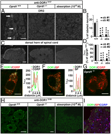
Images: Distinct distribution patterns of DORs in subsets of DRG neurons of mice. Immunostaining with antibodies against DOR13–17 [A: 1:30,000, antibody 1 (ab #1); DiaSorin and C: antibody 2 (ab #2); Neuromics] shows DORs in small DRG neurons and afferent fibers in spinal laminae I–II. This immunostaining pattern is abolished by the antiserum preabsorption or the deletion of Oprd1 exon 1. Reduction in immunostaining is quantitatively assayed by determining the percentage of positive DRG neurons (B; n = 6) and fluorescence intensity (Ifluo.) in the laminae I–II (D; n = 5). **P < 0.01; ***P < 0.001. (Scale bars: A and C, 40 μm.). DOR labeling (anti-DOR13–17, 1:30,000; DiaSorin) associated with vesicles in peptidergic small DRG neurons (E and F) is absent in Oprd1 exon 1-deleted mice (G). Colocalization of DORs and neuropeptides is shown by correlated peaks of Ifluo. measured along lines. (Scale bar: 8 μm.) (H) Immunostaining with antibodies against DOR12–18 (1:60,000; Alomone) shows the presence of DORs on the cell surface of large DRG neurons of mice. (Scale bar: 25 μm.) This staining pattern is abolished by preabsorption and is absent in Oprd1 exon 1-deleted mice. (Scale bar: 80 μm.) (I) Triple-immunostaining shows that DOR+ large DRG neurons contain neither SP nor CGRP. (Scale bar: 80 μm.)

Immunoblotting.The samples were processed for SDS/PAGE, transferred, probed with Rb antibodies against MOR (1:500; Neuromics), phospho-DOR1 (1:1,000; Neuromics), phospho-MOR (1:1,000; Neuromics), Myc (1:500; DSHB), Flag (1:1,000; Sigma), or actin (1:50,000; Chemicon) and visualized with enhanced chemiluminescence (19).

Featured and Related Reagents:

Mu Opioid Receptor-Rabbit


Mu Opioid Receptor-Guinea Pig


Delta Opioid Receptor 3-17


Delta Opioid Receptor 358-372


Delta Opioid Receptor 358-372


Kappa Opioid Receptor


phospho-Mu Opioid Receptor (Ser375)


MOR-1C
 

ORL 1-Pure


ORL1-Whole Serum


All Opioid Receptor Antibodies


Pain and Inflammation


Neurotransmission Research Antibodies
-GPCRs, Ligand Gated Ion
Channels, Biogenic Amines and more

i-Fect Transfection Kit
-gene silencing
of DOR, NaV1.8 tetrodotoxin-resistant sodium channel, NTS2 and more
in-vitro and in vivo

Primary Neurons and Astrocytes
-Primary
human, rat and mouse neurons and astrocytes