Sunday, April 26, 2015

Bioactive FGF Basic Recombinant Protein in Action

Maintaining Sheep Mesenchymal Stem Cell Multipotency

We have been very selective in the Bioactive FGF2 or FGF-basic Recombinant Proteins we make available to Stem Cell Researchers. They are many options available so we are always encouraged when we see ours referenced in publications.

Here researchers used our FGF2 to expand Sheep Bone Marrow Stem Cells (BMSCs) in culture: Troy D Bornes, Nadr M Jomha, Aillette Mulet-Sierra and Adetola B Adesida. Hypoxic culture of bone marrow-derived mesenchymal stromal stem cells differentially enhances in vitro chondrogenesis within cell-seeded collagen and hyaluronic acid porous scaffolds.Stem Cell Research & Therapy 2015, 6:84 doi:10.1186/s13287-015-0075-4.

These cells were used for chondrogenesis studies.

Expansion of BMSCs: Bone marrow aspirate collections containing 8 x 10⁷ MNCs were seeded within each 150-cm2 tissue culture flask. Culture medium composed of alpha-minimal essential medium (α-MEM) supplemented with 10% v/v heat-inactivated fetal bovine serum (FBS), penicillinstreptomycin-glutamine, 4-(2-hydroxyethyl)-1-piperazineethanesulfonic acid (HEPES), and sodium pyruvate (all from Life Technologies, Burlington, Canada) was pipetted into each flask. Fibroblast growth factor-2 (FGF-2; Neuromics Inc., Edina, USA) was added at a concentration of 5 ng/ml in order to maintain cell multipotency. Nucleated cells were allowed to adhere and grow for seven days before the first media change under normoxia (ambient 21% O2) or hypoxia (low 3% O2) at 37°C in a humidified incubator containing 5% CO2. Flasks from the hypoxic incubator experienced short periods (less than 5 minutes) of normoxic exposure during media changes. Thereafter, the media were changed twice per week until 80% cell confluence was obtained. Adherent BMSCs were detached using 0.05% w/v trypsin-ethylenediaminetetraacetic acid (EDTA; Sigma-Aldrich Corp., Oakville, Canada) and expanded under the same oxygen tension (normoxia or hypoxia) as during isolation until P2 prior to scaffold seeding. Hereafter for brevity, BMSCs described by expansion oxygen tension alone (normoxia-expanded and hypoxia-expanded BMSCs) will refer to BMSCs that were isolated and expanded under normoxia and hypoxia, respectively. The time taken from plating of nucleated cells (P0) to reach approximately 80% confluence at P2, before experimental use, varied from three to four weeks.

I anticipate more publications on these important bioactive reports as demand for them has been growing.

Tuesday, April 14, 2015

Markers for Oligodendrocyte Progenitor Cells (OPCs)

Improving OPC Expansion

Researchers reference use of Neuromics' GFAP and PDGF R Alpha/CD140A for OPC Selection

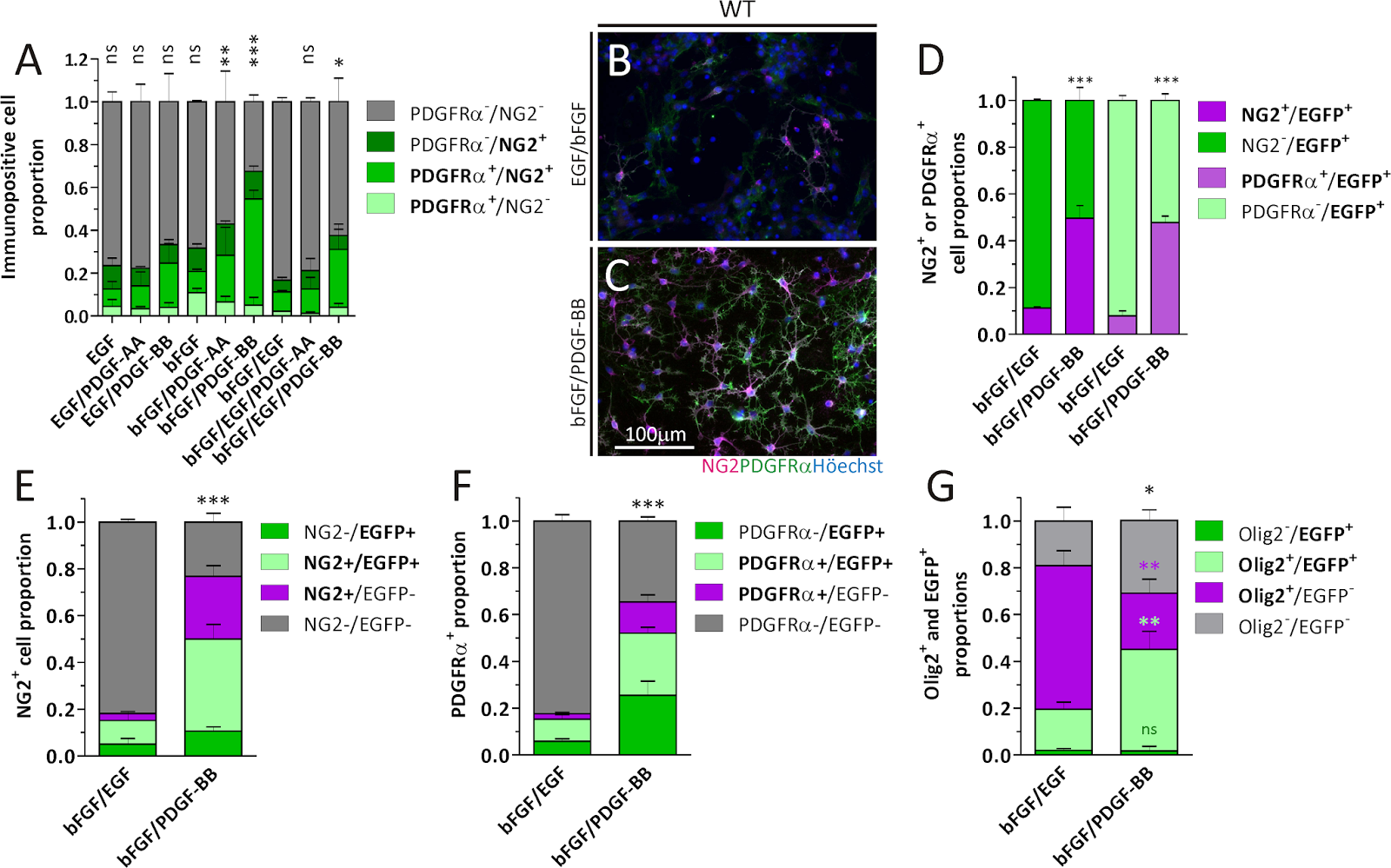
The data provided in this publication demonstrates that the OPC yield from SVZ-derived cell cultures can be improved with the PDGF-BB isoform in comparison to classical bFGF-EGF, or PDGF-AA-based protocols. Additionally, it would be expected that the OPC-enriched cultures obtained from NSC/NPC exposure to PDGF-BB and heparin generate cells suitable for cell transplantation for treating demyelinating diseases: Paula G. Franco, Juana M. Pasquini, Lucas Silvestroff. Optimizing Culture Medium Composition to Improve Oligodendrocyte Progenitor Cell Yields In Vitro from Subventricular Zone-Derived Neural Progenitor Cell Neurospheres. Published: April 2, 2015DOI: 10.1371/journal.pone.0121774:

Images: Detection of OPC markers by ICC on NS cells. A) Quantitation of NG2+ and/or PDGFRα+ cell proportions in WT mice NS generated in the presence of different growth factor combinations. Data belong to three independent experiments for each condition. Data for NG2-/PDGFRα- was analyzed with a One-way ANOVA plus Dunnett´s post test, where bFGF/EGF was set as the control. B, C) Representative images of NG2+ and PDGFRα+ cells generated from WT mice cultures in the presence of either bFGF/EGF or bFGF/PDGF-BB. D) Comparison of NG2+ or PDGFRα+ cell proportions in NS cultures generated from Act::EGFP mice in the presence of bFGF/EGF or bFGF/PDGF-BB. Data for NG2+ (dark magenta) or PDGFRα+ (light magenta) cells was analyzed separately with Student´s t test. E, F) NG2 and PDGFRα immunodetection in CNP::EGFP derived cultures. Gray bars in each graph were analyzed with Student´s t test. G) Olig2 expression in WT mice NS cultures. Asterisks are colour coded to indicate the pairs of bars compared and analyzed with Student´s t test. Cell proportions in A, and D-G are expressed as a fraction of the total cell nuclei counted for each condition. Error bars represent the SD for all bar graphs.. doi:10.1371/journal.pone.0121774.g001.

Neuromics has an excellent catalog of Stem Cell Selection Markers.

Monday, April 06, 2015

The Importance of in vivo Like Astroglial-Neuron Co-Cultures

What We Have Learned

I have many posting on both Neuron and Astroglial Cultures. We have now evolved our solution set to include in vivo like co-cultures. These Astroglial Neuron Co-cultures are designed to mimic in vivo like behaviors. They are potent, pure and proven to work in our clients' unique neurodegenerative disease and toxicity drug discovery assays.
Why is this important? The mix of Astrocytes, Glia and Neurons in your co-cultures can impact your data endpoints and lead to inaccurate conclusions. Here're examples as to how much your data can fluctuate. These are data generated from toxicity assays.
We stand ready to serve you and your team. Questions? Don not hesitate to call 612-801-1007 or e-mail me pshuster@neuromics.com. Pete Shuster, CEO and Owner, Neuromics.