Thursday, December 21, 2006
Customer Data-GFAP IHC
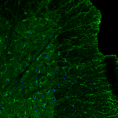
Image: GFAP (Catalog#: CH22102) Staining of of Astrocytes in the Ventral horn and Funiculus of Adult Rat Spinal Cord.
Dilution 1:500
Wednesday, December 13, 2006
NP Y2 Image and Publication
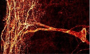
NPY Y2 Receptor-Catalog#: RA14112 immunoreactivity in the rat hippocampus.
Related Publication:
B Tu, Y Jiao, H Herzog, JV Nadler. Neuropeptide Y regulates recurrent mossy fiber synaptic transmission less effectively in mice than in rats: correlation with Y2 receptor plasticity. Neuroscience, 143, 1085-1094 (2006)
Wednesday, November 29, 2006
P2Y2-It is baaack!
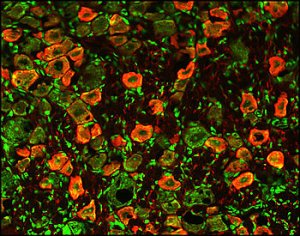
Image: P2Y2 Staining of Rat DRG.
Related Antibodies to Consider:
P2X1
P2X2-Guinea Pig
P2X2-Rabbit
P2X3-Rabbit
P2X3-Guinea Pig
Neuropeptides
Opioids
Opioid Neuropeptides
TRPV1s
Vision and Retina
Cancer
Friday, October 27, 2006
Excellent TUJ-1 IHC
Tuj-1 Catalog#: MO15013 staining of
staining of mouse cochlea (whole mount, ~50um).
Tuj-1 Rabbit Polyclonal
All Neuron-Glial Markers
All Stem Cell Reagents
Monday, October 16, 2006
P2X3 Western Blot
Here's an excellent publication showing both WB and IHC for this antibody.
P2X2 and P2X3 receptor expression in postnatal and adult rat urinary bladder and lumbosacral spinal cord Simon Studeny, Ali Torabi and Margaret A. Vizzard.
Figure: Western blot of whole urinary bladder (40 mg) for P2X3 receptor expression in postnatal (P0-P36) and adult (A) rats. Erk staining was used as a loading control. B. Preabsorption of P2X3 antibody with immunogen (1 mg ml−1) eliminated the band at 75kD. C. Histogram of relative P2X3 band density in all groups examined normalized to erk-1 staining. P2X3 receptor expression in urinary bladder is significantly increased from P14-P21 compared to adult rat bladder. *, p ≤ 0.005, #, p ≤ 0.01.
Friday, September 29, 2006
ESC to Neural Progenitors-Role of MOR-1C
Eunhae Kim, Amy L. Clark , Alexi Kiss, Jason W. Hahn, Robin Wesselschmidt. MU AND KAPPA OPIOIDS INDUCE THE DIFFERENTIATION OF EMBRYONIC STEM CELLS TO NEURAL PROGENITORS
JBC Papers in Press. Published on September 1, 2006 as Manuscript M603862200
Image: A-D-Staining of Differentiated Stem Cells. D3 ES cells were maintained in serum/LIF/ME induced self-renewal conditions (undifferentiated cells, D3). Upon treatment with 1 µM RA they undergo differentiation (dD3). Cells were fixed and permeabilized. Treatments were followed by overnight incubation with polyclonal MOR-1 (C-terminus, 1:2500)
Tuesday, September 19, 2006
New TRPV1-C Publication
Figure: WIN activates calcineurin in TG neurons and dephosphorylates TRPV1. (A) Calcineurin subunits are coexpressed with TRPV1. The colocalization of calcineurin A subunit (Upper) and B subunit (Lower) with TRPV1isdemonstratedin the respective panels.(B) The effect of various treatments on the nuclear translocation of NFATc4 (calcineurin activation). Cultured TG neuronswere exposed to vehicle, ionomycin (1 M), capsaicin (1 M), WIN (25 M), or WINCAIP(25 M50 M), and immunohistochemistry was performed by using an antibody againstNFATc4. (C) Graphical representation of the percent of NFATc4 positive neurons showing nuclear translocation of NFAFc4 after treatment with VEHVEH, VEHWIN (25 M), CAIP (50 M)WIN, VEHCAP (1 M), or CAIP(50 M)CAP (n 4 independent cultures assessed by blinded observer; n 152–180 cells per condition; **, P<0.01,anovawith n="3">
Saturday, September 09, 2006
4 New Interleukins
IL-1Alpha/IL-F1
Image: IL-1α staining of blood lymphocytes. Cells were stained using anti-gat-Cy3 (red) and counterstained with Fluoro Nissl Green
IL-1Beta
Image: IL-1 beta staining (red color) in mouse splenocytes stimulated with Con A. Cell nuclei counterstained green.
IL-1 R1
Image: IL-1R staining of mouse thymus. Staining was done using HRP-DAB (brown) detection. Tissue was counterstained with haematoxylin (blue).
IL-6
Image: IL-6 staining of mouse T cells. Cells were stained with goat anti-rat IL-6 (red) and nuclei counterstained with Fluoro Nissl Green.
Thursday, August 31, 2006
Monoclonal Ubiquitin Antibodies-Great IHC
Ubiquitin-Catolog#:19005
Image: ABC-HRP staining with Ubiquitin monoclonal antibody (diluted 1:1,000) of Alzheimer’s disease cerebral cortex showing dystrophic neuritis, which contain ubiquitinated tau. The inclusions are detected by the Ubiquitin antibody. Normal cortex does not contain inclusions and would be white in color.
Ubiquitin-Catalog#: MO18001
Confocal images of hippocampal CA1 sections from rats, (A) mock-treated or (B) subjected to 15 minutes ischemia followed by 24 hours of reperfusion, using ubiquitin mouse antibody (green) and propidium iodide (red). Data courtesy of Cell Signaling Technology, Inc.
Wednesday, August 02, 2006
New ChAT IHC Data
ChAT (Choline Acetyltransferase-Catalog#: CH23000)
Image: IHC Staining in Rat Brainstem
Other Reagents to Consider:
Neuron-Glial Markers-Chickens:
CNPase (CH23013)
GAP 43 (CH23004)
GFAP (CH23001)
GFAP (CH22102)
MAP2 (CH22103)
NSE (CH23003)
Nestin (CH23001)
Netrin-1 (CH23002)
NF-H (CH22104)
NF-L (CH22105)
NF-M (CH22106)
NF-M (CH23104)
PLP (CH23008)
Po (CH23009)
S-100 (CH23012)
Tuj 1 (CH23005)
All Neuron-Glial Markers
Stem Cell Reagents
Saturday, July 29, 2006
New P2X3 Data
Data Courtesy of Dr. Yan Sun University of Maryland School of Medicine Baltimore
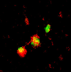
Image: Double immunofluorescence staining in cultured human bladder urothelial cells (BUCs). BUCs show cytokeratin (red areas) and P2X3 (green areas) expression.
Figure: Flow cytometry and FACS analysis of P2X3 expression in human BUCs. P2X3 expression in BUCs were compared among in vitro stretched samples and unstretched samples in both normal and interstitial cystitis (IC) patients. A, unstretched normal BUCs. B, stretched normal BUCs. C, unstretched IC BUCs. D, stretched IC BUCs. M1 region represents P2X3 localization detected.
Monday, July 24, 2006
New AD Research Antibodies
BACE-1
Cleavage of amyloid precursor protein (APP) into 40 or 42 amino acid amyloid β (aβ) peptides is a result of the action of two proteases β- and γ-secretase. The beta-site APP cleaving enzyme, or BACE-1, is a transmembrane aspartic protease that is localized in the golgi and endosomes. Low levels of BACE-1 mRNA are found in most adult tissues with a higher level of expression in pancreas.
Image: Detection of BACE-1 in human cerebellum. Stained with HRP-DAP (brown) and counter-stained with hemtoxylin (blue).
Abeta 40 (beta amyloid 1-40) and Abeta 42 (beta amyloid 1-42)
Amyloid beta-protein (Abeta) is associated with neuronal injury and death in Alzheimer's disease. These proteins are the main components of amyloid plaques. Abeta 42 plays a bigger role in deneurogeneration and is more strongly associated with Alzheimer's disease than Abeta 40.
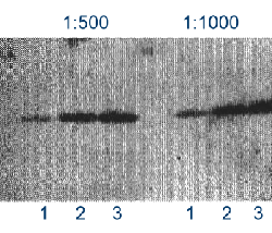
Image: Detection of Abeta 40 on 5 ng of peptide per lane using NB 300-225. Lane 1: Abeta-40, lane 2: Abeta-42, lane 3: Abeta-40 and -42 mix
Saturday, July 22, 2006
TRPV1-N and C Data
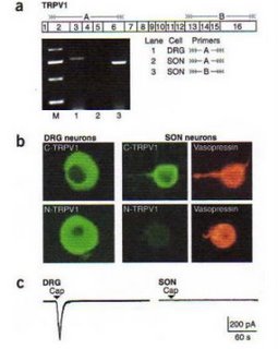
Sharif Naeini R, Witty MF, Seguela P, Bourque CW (2006) An N-terminal variant of Trpv1 channel is required for osmosensory transduction. Nature Neuroscience. (1):93-8.
Figure: SON Neurons express an N-terminal variant(s) of TRPV1 (a) Simplified structure of mRNA (numbered boxes show exons of TRPV1 gene) encoding TRPV1 (above). Gray arrow tails and lines illustrate mRNA regions detected by primer sets A and B. RT-PCR analysis of mRNAs expressed in DRG and SON (below). Note the lack of signals for the N terminus of TRPV1 (lane 2, exons 2-6, primer set A) in SON. (b) Immunocytochemical staining confirmed the presence of full length TRPV1 in DRG neurons. In contrast, only the C-terminus of the TRPV1 protein (C-TRPV1) was detected in the AVP positive SON neurons. (c) Whole-cell recordings reveal that DRG neurons are sensitive to capsaicin ('Cap,' 10 uM, 5s) but that SON neurons are insensitive.
Sunday, June 25, 2006
New Publication-Neuromic's E-18 Primary Hippocampal Neurons
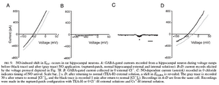
Hippocampal cultures were made from 18-day-old rat (Fischer 3440) embryos. Micro-dissected hippocampal tissue was obtained from Neuromics (Bloomington, MN) and prepared and maintained according to the supplier’s protocols. Recordings from single hippocampal neurons were made after 4–7 days in culture. No physiological assessments of the relative maturity of these cells have been made. According to their developmental timeline alone, they would be considered embryonic.
Thursday, June 15, 2006
New Goat VR1 (TRPV1) and Rabbit NK1 Antibody
Catalog #: GT15129
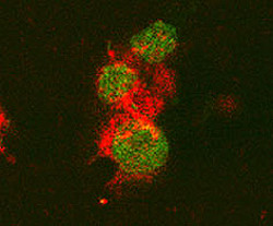
Image: VR1 staining in Rat DRG: red color - TRPV1 labeling, green-counterstain.
Description: VR1, also known as TRPV1 (Transient Receptor Potential Vanilloid 1) and capsaicin receptor, is a 115 kDa integral membrane ion channel that has six transmembrane domains and contains intracellular N-and C-termini. It contributes to normal pain and temperature sensation and also has a "sensory-effector" function. By alternative splicing, at least three VR1 isoforms are known. The sequence of residues 4 - 21 of rat VR1 is 78% and 28% identical to that of mouse and human VR1, respectively.
Catalog #: RA25001
Image: NK 1 staining of lamina III neuron in the spinal cord of a rat.
Description: The tachykinins belong to an evolutionary conserved family of peptide neurotransmitters that share the c-terminal sequence Phe-X-Gly-Leu-Met-NH2 and have an established role in neurotransmission. The mammalian tachykinins include substance P, neurokinin A (NKA) and neurokinin B (NKB) which exert their effects by binding to specific receptors. Tachykinin peptides are important in the mediation of many physiological and pathological processes including inflammation, pain, migraine headache and allergy induced asthma. Three tachykinin receptor types have been characterized, NK-1, NK-2 and NK-3 which have preferential affinities for SP, NKA and NKB respectively. All three receptors share a high degree of sequence homology, have seven transmembrane spanning domains and similar signal transduction mechanisms (e.g. G-protein coupled activation of phospholipase C).
Wednesday, June 07, 2006
Tuesday, June 06, 2006
New SHANK1a C-terminus IHC Image
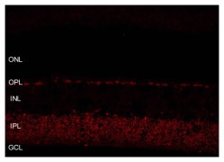
SHANK1a C-terminus Staining in Mouse Retina. Image Courtesy of Sal Stella, UCLA
Antibody Protocol: on vertical sections of rat and mouse retina
Wednesday, May 24, 2006
New BDNF and NT-3 IHC Images
Monday, May 08, 2006
You've Been Patient-Now We Have It!
Friday, April 14, 2006
The Neuromics-AlphaGenix alliance
Neuromics and AlphaGenix work closely with Dr. Mark Behlke, Vice President of Molecular Genetics at Integrated DNA Technologies to develop optimum strategies for the design of RNAi molecules.
This is followed by developing in vitro studies on primary cells, which are required in order to design in vivo strategies to inhibit targeted gene expression. While transfection studies enhance our understanding of RNAi technology, we believe that it is vital to demonstrate validation by including sufficent controls as well as demonstrating knockdown of endogenous message.
We will be routinely publishing new publications to this blog. In this way, researchers are able to get leading papers and reviews in these fields of research as well as having the ability to bring their own expertise to the table. Indeed, science is a collective effort and we encourage you as colleagues to help all of us keep on the cutting edge.
Best regards,
Stephen Hall, Ph.D.
Thursday, April 13, 2006
RNAi Transfection Alliance
Neuromics and Alphagenix would like to announce our RNAi Transfection Alliance. We will be partnering with IDT to develop and commercialize better ways to deliver RNAi to Glia, Neurons and the Nervous System.
Here are some related publications:
Progress Towards in vivo Use of siRNAs
siRNAi: Applications in Functional Genomics and Potential as Therapeutics
Assembly and Function of RNAi Silencing Complexes
Synthetic dsRNA Substrates Enhance RNAi Potency and Efficacy
In vivo Publications:
Luo MC, Zhang DQ, Ma SW, Huang YY, Shuster SJ, Porreca F, Lai J (2005). An efficient intrathecal delivery of small interfering RNA to the spinal cord and peripheral neurons. Molecular Pain 2005, 1:29.
Priti Kumar, Sang Kyung Lee, Premlata Shankar, N. Manjunath (2006) A Single siRNA Suppresses Fatal Encephalitis Induced by Two Different Flaviviruses. PLOS Medicine, Volume 3 Issue 4 APRIL 2006
Monday, April 03, 2006
New Article with RNAi and i-Fect in vivo Results
Priti Kumar1, Sang Kyung Lee2, Premlata Shankar1*, N. Manjunath1*
1 The CBR Institute for Biomedical Research and Department of Pediatrics, Harvard Medical School, Boston, Massachusetts, United States of America, 2 Department of Bioengineering, Hanyang University, Seoul, Korea
ABSTRACT
Background
Japanese encephalitis virus (JEV) and West Nile virus (WNV) are neurotropic flaviviruses that can cause acute encephalitis with a high fatality rate. Currently there is no effective treatment for these infections.
Methods and Findings
We tested RNA interference (RNAi)-based intervention to suppress lethal JE and WN encephalitis in mice. To induce RNAi, we used either lentivirally expressed short hairpin RNA (shRNA) or synthetic short interfering RNA (siRNA). As target, we selected the cd loop-coding sequence in domain II of the viral Envelope protein, which is highly conserved among all flaviviruses because of its essential role in membrane fusion. Using as a target a species-specific sequence in the cd loop that is conserved only among the different strains of either JEV or WNV, we could achieve specific protection against the corresponding virus. However, by targeting a cross-species conserved sequence within the cd loop, we were able to protect mice against encephalitis induced by both viruses. A single intracranial administration of lentivirally delivered shRNA or lipid-complexed siRNA before viral challenge or siRNA treatment after viral challenge was sufficient for protection against lethal encephalitis.
Conclusions
RNAi-based intervention affords near complete protection from both JEV- and WNV- induced encephalitis in mice. Our results show, to our knowledge for the first time, that siRNA can be used as a broad-spectrum antiviral agent for treating encephalitis caused by multiple related viruses.
Funding: This work was supported by NIH grant U19 AI 056900 to NM and PS.
Competing Interests: The authors have declared that no competing interests exist.
Academic Editor: Clifford Lane, National Institutes of Health, United States of America
Received: September 20, 2005; Accepted: December 16, 2005; Published: February 14, 2006
DOI: 10.1371/journal.pmed.0030096
Copyright: © 2006 Kumar et al. This is an open-access article distributed under the terms of the Creative Commons Attribution License, which permits unrestricted use, distribution, and reproduction in any medium, provided the original author and source are credited.
Relevant to i-Fect (TM)
"Although there is a report of the successful use of naked siRNA targeting the pain-related cation channel P2X3 to treat chronic neuropathic pain in a rat model [28], other studies suggest that naked siRNA is poorly taken up by brain parenchymal cells [29,30]. Recently, a cationic lipid formulation, i-Fect (Neuromics) has been found to deliver siRNA into neuronal cells without toxicity (Dr. Josephine Lai, University of Arizona Health Sciences Center, personal communication). We tested if synthetic FvEJ siRNA (siFvEJ) complexed with i-Fect can protect mice against viral encephalitis. After confirming that i-Fect can transduse siFvEJ siRNA as efficiently as lipofectamine to inhibit JEV infection in Neuro 2a cells (Figure 3A), we infected mice by IC injection with JEV and, after allowing 30 min for viral adsorption, injected the synthetic siFvEJ or control luciferase siRNA (siLuc) complexed with i-Fect at the same site. All mice injected with siLuc died by day 5, whereas all of the siFvEJ-injected mice survived indefinitely (Figure 3B), suggesting that i-Fect can deliver siRNA into neuronal cells and result in protection that is similar to the lentivirally delivered shRNA."
Saturday, April 01, 2006
More Customer Data-Ret and SHANK 1a-C-terminus
Thank you Kim Carnes, UIUC, for the image and protocol for the Ret staining of spermatagonial stem cells.
Protocol
Depraffinize and hydration
Peroxidase blocking - 30 minutes in 0.06% H2O2 in methanol.
Antigen retrieval - 0.01M Citrate buffer (pH6.0) in microwave (10 minutes, power level 7)
Serum blocking - horse serum (unfiltered normal, diluted1:100) for 30 minutes.
Primary antibody - Neuromics Ret antibody (1:200), overnight at 4º C in a moisture chamber.
Secondary Ab - this was a horse universal biotinylated ab that came in a kit (with the above horse serum) diluted 1:50 with PBS and horse serum (also 1:50) for 60 minutes at room temp.
ABC incubation (Vector ABC Elite kit) - 30 minutes.
Colorization with DAB, counterstain with hematoxylin followed by dehydration.
We use PBS rinses between steps.
Thank you Alex Vila, UCLA, for your image and protocol for the SHANK 1a C-terminus staining of retinol tissue
Protocol
Species: Mouse C57B6
Fixation: 15 minute 4% paraformaldehyde fixation
Antibody dilution: 1:800 overnight.
Secondary Alexa 568 1:500 for 45 minutes
Tissue preparation: Blocked for 2 hrs at room temp in 10% normal goat serum and
Triton X-100 (0.5%) in 0.1 M Phosphate buffered saline (PBS)
Washed: 3X 0.1 M PBS between primary and secondary step.
Mounted tissue with Paramount.
Confocal: Images are 2048 X 2048, 12 bit, collected with a 40X water objective, 10 stacks 0.55 um per stack
Wednesday, March 01, 2006
New Park7 Data-Thank you Amanda Ashley
Click to see full sized image.
WB: Wild type and PARK7 (DJ-1) KO animals
Data courtesy of Amanda Ashley and Dr. Marie Legare, Colorado State University.
Western conditions:
– 25mg protein from mouse brain lysate
– 1:2000 Park7 (DJ-1) antibody (Cat # RA19006)
– 12% gel
– Electrotransferred onto PVDF
– Blocked with 5% BSA in TBS-Tween
– Detected with ImmunStar HRP (BioRad)
– Dually probed for beta-actin (1:1000, Sigma)
Tuesday, February 07, 2006
News Release from Neuromics RNAi Collaborators
... of Dr. Frank Porreca and Dr. Josephine Lai at the University of Arizona
evaluated siRNAs that target the gene for NPY, a pain-associated peptide. ...
Friday, February 03, 2006
Annual SFN Chicago Meeting-March 3rd 2006
We are pleased that Dr Tomas Hokfelt, a friend and key Neuromics' collaborator, will be presenting:
Three Decades of with Three Swedish Peptides: Substance P, NPY and Galanin
“Working with Neuromics has truly been a collaborative effort, from the initial antibody generation strategy to its development and characterization. This interaction between our groups was an important factor in our successful generation of a novel NPY Y2 antibody.” Dr. Tomas Hokfelt, Karolinska Institute
More specifics to follow...
Wednesday, February 01, 2006
PDF to Walter Low's Stem Cell Data
Walt Low's PDF Report on Neuromics' Rat NSCs
Neural Stem Cell Data Was Generated Using Neuromics' Neural Stem Cell Kit (Cat#: NP37100)
New Antibody from The David Holtzman Lab
ABCA1
By Western blot mouse anti-ABCA1 (MO13101; clone HJ1) recognizes rat (left image), mouse (right image) and to a lesser extent human (left image) ABCA1.
Dr. David Holtman Lab-Washington University School of Medicine
Tuesday, January 31, 2006
Finding a Better Way
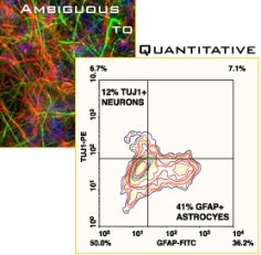
FACS/Phenotyping-A constant challenge working with neural stem and progenitors cells is to determine the percentage of cells of a given phenotype within the culture. Phenotypic characterization of the entire cell population is of the utmost importance in determining the efficacy of your cell culture conditions to generate the neural or glial cell of interest. Immunocytochemistry can get you a long way to understanding the types of cells you obtained in your culture, but it is both time consuming and qualitative in nature.
A far better approach is to analyze your cells by FACS. Although routinely employed within many branches of biology and medical technology, FACS is not a common technique used in Neuroscience. It is our hope that by offering the IntraCtyeTM kits, intracellular FACS analysis will be more accessible to and used more often by neuroscientists. We are currently offering several basic kits as well as a kit designed for rat neural progenitor cell phenotyping (IntraCyte-rNSC). The kit has been tested with our rat Neural Stem Cell Kit and is a powerful analytic tool to analyze the neural and glial cell within your culture.
The IntraCyte kit technology and the precision and accuracy of flow cytometry, enables you to get statistically significant, multi-parameter data at the single-cell level, on thousands of cells per sample in seconds. Even though we enjoy nothing more than looking at beautifully labeled neuronal cultures, we can do without the tedious and inaccurate counting of these cells in the dark!
Wednesday, January 18, 2006
RNAi and Huntington's
Excellent Neural Stem Cell Data and images
Figure1 and Figure2
Customer Data generated using Neuromics' Neural Stem Cell Kit Catalog #: NP37100. Data Courtesy of Walter Low and Jing Xiao, University of Minnesota
Neural progenitor cells were plated and maintained in Proliferation Media (Figure 1). For differentiation, cells were added to 4 well chamber slides, grown in proliferation media for 24 hours and then switched to Commitment Media with various growth factors added. Cells were cultured for 14 days and then processed by immunocytochemistry. Figure 2 shows representative data from 1 experiment using bFGF/EFG (figure 2B) for differentiation.
Wednesday, January 04, 2006
Post Comments
Sam Shuster
CEO and CSO
Stem Cell to Neurons
Small synthetic molecule initiates differentiation of adult neuronal rat brain cells to neurons
12/28/2005 - Adult neuronal stem cells have the potential to mature into functional nerve cells (neurons) and neuron support cells (astroglia). The mechanisms for this process are thus far not well understood. Res...
http://www.bionity.com/news/e/51090/01065/137777
Sunday, January 01, 2006
http://www.nature.com/nbt/journal/v23/n2/abs/nbt1051.html
Dong-Ho Kim, Mark A Behlke, Scott D Rose, Mi-Sook Chang, Sangdun Choi & John J Rossi. Synthetic dsRNA Dicer substrates enhance RNAi potency and efficacy. Nature Biotechnology 23, 222 - 226 (2004).