Monday, January 28, 2019

Neuromics' Islet-1 Used to Study Avian Heart Morphology

Evolution of High-Performance Hearts

Our Islet-1 Antibody showed its versatility a comparative morphology of avian hearts.

Figure: Sinuatrial node in Mallard (a–c), chicken HH42 (d–g), Lesser redpoll (h–j), and Jackdaw (k–m). All sections are in the transverse plane. The boxed areas in the left-hand column images indicate the areas of the images of the middle and right-hand columns. (a–c) In the Mallard, a nodal structure at the base of the right leaflet of the sinuatrial valve (black arrowhead in [a]) expressed Isl1 (b, c) and had a large coronary artery (white arrowhead in [b]). Sections 271 (cranial) to 1201 (caudal) encompassed the atria and the sections shown are 691 (a), 692 (b), 762 (c). (d–g) In the chicken HH42, the sinus venosus (SV) expressed the myocardial marker cTnI and this expression was relatively weak at the base of the right leaflet of the sinuatrial valve (black arrowhead in [e]). (f–g) The base of the right leaflet of the sinuatrial valve expressed Bmp2 (red arrowheads in [f]) and Isl1 [g]). Sections 17 (cranial) to 297 (caudal) encompassed the atria and the sections shown are 80 (d-e), 78 (f ), 88 (g). The sections were from the mid-height of the atria. (h–j) In the lesser redpoll, Isl1 was expressed in the base of the right leaflet of the sinuatrial valve. There was no nodal structure but the Isl1 expressing wall was thicker than the surrounding walls and contained a large coronary artery (white arrowhead in [i]). Sections 321 (cranial) to 621 (caudal) encompassed the atria and the sections shown are 401 (h), 402 (i), 422 (j). (k–m) In the Jackdaw an Isl1 positive area was seen in the left sinus venosus myocardium (l) in which a coronary artery was visible (white arrowhead in (l). At the base of the right sinuatrial leaflet no positive Isl1 cells could be seen (m). Sections 122 (cranial) to 332 (caudal) encompassed the atria and the sections shown are 190 (k) and 191 (l–m). Ao = aorta; LA = left atrium; PA = pulmonary artery; RA = right atrium; SAJ = sinuatrial junction. In the picro-sirius red images blood has been painted over with white for clarity. DOI: 10.1002/jmor.20952

We have an extensive catalog of high-performance antibodies.

Friday, January 18, 2019

Markers for the Study of Diabetic Neuropathy

Widely Used and Frequently Published
Our Pain and Inflammation Research Antibodies have proven valuable for the study of neuropathic, nociceptive and inflammatory pain.

This recent publication is focused on Diabetic Neuropathy. José Eduardo Roa-Coria, Jorge Baruch Pineda-Farias, Paulino Barragán-Iglesias, Geovanna Nallely Quiñonez-Bastidas, Ángel Zúñiga-Romero, Juan Carlos Huerta-Cruz, Juan Gerardo Reyes-García, Francisco Javier Flores-Murrieta, Vinicio Granados-SotoView ORCID ID profile and Héctor Isaac Rocha-González (2019). Possible involvement of peripheral TRP channels in the hydrogen sulfide-induced hyperalgesia in diabetic rats. BMC Neuroscience 201920:1 https://doi.org/10.1186/s12868-018-0483-3.

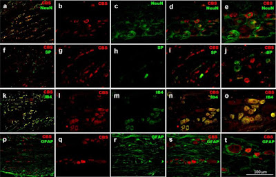
The results show Hydrogen Sulfide (H2S) catalyzes hyperalgesia in rats via TRP Channels. Our Substance P is used in the study.


Figure: Immunolocalization of cystathionin-β-synthase enzyme (CBS, red) with a–e neuronal nuclear antigen (NeuN)-, f–j substance P (SP)-, k–o isolectin B4 (IB4)- and p–t glial fibrillary acidic protein (GFAP)-positive dorsal root ganglion neurons of diabetic rats (green). a, f, k and p show a representative 16 µm-slice from dorsal root ganglia. b, g, l and q show a representative CBS staining with Cy3 from dorsal root neurons, whereas c, h, m and r show NeuN, SP, IB4 and GFAP representative staining with Cy2 in dorsal root neurons, respectively. d, i, n and s show the merged image from Cy3 and Cy2 signals, the overlapping between CBS and neuronal markers is shown in a yellow-orange color. e, j, o and t show a magnification of d, i, n and s, respectively.


The authors conclude that H2S leads to hyperalgesia in diabetic rats through activation of TRPV1, TRPA1 and TRPC channels and, subsequent intraepidermal fibers loss. CBS enzyme inhibitors or TRP-channel blockers could be useful for the treatment of painful diabetic neuropathy.

Wednesday, January 09, 2019

Rat Hippocampal Neurons

Used to Study Apoptosis Induced by Brain Insults
Although our primary human cells are increasingly being used in drug discovery and toxicology assays versus animal cells, we are always pleased to see the success of the latter in sophisticated studies.

This publication references use of our e18 rat hippocampal neurons. These cells continue to be easy to culture, pure and potent. Chiara Porro, Antonia Cianciulli, Teresa Trotta, Dario Domenico Lofrumento, Rosa Calvello and Maria Antonietta Panaro. (2019). Formyl-methionyl-leucyl-phenylalanine Induces Apoptosis in Murine Neurons: Evidence for NO-Dependent Caspase-9 Activation. Biology 8(1), 4; doi:10.3390/biology8010004.

Highlights
Formyl-methionyl-leucyl-phenylalanine (fMLP) may be present in the brain in the course of some infectious diseases of the central nervous system (CNS), although little is known about its role. This investigation was performed to study the effect of fMLP on neuron apoptosis. The results showed that fMLP treatment of primary cultures of neurons was able to induce morphological features of apoptosis in cell cultures, as well as activation of the intrinsic apoptotic pathway, through the upregulation of caspase-9 and caspase-3.

Figure. (A) Western blot analysis was performed on membrane-enriched cell extracts (25 µg lysate) of primary neurons. The blots were probed with formyl peptide receptor 2 (FPR2) antibody (Ab) and detected by chemiluminescence. A ~41 kDa band corresponding to FPR2 was observed as compared to the positive control. Lane 1: Marker; lane 2: Positive control, lane 3: Primary neuron lysate. (B) Immunofluorescence identification of FPR. Double staining shows the expression of the FPR receptor on the cell membrane and neurofilaments. FPR2 expression (green); skeleton protein staining of neuron-specific neurofilament 68 (red); DAPI nuclear staining (blue); cells stained by both neurofilament 68, FPR2 and DAPI (merged). Scale bar: 100 μm. 1): neurofilaments stain; 2) FPR2 expression; 3) DAPI stain; 4) merged.

The present study emphasizing the potential role of infectious agents, such as N-formyl peptides, in neurodegenerative diseases may help to promote the development of new therapies able to modulate the expression of the N-formyl peptide receptors.

Tuesday, January 01, 2019

Human Primary Cells Trifecta

Tri-culture of Neuromics' Human Primary Cells in 3-D BBB Model

I mentioned in an earlier post that we take Researchers questioning the validity of our Human Primary Cells very seriously. We follow up with our clients to make sure our cells are working as expected. If they are not 100% happy, we offer a free replacement or full refund.

...So it is encouraging to see Researchers using our Human PericytesBrain Microvascular Endothelial Cells (HBMES) and Astrocytes to build a static 3-D Blood-Brain Barrier Model.

Ece Bayir, M. Mert Celtikoglu, Aylin Sendemira. The use of bacterial cellulose as a basement membrane improves the plausibility of the static in vitro blood-brain barrier model. https://doi.org/10.1016/j.ijbiomac.2018.12.257.
Image: Pericytes, HBMECS, and Astrocytes 5 days after culturing. Live cells (green) and dead cells (red).

The investigators concluded, "Caffeine and sucrose permeability values obtained from all models were close to literature data and physiological values."