Here researchers study: Yongheng Cao1, Masanori Nakata, Shiki Okamoto, Eisuke Takano, Toshihiko Yada, Yasuhiko Minokoshi, Yukio Hirata, Kazunori Nakajima, Kristy Iskandar, Yoshitake Hayashi, Wataru Ogawa, Gregory S. Barsh, Hiroshi Hosoda, Kenji Kangawa, Hiroshi Itoh, Tetsuo Noda, Masato Kasuga, Jun Nakae.PDK1-Foxo1 in Agouti-Related Peptide Neurons Regulates Energy Homeostasis by Modulating Food Intake and Energy Expenditure. PLoS ONE 6(4): e18324. doi:10.1371/journal.pone.0018324.
 |
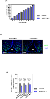
Images: Functional defects of AGRP neurons in AGRPPdk1−/− mice. (A) Numbers of AGRP positive cells in the arcuate nuclei of AGRPPdk1+/+ (gray bar) and AGRPPdk1−/− (blue bar) mice. AGRP cell counts indifferent regions of the arcuate nucleus showed different numbers of AGRP neurons in AGRPPdk1+/+ (n = 3) and AGRPPdk1−/− mice (n = 3). (B) Representative Immunofluorescence images of AGRP in the hypothalamic regions of AGRPPdk1+/+ (left panel) and AGRPPdk1−/− mice (right panel). Green, AGRP; blue, DAPI. Scale bars indicate 100 µm. (C) Expression in the fed-state of hypothalamic neuropeptide genes in control (gray bar) and AGRPPdk1−/−(blue bar) mice. Data were normalized to β-actin expression and represent the mean ± SEM of six mice per genotype.
doi:10.1371/journal.pone.0018324.g002
doi:10.1371/journal.pone.0018324.g002
Key Findings:PDK1 and Foxo1 signaling pathways play important roles in the control of energy homeostasis through AGRP-independent mechanisms. data indicated that PDK1 was indispensable for the orexigenic activity of AGRP neurons. Hypothalamic AGRP neurons express AGRP, NPY, the neurotransmitter GABA, and potentially other undiscovered molecules. In AGRPPdk1−/− mice, the expression of Agrp and Npy tended to be lower than that observed in control mice although the difference was not significant. Interestingly, the Δ256Foxo1AGRPPdk1−/− mice exhibited significantly increased food intake compared to AGRPPdk1−/− mice in spite of significantly decreased expression of Agrp and Npy. Therefore, changes in the expression levels of Agrp and Npy may not explain the changes in food intake in AGRPPdk1−/− mice.


1 comment:
Hello,
This is the perfect blog for anyone who wants to know about this topic. Energy homeostasis requires the coordination of several metabolic fluxes, these fluxes comprise the use of nutrients as metabolic fuels...
Drug Discovery
Post a Comment