Ubiquitin-Catolog#:19005
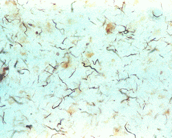
Image: ABC-HRP staining with Ubiquitin monoclonal antibody (diluted 1:1,000) of Alzheimer’s disease cerebral cortex showing dystrophic neuritis, which contain ubiquitinated tau. The inclusions are detected by the Ubiquitin antibody. Normal cortex does not contain inclusions and would be white in color.
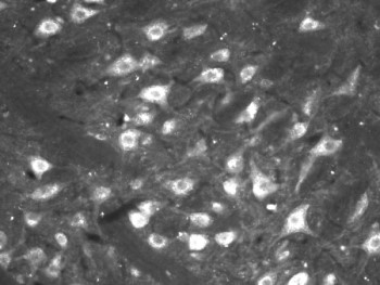
Ubiquitin-Catalog#: MO18001

Confocal images of hippocampal CA1 sections from rats, (A) mock-treated or (B) subjected to 15 minutes ischemia followed by 24 hours of reperfusion, using ubiquitin mouse antibody (green) and propidium iodide (red). Data courtesy of Cell Signaling Technology, Inc.