Tuesday, January 31, 2017

Desperately Seeking Data

Answering the Bell
We continue to seek data using our cells. We offer a reward of 25 USD Starbucks' Gift Card.

We were pleased to receive a recently published study from Dr. Mahendran Subramanian of Keele University. In this study, researchers showed that oscillating nanomagnetic gene transfection could be used to successfully transfect SH‐SY5Y cells as well as our primary hippocampal and cortical neurons on different days in vitro. This novel technique was used to effectively deliver genetic material into various cell types, resulting in high transfection efficiency and viability. Mahendran Subramanian, Aimee‐Jayne Tyler, Eva Maria Luther, Elena Di Daniel, Jenson Lim and Jon Dobson. Oscillating Magnet Array−Based Nanomagnetic Gene Transfection: A Valuable Tool for Molecular Neurobiology Studies. Nanomaterials 2017, 7, 28; doi:10.3390/nano7020028...Primary rat hippocampal and cortical neurons were obtained from Neuromics (Edina, MN, USA) and disassociated using papain disassociation kit (Worthington, NJ, USA) according to the manufacturer’s instructions. Isolated neurons were maintained using neurobasal medium supplemented with 5% FBS, 0.5 mM Glutamax, 2% B27 supplement, 25 μM L‐glutamine and seeded onto poly‐D‐lysine–coated cells culture plates...
Figure 1. Oscillating magnet array−based nanomagnetic gene transfection experimental setup. (A) Representation of a 96‐well oscillating magnet array–based nanomagnetic transfection setup using NdFeB magnetic array (nanotherics); (B) Dimensions of the permanent magnets and magnetostatic (vectorpotential) algorithm based magnetic field density |B| distribution (T) contour plot for the NdFeB magnetic array.

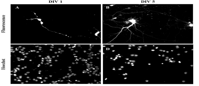
Figure 2. Gene delivery by oscillating nanomagnetic gene transfection in primary cortical neurons. Images of pmaxGFP plasmid expressed in primary neurons using fluorescence microscopy and its corresponding Hoechst 33,342 stained counterpart of transfected DIV 1 (A,C) and DIV 5 (B,D) mature neurons were taken 48 h post transfection.

If you have data to share email it to me, pshuster@neuromics.com and we'll email you a 25 USD gift card. Thank you. Pete Shuster, CEO & Owner.

Saturday, January 28, 2017

mCherry and GFP

Excellent Markers

These markers are stout fluorescent tracers for transfection and transgenic experiments. Here is data showing our mCherry Mouse Monoclonal Antibody Staining transfected HeLa Cells.

Image: Blot of HEK293 cells transfected with pFin-EF1-mCherry vector, courtesy of the Semple-Rowland lab at the University of Florida. There is a strong clean band at about 30 kDa. HEK293 cells which were not transfected with this vector show no protein bands.

As with all our products, if you are not 100% satisfied, we refund your purchase with no questions asked.

Saturday, January 21, 2017

Leptin and Remyelination

Leptin Promotes Proliferation of OPCs

Demyelination occurs in many diseases of the Central Nervous System (CNS) including Multiple Sclerosis and Parkinson's Disease. Here researchers show that Leptin plays a role in the proliferation of Oligodendrocyte Precursor Cells (OPCs): These cells are critical for keeping the myelin sheath on Neurons of the CNS healthy and happy.  Ken Matoba, Rieko Muramatsu & Toshihide Yamashita. Leptin sustains spontaneous remyelination in the adult central nervous system. Scientific Reports 7, Article number: 40397 (2017) doi:10.1038/srep40397.

Our LepRB Antibody is used in this study to stain OPCs.


Figures: (a) Representative image of cultured OPCs stained with antibodies against LepRb (green) and PDGFRα (red). Scale bar: 25 μm. (b) Relative BrdU incorporation into the OPC obtained from the brain (left graph) and spinal cord (right graph). Cells were treated with recombinant leptin for 48 h (n = 4). (Left graph) P = 0.005993 (control vs 10 ng/mL), 0.045616 (control vs 100 ng/mL), (Right graph) P = 0.004456 (control vs 10 ng/mL), 0.017859 (control vs 100 ng/mL). (c) Relative BrdU incorporation into the OPC after leptin stimulation (10 ng/ml) with U0126 (20 μM), a MEK inhibitor (n = 4 for brain OPCs, n = 3 for spinal cord OPCs). (Left graph) P = 0.019753 (control vs leptin), 0.039433 (leptin vs leptin + U0126), (Right graph) P = 0.045545 (control vs leptin), 0.04486 (leptin vs leptin + U0126). (d) Representative images of western blotting (upper panels) and quantitative analysis of ERK phosphorylation (lower graph) are shown. OPCs were treated with leptin (10 ng/ml) under indicated periods (n = 3). P = 0.006352 (2 min), 0.016571 (5 min), 0.017675 (10 min), 0.024100 (15 min), 0.081342 (30 min).
We are in the process of looking for Labs to sponsor Neuromics' to isolate and purify adult human OPCs in return for receiving 2,000,000 cells. Stay tuned.

Friday, January 13, 2017

Rising FBS Prices Raising Your Costs?

Give us a shot

Fetal Bovine Serum (FBS) is the most highly used growth factor in cell culture due to its high level of nutrients. Our FBS is produced to the highest standards in order to reduce risk and product variability and to ensure product safety and performance. Our FBS is 9CFR-tested, meeting FDA and USDA requirements. Fetal bovine serum products can also be tested to meet EMEA requirements.

Note: We test each lot of FBS on our primary human cell cultures enabling us to choose lots yielding the best results...Testimonial: "We have used the FBS from Neuromics in feeding media for primary mouse astrocytes as well as for some cell lines. Your product is good and we plan to continue using it.” - Svetlana Vidensky, MS, Senior Research Specialist in Dr. Jeffrey Rothstein lab, Department of Neurology, Johns Hopkins University


100 ml trial size is available for only $149. Contact me at 612-801-1007 or pshuster@neuromics.com to order the trial size. We also offer 100% money back guarantee should we not meet your expectations. Pete Shuster-CEO and Owner Neuromics

Monday, January 02, 2017

3-D BBB Penetration Assays

Making 2017 the Year of Tissue-Like, 3-D Models

There is growing interest in our 3-D BBB Model for testing penetration of compounds, small molecules, peptides and oligos through the BBB into our tissue like brain side.


 I would like to present related data:
We plan on adding a pancreatic model in January so stay tuned.