Saturday, November 28, 2015

Neuron Astrocyte Glial Markers

Potent and Frequently Published!

We have a stout catalog of Neuron-Astrocyte and Glial Markers.

They are widely used and frequently published. Here're some recent examples:
Mouse Monoclonal GFAP: Chelsea M. Larabee, Constantin Georgescu, Jonathan D. Wren and Scott M. Plafke. Expression profiling of the ubiquitin conjugating enzyme UbcM2 in murine brain reveals modest age-dependent decreases in specific neurons. BMC Neuroscience201516:76 DOI: 10.1186/s12868-015-0194-y© Larabee et al. 2015.

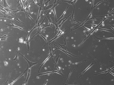
Image: Mixed neuron-glial cultures stained with Mouse Monoclonal GFAP, and Chicken Polyclonal Neurofilament-NF-L (green). The GFAP antibody stains the network of astrocytes in these cultures, while the NF-L antibody stains neurons and their processes. The blue channel shows the localization of DNA. This antibody also works on formalin fixed paraffin embedded brain tissues. Protocol on Datasheet.

Tuj-1: Gaoying Sun, Wenwen Liu, Zhaomin Fan, Daogong Zhang, Yuechen Han, Lei Xu1, Jieyu Qi, Shasha Zhang, Bradley T. Gao, Xiaohui Bai,Jianfeng Li,Renjie Chai, Haibo Wang. The three-dimensional culture system with matrigel and neurotrophic factors preserves the structure and function of spiral ganglion neuron in vitro.

Need more assurances? Here're some feedback highlights.

STEPHEN C. Nov 19, 2015 Staining (Tuj1) worked well on human neural progenitor cells. Product Name: Tuj 1, Mouse – (Cat# MO15013-100) http://bit.ly/1Qx4ASc Organization: UCONN
GINA D. Oct 07, 2015 Very nice antibody and ordering is very easy using the website. Product Name: proDynorphin (rat), Guinea Pig – (Cat# GP10110) http://bit.ly/Sw4RJ9 Organization: Rosalind Franklin University
CAROL Feb 05, 2015 We got antibodies and received them fast with a correct temperature. Product Name: Coronin 1A, Chicken – (Cat# CH23017) http://bit.ly/1AxduYvProduct Name: Integrin alpha-M, Chicken – (Cat# CH23021)http://bit.ly/16lMihU Organization: UCSF

We have a full money back guarantee so do not hesitate to consider these markers for your assays.

Friday, November 20, 2015

Staining Neurons and 3-D

Tuj-1 or Beta Tubulin Antibody in Action!

Our potent Neuron-Glia Markers are widely used and frequently published. We are also pleased with the many positive reviews.

In this study researchers use our Tuj-1/Beta Tubulin Antibody to stain neurons in 3-D Cultures: Gaoying Sun, Wenwen Liu, Zhaomin Fan, Daogong Zhang, Yuechen Han, Lei Xu1, Jieyu Qi, Shasha Zhang, Bradley T. Gao, Xiaohui Bai,Jianfeng Li,Renjie Chai, Haibo Wang. The three-dimensional culture system with matrigel and neurotrophic factors preserves the structure and function of spiral ganglion neuron in vitro...Tuj-1-β-tubulin (1:1000, Neuromics, USA)...

Images: Morphology of the SGNs growth cone cultured in 2D and 3D systems. Phalloidin, green; β-tubulin, red.

I will continue to post positive developments concerning use of our Neuron-Glia Markers.

Wednesday, November 11, 2015

Reconstructing 3D Living Biomaterials

Hydrogel Solutions for Tissue Reconstruction and Repair

Alphabioregen/Neuromics are pleased to announce the use of our Hydrogels for reconstructing living biomaterials for regenerative biology and medicine: Suwan N. Jayasinghe,Jensen Auguste, Chris J. Scotton. Platform Technologies for Directly Reconstructing 3D Living Biomaterials. DOI: 10.1002/adma.201503001.

In this study, researchers use our  Collagen Hydrogel, Collagen Hydrogel Soft and Collagen Hydrogel Soft+  to describe  in vivo applications using a murine model to interrogate biocompatibility and cellular behavior post-transfer.

Luc macrophages (CC or BES) were then incorporated into a fl uid mixture containing one of three alternative collagen-based hydrogels, namely, Collagen Hydrogel, Collagen Hydrogel Soft, or Collagen Hydrogel Soft+ (resulting in six possible combinations of macrophages and biopolymer).

Overview of Results


Figure: Bioluminescent imaging of implanted macrophage–biopolymer mixes. IC-21-Luc macrophages (CC or BES) were mixed with biopolymer (Hydrogel, Soft, or Soft+) and subcutaneously injected into the dorsal fl anks of C57Bl/6 mice (three mice per hydrogel, with CC on the left fl ank and BES on the right). Following intraperitoneal injection of delta-luciferin, macrophage bioluminescence was detected using an IVIS Lumina II imaging system. A representative image from day 1 post-implantation (and 25 min post luciferin injection) is shown, indicating the peak detectable radiance (photons s −1 cm −2) in identically sized regions of interest. The CE results were very similar to the BES implants.


Figure:  Histological analysis of the macrophage–biopolymer implantation site (MSB staining). At day 4 post-implantation, skin was harvested from each dorsal fl ank, and processed for histology. Where possible, serial sections directly adjacent to those shown in Figure 4 were stained with a modifi ed trichrome stain. Mature fi brillar collagen within the dermis appeared dark blue, while the collagen within the hydrogel was generally lighter blue in appearance, indicating a less highly cross-linked or fi brillar form of collagen. Scale bar: 200 µm.

These Collagen Hydrogels are proving easy to use and effective in the hands of our customers. If you are looking for solutions for tissue reconstruction/repair or 3-D in vivo like cell based assays, do not hesitate to contact me @ direct phone: 612-801-1007 or pshuster@neuromics.com. Thank you, Pete Shuster, CEO and Owner, Neuromics.

Saturday, November 07, 2015

Human alpha-Sensory Neurons

Put Them to Work!

We continue to add unique primary Human Neurons to our offerings.

We have discounted our new alpha-Sensory Neurons to make it easier to buy and try-only 850 USD/1,000,000 cells.


Just thaw and culture. Simply culture on ECM- Laminin or Fibronectin coatings with our alpha Motor Neuron Maintaining Media. Questions? Please contact me directly 612-801-1007 or pshuster@neuromics.com. Pete Shuster, CEO and Owner, Neuromics.