Monday, October 27, 2014

100% Serum-Xeno Free Media for Expanding hMSCs

Works as well as our Serum Containing Media

 Selection of Growth and Differentaition media for Mesenechymal Stem Cell Assays is important for the ultimate performance of your cell based assays. The better the media the better the cutures & the lower your costs.


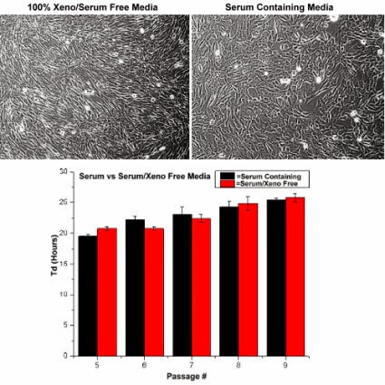
Images: Human mesenchymal stem cells (Catalog no. SC00A1) were plated at 5,000/cm² in a Falcon BD TC-coated T-25 flasks and maintained in serum containing media (Cat. No. SC00B1) and serum-free/xeno-free media (Cat. No. SC00B3) in reduced O2 environment (1% O2, 5% CO2, 90% N2) at 37°C in a humidified chamber. When cells became 80-90% confluent, they were subcultured, counted on a Beckermen Z2 particle counter (range 10-30uM), and passaged. Doubling time of 20-25hrs were calculated in each pass using ln(2*dT)/ln(Cf/Ci), where dT is the time, in hours, from inoculation to detachment; Ci is the initial number of cells plated and Cf is the final number of cells recovered from subculture. There were no apparent differences in doubling time with TC-coated flasks and Laminin/Fibronectin treated flasks.

Testimonial: “We tested the effects of MSCGroTM defined medium using several different lots of human adult primary stem cells and found that MSCGro supports a more robust proliferation rate than normal undefined media. This provided shorter doubling times and increased cellular yield, and maintained the cells in an undifferentiated state. We also found that MSCGro medium is stable under normal laboratory conditions for an extended time period compared to other defined media.” Ben Buehrer, VP and CSO, Zen-Bio

Tuesday, October 21, 2014

Neurite Outgrowth Assays

Cells, Media and Markers

I have previously posted use of our Neurons in Live Content Assays for the study of Neurite Outgrowth: Neurons-Live Content Assays. These assays are critical for the study of repair and regeneration.

A recent publication featured several of our Neuron MarkersSerena Quarta, Bastian E. Baeumer, Nadja Scherbakov1, Manfred Andratsch, Stefan Rose-John, Georg Dechant3, Christine E. Bandtlow, and Michaela Kress: Peripheral Nerve Regeneration and NGF-Dependent Neurite Outgrowth of Adult Sensory Neurons Converge on STAT3 Phosphorylation Downstream of Neuropoietic Cytokine Receptor gp130. The Journal of Neuroscience, 24 September 2014, 34(39): 13222-13233; doi: 10.1523/JNEUROSCI.1209-13.2014.
Live labeling of neuron cultures: After 20 or 48 h, neurons were live-labeled with α-gp130 antibody diluted in cold TNB medium for 30 min on ice. After washing, neurons were incubated with the secondary antibody diluted in cold TNB medium for 30 min and washed with PBS. Cells were fixed either with 4% PFA for 20 min at room temperature (RT) or with methanol at −20°C for 2 min. After permeabilization with 0.01% TX-100 (Pierce) unspecific binding was blocked for 30 min with 10% normal goat serum (Sigma-Aldrich) in PBS. Cells were incubated with the first antibody for 1 h, washed three times for 10 min with PBS and incubated with the appropriate secondary antibody for 30 min, counterstained with 4′, 6-diamidino-2-phenylindole (1:10,000; Sigma-Aldrich) and embedded in Mowiol (Calbiochem). As primary antibodies, α-gp130 (1:50; Neuromics), α-β-III-tubulin clone TuJ-1 (1:1000; R&D Systems), and α-neurofilament-H (α-NF-H; 1:200; Neuromics) were used. Secondary antibodies used were α-goat AlexaFluor 594 (1:1000; Invitrogen), chicken α-mouse AlexaFluor 594 or donkey α-mouse AlexaFluor 488 (1:1000; Invitrogen), and goat α-chicken AlexaFluor 568 (1:10,000; Invitrogen) for fluorescence microcopy.

Images: Reduced density of TuJ-1+ nerve endings in the epidermis in SNS-gp130−/− mice after lesion. A, Representative cross sections of hindpaw glabrous skin of naive and 12 dpl gp130fl/fl and SNS-gp130−/− mice stained with the pan neuronal marker TuJ-1. The dotted line indicates the border between dermis and epidermis. Scale bar, 40 μm. B, Quantification of the total number of TuJ-1+ fibers (NE) per 1000 μm2 of epidermal area shows a significant decrease in density in SNS-gp130−/− mice after lesion compared with control animals (*p < 0.05; n = 4 for each group). Data are presented as mean ± SEM and analyzed by Mann–Whitney U test. C, 3D reconstruction of the deeper layer of the dermis shows fewer nerve bundles in SNS-gp130−/− dermis compared with controls. D, No NF-H+ proprioceptive fibers were detectable in the epidermis of gp130fl/fl animals at 12 dpl. Scale bar, 40 μm.

If you want to learn more about our Neuron-Glial-Astrocyte based assay solutions do not hesitate to contact me (612-801-1007) or pshuster@neuromics.com. Pete Shuster, Owner and CEO, Neuromics.

Sunday, October 05, 2014

The Growing Value of Cancer Associated Fibroblasts or CAFS

Balancing Supply and Demand

There is reciprocal communication between Cancer Associated Fibroblasts (CAFs) and tumor cells. They are now recognized as playing a key role in promoting the malignant process and blocking immune surveillance  They are becoming increasing important in Cancer Drug Discovery Programs.

Their growing value is making it harder for us to source resected tumors. We have been receiving advanced orders from clients who need to insure they have a stock. We have ordered tumors for the below CAFs and will be shipping to those who have place advance orders by the end of November.

NameCatalog #TypeSpeciesApplicationsSizePrice
Human Colorectal Tumor CAFs KitCAF05Human Primary CAFsHCell Assays1,000,000 cells$799
Human Endometrial Adenocarcinoma CAFsCAF01Human Primary CAFsHCell Assays1,000,000 cells$799
Human Ovarian Serous Tumor CAFsCAF02Human Primary CAFsHCell Assays1,000,000 cells$799
Human Lung Adenocarcinoma CAFsCAF07AHuman Primary CAFsHCell Assays1,000,000 cells$799
Human Lung Squamous Cell Tumor CAFsCAF07SCHuman Primary CAFsHCell Assays1,000,000 cells$799
Human Immortalized Pancreatic CAF-Stellate CellsCAF08Human Primary CAFsHCell Assays500,000 Immortized Cells$4,335
Human Pancreatic Tumor Cells KitPXPC3A1Cells+Media KitHCell Assays1,000,000 cells$499
Conditioned Media for Ovarian Serous Tumor CAFSCAF04Cell Growth MediaHCell Assays5 ml$250
Conditioned Media from Endometrial CAFSCAF03Cell Growth MediaHCell Assays5 ml$250
VitroPlus III, low serum, completePC00B1-100Cell Growth MediaHCell Assays100 ml
500 ml
$65
$165
Image: Endometrial CAFs courtesy of Tiana Tonrey, Vitro Biopharma

Should have questions about availability and ship dates for any of these please call (612-801-1007) or e-mail: pshuster@neuromics.com. Thank you.
Pete Shuster, CEO, Neuromics