Friday, September 27, 2013

From Zero to 3-D Cell Based Assays in 15 Minutes

Collagel Hydrogels are Designed to Match Your Cell Types.

We are pleased to add Collagel Hydrogels to our 3-D Cell Based Assay Solutions.

The are 3 different gel types that mimic the different in vivo extracellular environment that your cells experience. You will be able to get really nice adipogenesis with MSCs using our CollaGel Hydrogel Standard. CollaGel Hydrogel Soft is an ideal matrix for growing fibroblast; primary hepatocyte cultures and great for growing smooth muscle cells. CollaGel Hydrogel Soft+ is an ideal matrix for growing nervous cells, veins cells and Hydroxyapatite crystals.

Image: Primary hepatocyte culture in 3D model using our CollaGel Hydrogel. 

Using these gels, you can now form small tube-like structures in combination with Human Umbilical Vein Endothelial Cells (HUVEC) in a 3D model. Our CollaGel Hydrogel can also be used for 3D printing without worrying about the needles in your machine been broken due to the fast gelling process. Collagel Hyrogels can be used for: Stem Cell Behavior Studies Fibrosis Studies,  Cosmetic Toxicology, Hepatocyte Assays, Neuronal Branching, Wound Healing Assays, Cell Invasion Assays, Migration Assays, Cancer Cell Phenotyping and  Bioprinting.

I will be posting Collagel Hydrogel related data and images provided by our customers.

Thursday, September 19, 2013

New Astrocyte-Glial Markers

More Options!

We get a lot of request for markers that will more specifically stain astrocytes, glia and microglia. Let's say, for example, you want to pinpoint these cell types in a mixed neuron-glial culture. You can now do a dual label and generate these results.

Image: Neuron-glial cell mixture cultures stained with  ALDH1L1 (red) and our monoclonal antibody against GFAP (green). Blue is a DNA stain. ALDH1L1 stains astrocytes and excludes from neuron cells. ALDH1L1 stains the astrocytes cell body and processes, whereas GFAP labels the intermediate filament of the cytoskeleton in subset of astrocytes. Astrocytes are positive for both ALDH1L1 and GFAP appear yellow. ALDH1L1 also labels many astrocytes not labeled by GFAP, which appear as red. Inset:Blot of rat liver tissure homogenates blotted with ALDH1L1. The antibody binds strongly a band at ~100 kDa.

Or how about these results:
Image: Mixed neuron-glial cultures stained with Mouse Monoclonal GFAP, and Chicken Polyclonal Neurofilament-NF-L (green). The GFAP antibody stains the network of astrocytes in these cultures, while the NF-L antibody stains neurons and their processes. The blue channel shows the localization of DNA. This antibody also works on formalin fixed paraffin embedded brain tissues. Inset: Strip blot of rat spinal cord protein extract stained with GFAP. A prominent band at about 55 kDa corresponds to the major isoform of GFAP.

We will continue to post new additions to our Neuronal-Glial Markers.

Sunday, September 15, 2013

Antibody Array-ELISA Kits for Cancer Researchers

We continue to build our catalog of Antibody Arrays & ELISA Kits
for Cancer Researchers. Our RayBio® C-Series Ctyokine Arrays
enable you to analyze the expression of up to 120 Cytokines for about
the price of a single ELISA (585 USD).

 Antiboy Array 5
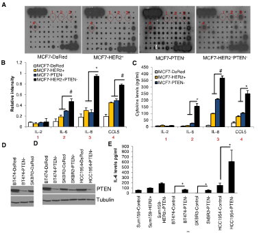
Figure 3. PTEN Downregulation in HER2-Overexpressing Cells Activates an IL6/NF-kB-Mediated Inflammatory Feedback Loop (A–E) MCF7-HER2+PTEN cells secreted 3- to 5-fold higher levels of IL6, IL8, and CCL5 compared to MCF7-HER2+ or MCF7-PTEN cells as determined by RayBio human cytokine antibody Array 5 (A). The intensity of each blot compared to control was determined by Kodak image analyzer (B) and confirmed by ELISA (C). Downregulation of PTEN in HER2-amplified breast cancer cells, BT474, SKBR3, HCC1954, and Sum159-HER2+ (D) results in increased levels of these cytokines in vitro (E). http://dx.doi.org/10.1016/j.molcel.2012.06.014

Array Capabilities:
  • Detects expression of 96 secreted proteins in a single sample, in a single day. 
  • If you can do a Western, you can use a C-Series Antibody Array. And get excellent results the first time!  
  • Sandwich ELISA pairs give C-Series arrays high sensitivity and specificity.  
  • C-Series arrays are compatible with practically ANY liquid sample. 
References have cited using membrane-based C-Series Cytokine Antibody Arrays with nearly every liquid sample type imaginable, including: cell-cultured and co cell cultured media, cell and tissue lysates, tissue/organ perfusates, serum, plasma, urine, and many other body fluids, including cyst, interstitial, synovial, blister, cerebrospinal, prostatic and amniotic fluids, as well as abscesses, broncho-alveolar lavage, sputum, saliva, tears, breath condensates, and even human milk and colostrum. Publications referencing use of our Antibody Arrays.

I will be posting more new developments here.

Monday, September 09, 2013

Stem Cell Markers

Our Stem Cell Reagents our widely used and frequently published. We are proud of the positive feedback on our Stem Cell Markers.
Image: Tuj-1 staining of Neuron-specific class III β-tubulin in differentiated human neural progenitor cells. Cells were stained using goat anti-mouse Alexa Fluor 488 (green) secondary antibody (Molecular Probe, A-11001) and counterstained with PI (red).
Image: Neural progenitors were labeled with anti-rat Nestin polyclonal antibody (Cat#:GT15114) and stained with conjugated donkey anti-goat secondary anti-body (green). Differentiated neurons were labeled with neuron-specific mouse anti-β-III tubulin/ Tuj1-(Cat#MO15013 monoclonal antibody (red). Nuclei were stained with DAPI (blue).

Tuj-1: Customer Publications and Data.

I will continue to post updates.