Monday, April 29, 2013

Neuron-Glial Cultures-Setting a Higher Bar!

Improved Methods for Long Term, High Denisty Cultures
 
Dr. Randen Patterson and his team at UC Davis have developed new culturing techniques using our e18 Rat Primary Hippocampal Neurons. They have developed a protocol that allows for culturing of E18 hippocampal neurons at high densities for more than 120 days. These cultured hippocampal neurons are (i) well differentiated with high numbers of synapses, (ii) anchored securely to their substrate, (iii) have high levels of functional connectivity, and (iv) form dense multi-layered cellular networks. We propose that our culture methodology is likely to be effective for multiple neuronal subtypes–particularly those that can be grown in Neurobasal/B27 media. This methodology presents new avenues for long-term functional studies in neurons. This is good news indeed: Todd GK, Boosalis CA, Burzycki AA, Steinman MQ, Hester LD, et al. (2013) Towards Neuronal Organoids: A Method for Long-Term Culturing of High-Density Hippocampal Neurons. PLoS ONE 8(4): e58996. doi:10.1371/journal.pone.0058996.
 
Protocol Highlights: 
Substrate Preparation
1. On the day of plating, prepare 25 mm coverslips by removing them from 70% ethanol storage solution and propping them up at an angle in each well of a 6-well culture plate to allow drying. [No more than 5 plates (30 coverslips) should be dried simultaneously for 15–25 minutes in culture hood to avoid over-drying.]
2. Once dry, shake slips down flat into their respective wells and coat with 1 mL 0.1% poly-D-lysine, taking care to form a liquid meniscus on each slip. Carefully transfer coverslips into incubator, taking care to preserve meniscus.
3. Incubate for 1 hr at 37°C. [Keeping poly-D-lysine meniscus on top of coverslip is important; this serves to avoid poly-D-lysine coating under coverslip surface that may lead to problematic flotation of coverslip.] After incubation, remove poly-D-lysine and rinse each coverslip three times with 2 mL sterile deionized water. Take care to ensure coverslips do not completely dry at any point during the rinse. After the third and final rinse, leave coverslips in 2 mL sterile deionized water for at least 1 hr. Remove water just before plating, again, make sure to avoid over-drying. [This critical step requires attention. Take care to aspirate off all sterile water remaining from the final rinse, but also use caution as not to over-dry the coverslips. Ultimately, the coverslip must be mostly dry as to allow for the meniscus formation during plating (Fig. 1, Step 12), whereas over-drying can result in the neurons peeling off the glass coverslips days to weeks after plating.]
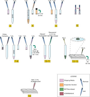
Fig 1: Neuron-Glial Culturing Steps
Preparation of Isolated Neurons (Numbers in Fig. 1 correspond to numbers below).
1. Store tissue at 4°C until ready to use. If dissecting your own cultures, upon isolation of the tissue, store in an appropriate storage media.
2. When ready to plate, make 2 mL of enzymatic solution without B27. In our case, we used Hibernate E-Ca, containing 4 mg (2 mg/mL) of papain. If making your own solution, use a commercially available papain dissociation kit. Make sure to sterile filter solution with 0.2 micron filter after adding papain if source of enzyme is not sterile.
3. Remove the storage media from the dissected tissue and transfer into sterile 15 mL screw-cap tube; be careful not to disturb or remove tissue from original tube. Save the storage media, do not discard.
4. Add 2 mL of media made in Step 2 to tissue (in our case, Hibernate E-Ca containing 2 mg/mL of papain). Incubate for 35 min at 37°C. [Be sure to add Hibernate E-Ca containing papain slowly as to avoid disturbing tissue.]
5. Remove enzymatic solution from tissue, again, take care not to disturb or remove tissue. Add back 1 mL of storage media saved in 15 mL tube.
6. Using a 1 mL pipettor with a sterile plastic pipette tip (tissue can adhere to glass pipettes), aspirate the tissue with the medium into the pipette and immediately dispense contents back into same container. Take care not to create bubbles. [This is another critical step that requires attention. Take care to make sure pipette tip remains in a stable position (as shown in Fig. 1, Step 6). Maintain slow, steady speed when both drawing in and re-dispensing media containing tissue.]
7. Repeat this trituration step 10–12 times or until most all the tissue is dissociated and the cells are dispersed. [Under close examination cell dispersion is highly visible. Stop pipetting immediately upon cell dispersion.]
8. Slowly transfer contents of the tissue tube into a new sterile 15 mL screw-cap tube.
9. Use the remainder of storage medium saved in Step 3 and rinse the interior of the tissue tube before adding it to the sterile 15 mL screw cap tube containing dispersed cells from step 7. [This step helps ensure minimal wastage, as any remaining cells should be saved with this extra rinse.]
10. Spin dispersed cells at 1,100 rpm (200Xg) for 1 min.
11. Discard the supernatant while being careful not to remove any of the cells from cell pellet.
12. Flick tube a few times to loosen the cell pellet. Re-suspend pellet in 2.4 mL of pre-warmed B27/Neurobasal/0.5 mM glutamine medium. Re-suspend by gently pipetting up and down. For E18 Hippocampus, medium includes 25 µM glutamate.
13. Plate cells within a meniscus (approx. 10 mm diameter) at a minimum of 40 µL per 25 mm coverslip. Take care not to disturb meniscus. [Periodically pipette up and down throughout plating process (no more than once every plate per 6 coverslips) to help maintain equal cell density. Again, plating with meniscus formation is critical.]
14. Incubate plated cells at 37°C with 5% CO2 and/or 9% or 20% oxygen for 1 hr.
15. Add 1.5 mL per well of pre-warmed 1:1 ACM/NbActiv4. [Slow and steady media addition rate and proper pipette position are necessary for successful plating density consistency. Position pipette tip at 45° angle along middle of 6-well interior sidewalls, dispense 1.5 mL as slowly and steadily as possible (see Steps 1–14).]
16. Incubate cells at 37°C with 5% CO2 and/or 9% or 20% oxygen.
17. Add Cytosine β-D-arabinofuranoside (Ara-C) to a final concentration of 5 µM, 5–6 days after plating to curb glial proliferation. [Remove 1/3 of media from each well and replace with equal volume containing final concentration of Ara-C]
18. After 4 days or longer, neurons are well differentiated. If further culture is desired, change 1/3 of medium with fresh, pre-warmed 1:1 ACM/NbActiv4 every 7–8 days.
Images: 40X Confocal Images of 30 DIV Hippocampal Cultures. Immunofluorescence detection of MAP-2 (green) and GFAP (red) in 30 DIV (A–C) cultured E18 hippocampal cells using a 40X objective. These images (A and B) clearly depict the intimate physical contact between glia processes and dendritic arbors. Under closer examination (CI and CII), it is clear that the dendrites have grown both bellow (blue arrows) and above (white arrows) glial processes, forming a highly interconnected three-dimensional network by 30 DIV. doi:10.1371/journal.pone.0058996.g004.
All Primary Neuron Assay Customer Publications
 
Related Content: If you have any questions on optimizing your cell cultures, do not hesitate to contact me @ pshuster@neuromics.com or 612-801-1007

Monday, April 22, 2013

Small Molecules-Peptides for Neuroscience Research

Agonists, Antagonists, Inhibitors and Ligands for Studying Neuromodulation

Our friends at R and D Systems/Tocris Bioscience have made available to us select Small Molecules/Peptides. Our focus will be on providing agonists, antagonists, inhibitors and ligands that complement our Neuroscience and Pain Research products and expertise.


We will be adding about 10 new molecules/peptides per month. Here's a sampling our our most recent additions:
NameTypeBioactivity
(±)-trans-ACPDAgonistPotent NMDA agonist. Also group II mGluR agonist
(S)-(-)-5-FluorowillardiineAgonistVery potent AMPA agonist
(S)-4-CarboxyphenylglycineAntagonistCompetitive group I mGluR antagonist/weak group II agonist
2-APBModulatorTRP channel modulator. Also IP3 receptor antagonist
2-Methylthioadenosine triphosphate tetrasodium saltAgonistP2 purinergic agonist
AM 404ModulatorVanilloid receptor agonist. Also anandamide transport inhibitor
BRL 52537 hydrochlorideLigandPotent and selective κ opioid receptor agonist
CNQXAntagonistPotent AMPA/kainate antagonist
Clocinnamox mesylateAntagonistIrreversible μ-opioid receptor antagonist
Endomorphin-1AgonistPotent and selective μ opioid receptor agonist
Endomorphin-2AgonistPotent and selective μ opioid receptor agonist
FITAgonistIrreversible δ opioid receptor agonist
GBR 13069 dihydrochlorideAgonistPotent dopamine uptake inhibitor
L-NIO dihydrochlorideInhibitorPotent eNOS inhibitor
L-Quisqualic acidAgonistVery potent group I mGluR agonist
N-Benzylnaltrindole hydrochlorideAgonistOpioid receptor selective non-peptide antagonist
NociceptinInhibitorEndogenous NOP agonist
O-Phospho-L-serineAntagonistGroup III mGluR agonist; enhances neuronal differentiation
Ro 51AntagonistPotent P2X3, P2X2/3 antagonist
cis-ACPDAgonistPotent NMDA agonist. Also group II mGluR agonist
We will be posting new additions and related data and publications

Sunday, April 14, 2013

P2X3 Receptors and Migraine

P2X3 Receptors of Trigeminal Sensory Neurons and Familial Hemiplegic Migraine Type 1 (FHM-1).

Our P2X Receptor Markers continue to be used in interesting and novel ways. Here researchers use our P2X3 Receptor Antibody to study expression using primary rat ganlia cultures: Swathi K. Hullugundi,Michel D. Ferrari, Arn M. J. M. van den Maagdenberg, Andrea Nistri. Andrea Nistri. The Mechanism of Functional Up-Regulation of P2X3 Receptors of Trigeminal Sensory Neurons in a Genetic Mouse Model of Familial Hemiplegic Migraine Type 1 (FHM-1). PLoS ONE 8(4): e60677. doi:10.1371/journal.pone.0060677

Abstract: A knock-in (KI) mouse model of FHM-1 expressing the R192Q missense mutation of the Cacna1a gene coding for the α1 subunit of CaV2.1 channels shows, at the level of the trigeminal ganglion, selective functional up-regulation of ATP -gated P2X3 receptors of sensory neurons that convey nociceptive signals to the brainstem. Why P2X3 receptors are constitutively more responsive, however, remains unclear as their membrane expression and TRPV1 nociceptor activity are the same as in wildtype (WT) neurons. Using primary cultures of WT or KI trigeminal ganglia, we investigated whether soluble compounds that may contribute to initiating (or maintaining) migraine attacks, such as TNFα, CGRP, and BDNF, might be responsible for increasing P2X3 receptor responses. Exogenous application of TNFα potentiated P2X3 receptor-mediated currents of WT but not of KI neurons, most of which expressed both the P2X3 receptor and the TNFα receptor TNFR2. However, sustained TNFα neutralization failed to change WT or KI P2X3 receptor currents. This suggests that endogenous TNFα does not regulate P2X3 receptor responses. Nonetheless, on cultures made from both genotypes, exogenous TNFα enhanced TRPV1 receptor-mediated currents expressed by a few neurons, suggesting transient amplification of TRPV1 nociceptor responses. CGRP increased P2X3 receptor currents only in WT cultures, although prolonged CGRP receptor antagonism or BDNF neutralization reduced KI currents to WT levels. Our data suggest that, in KI trigeminal ganglion cultures, constitutive up-regulation of P2X3 receptors probably is already maximal and is apparently contributed by basal CGRP and BDNF levels, thereby rendering these neurons more responsive to extracellular ATP.

Images: Examples of TNFR2 and P2X3 co-exexpression in (wildtype) WT and R192Q (knockin) KI neurons. Left panel shows P2X3 expression (green), and right panel shows TNFR2 staining (red). B, Histograms quantifying % of cells co-expressing TNFR2 and P2X3: both WT and KI cultures show similar TNFR2 and P2X3 co-expression. N = 3 independent experiments (6 mice). C, Representative traces of currents induced by application of α,β-meATP (10 µM, 2 s) to WT or R192Q KI neurons in control conditions or after 4 h TNFα application. D, Histograms show average peak amplitudes of P2X3 receptor-mediated currents: WT control (open bar), n = 30; WT TNFα (stippled bar), n = 38; KI control (grey bar), n = 34; KI TNFα (stippled gray bar), n = 34; ** = p<0 .006="" i="" nbsp="" p="">doi:10.1371/journal.pone.0060677.g001.

Understanding the interplay between TNFR2 and P2X3 could lead to a better understanding of the root causes of migraines. This could open up yet more potential drug targets for this insidious condition.

Check out these related reagent categories:
All Purinergic Receptor Antibodies
Pain and Inflammation Research Antibodies 
Neurotransmission Research Antibodies
Primary Neurons and Astrocytes-Primary human, rat and mouse neurons and astrocytes

Thursday, April 04, 2013

Science Behind Elecroacupuncture for Treating Shingles

Postherpetic neuralgia (PHN) or shingles, caused by caused by herpes zoster, causes nerve damage in the skin and results in abnormal electrical signals to the brain,  and may persist or recur for months, years or for life.

Electroacupuncture (EA) is effective in relieving pain in patients with PHN. Researchers in this study determined the beneficial effect of EA and the potential mechanisms in a rat model of PHN. They use our TRPV1 antibody to track expression in the Dorsal Root Ganglia (DRG) and Dorsal Horn (DH): Cai-hua Wu, Zheng-tao Lv, Yin Zhao, Yan Gao, Jia-qing Li, Fang Gao, Xian-fang Meng, Bo Tian, Jing Shi, Hui-lin Pan and Man Li. Electroacupuncture improves thermal and mechanical sensitivities in a rat model of postherpetic neuralgia. Molecular Pain 2013, 9:18 doi:10.1186/1744-8069-9-18


Conclusions: EA treatment improves thermal perception by recovering TRPV1-positive sensory neurons
and nerve terminals damaged by RTX. EA Also reduces RTX-induced tac tile allodynia by attenuating the damage of myelinated afferent nerves and their abnormal sprouting into the spinal lamina II. Our study provides new information about the mechanisms of the therapeutic actions of EA in the treatment of PHN.
Figure 2:  Effect of EA on RTX-induced deletion of TRPV1-immunoreactive neuron s in the DRG. A, Representative images showing TRPV1-immunoreactive neurons in the lumbar DRG of vehicle ( a ) , RTX ( b ), RTX plus 2 Hz EA ( c ), RTX plus 15 Hz EA ( d ), RTX plus 100 Hz EA ( e ), and RTX plus sham EA ( f ) groups. Scale bar, 50 μ m. B, Summary data show the number of TRPV1 immunoreactive neurons in different groups. Data are expresse d as means ± SEM (n = 6 rats in each group). *P < 0.05, compared with the vehicle group; # P< 0.05, compared with the sham EA group.
Figure 3: Effect of EA on RTX-induced deletion of TRPV1 immunoreactive central terminals in the spinal dorsal horn. A, Representative images showing TRPV1 immunoreactive central terminals of afferent fibers in the spinal dorsal horn of vehicle (a) ,RTX (b), RTX plus 2 Hz EA (c), RTX plus 15 Hz EA (d), RTX plus 100 Hz EA (e), and RTX plus sham EA (f) groups. Scale bar, 50 μm. B, Summary data show the area of TRPV1 immunoreactive central terminals in different groups. Data are expressed as means ± SEM (n= 6 rats in each group). *P < 0.05, compared with the vehicle group; # P < 0.05, compared with the sham EA group. 
I am always interested in how our Pain and Inflammation Research Markers are used to help understand the science behind pain therapies and also discovery of new therapies. There are multiple postings on these subjects with many more to come.